. 2013 Feb;9(2):e1003165.
doi: 10.1371/journal.ppat.1003165. Epub 2013 Feb 7.

Structural and functional analysis of the CspB protease required for Clostridium spore germination

Affiliations

Structural and functional analysis of the CspB protease required for Clostridium spore germination

Chloe M Adams et al. PLoS Pathog. 2013 Feb.

Abstract

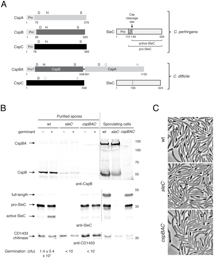
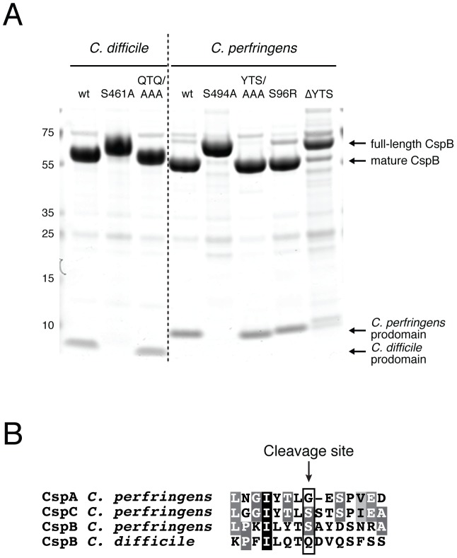
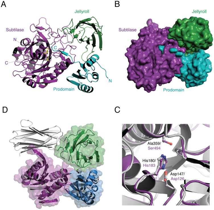
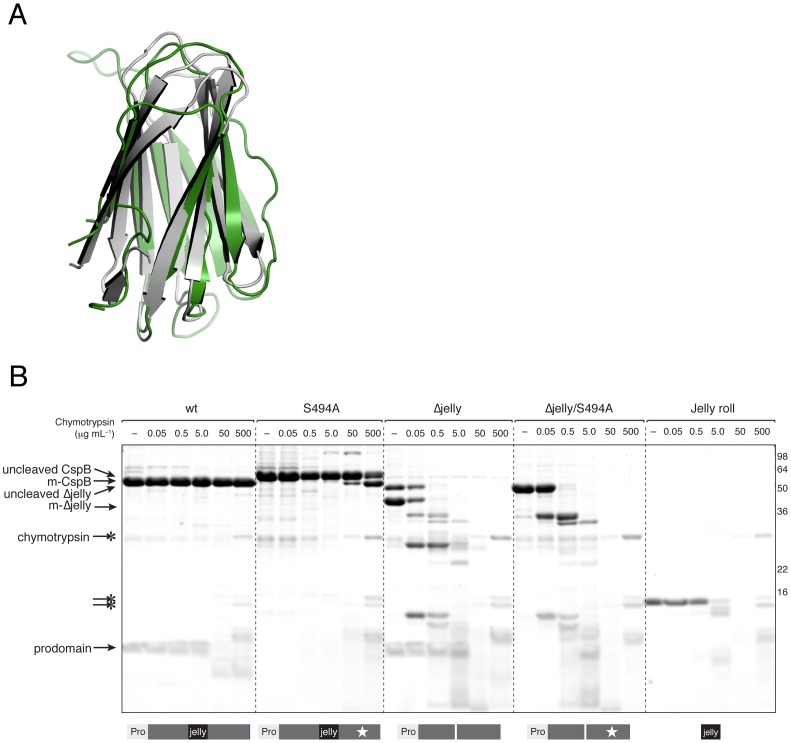
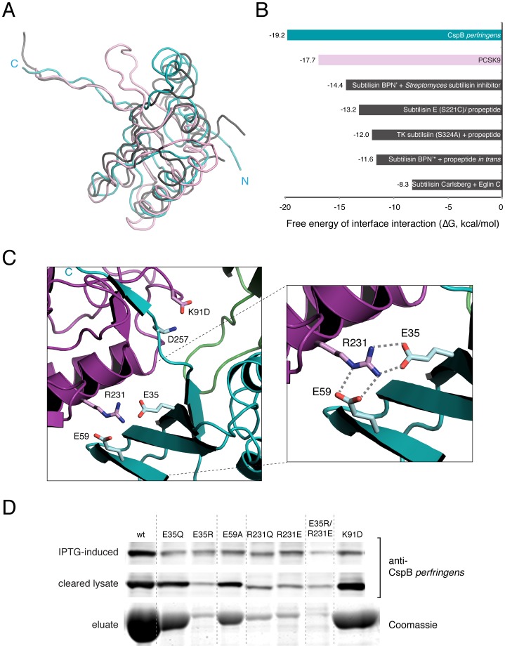
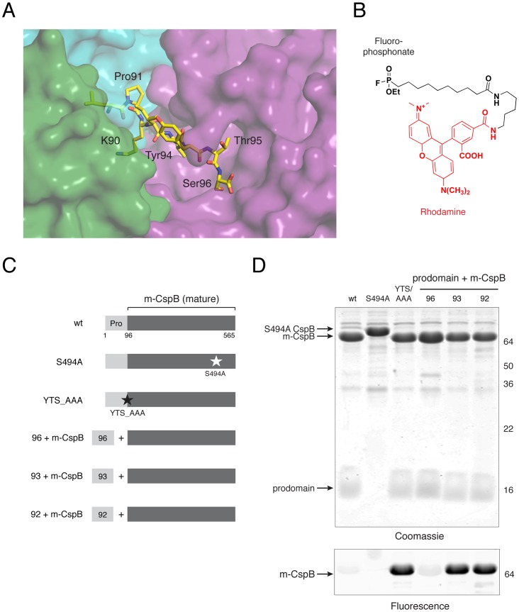
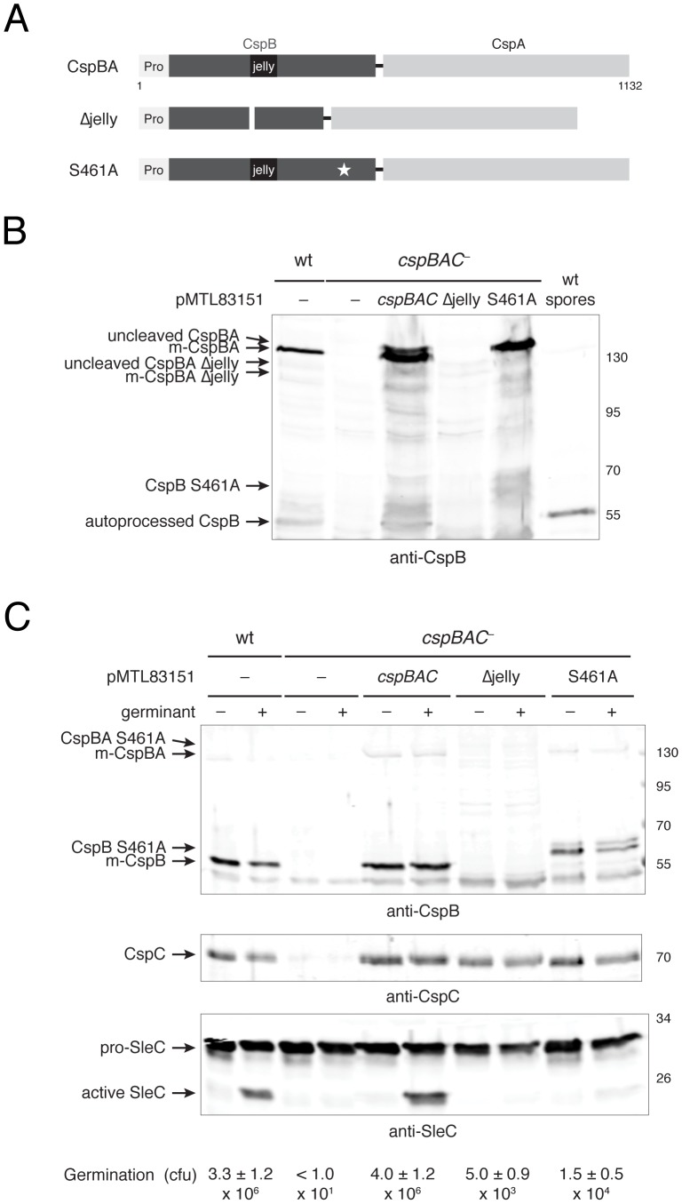
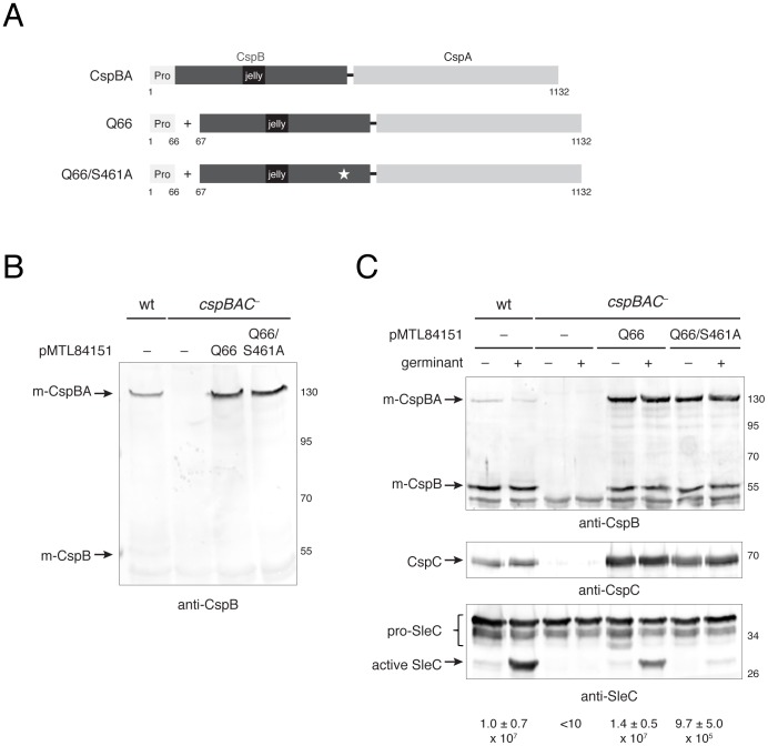
Spores are the major transmissive form of the nosocomial pathogen Clostridium difficile, a leading cause of healthcare-associated diarrhea worldwide. Successful transmission of C. difficile requires that its hardy, resistant spores germinate into vegetative cells in the gastrointestinal tract. A critical step during this process is the degradation of the spore cortex, a thick layer of peptidoglycan surrounding the spore core. In Clostridium sp., cortex degradation depends on the proteolytic activation of the cortex hydrolase, SleC. Previous studies have implicated Csps as being necessary for SleC cleavage during germination; however, their mechanism of action has remained poorly characterized. In this study, we demonstrate that CspB is a subtilisin-like serine protease whose activity is essential for efficient SleC cleavage and C. difficile spore germination. By solving the first crystal structure of a Csp family member, CspB, to 1.6 Å, we identify key structural domains within CspB. In contrast with all previously solved structures of prokaryotic subtilases, the CspB prodomain remains tightly bound to the wildtype subtilase domain and sterically occludes a catalytically competent active site. The structure, combined with biochemical and genetic analyses, reveals that Csp proteases contain a unique jellyroll domain insertion critical for stabilizing the protease in vitro and in C. difficile. Collectively, our study provides the first molecular insight into CspB activity and function. These studies may inform the development of inhibitors that can prevent clostridial spore germination and thus disease transmission.


Conflict of interest statement

The authors have declared that no competing interests exist.

Figures

Figure 1
Figure 1. The CspBA fusion protein undergoes processing during sporulation.
(a) Schematic of Csps and SleC in C. perfringens and C. difficile. Intact catalytic residues are black, while catalytic mutations are grey. The prodomain of C. perfringens Csps are shown in light grey, with their lengths indicated. The predicted prodomain of CspBA is also indicated. SleC is outlined in black, with the prepeptide (Pre), propeptide (Pro), and Csp cleavage site indicated for C. perfringens SleC , (b) Western blot analysis of sporulating C. difficile and purified spores. Purified spores of the indicated strain were either untreated (−) or exposed to 0.2% w/v sodium taurocholate (+, germinant) for 15 min at 37°C and analyzed by Western blotting and for germination efficiency via colony forming unit (cfu) determination. The processing products of CspB and SleC are indicated. CD1433 was previously shown to be a component of C. difficile spores and is used as a loading control ; the anti-CD1433 antiserum primarily recognizes the chitinase domain of CD1433. CspB levels were 3.5-fold lower in sleC spores relative to wildtype spores, despite containing similar amounts of CD1433. (c) Phase-contrast microscopy of sporulating C. difficile strains used in (b) showing equivalent levels of sporulation as measured by particle counting. The white triangles indicate mature phase-bright spores that have been released from the mother cell; the black triangles highlight immature forespores in the mother cell.
Figure 2
Figure 2. CspB undergoes autoprocessing in a position-dependent manner.
(a) Coomassie staining of recombinant C. perfringens and C. difficile CspB variants. 7.5 µg of each purified CspB variant was resolved by SDS-PAGE on a 4–12% Bis-Tris gel and visualized by Coomassie staining. The P3-P1 residues of the prodomain were mutated to Ala for the YTS/AAA and QTQ/AAA mutants, while the P3-P1 residues were deleted from CspB perfringens in the ΔYTS mutant. The products resulting from autoprocessing are indicated. (b) Sequence alignment of Csp prodomain cleavage sites mapped by Edman sequencing; the Csp perfringens cleavage sites were mapped in a previous study . Completely conserved identical residues are blocked in black with white text, conserved identical residues in grey with white text, and conserved similar residues in light grey.
Figure 3
Figure 3. Overall structure of CspB perfringens.
(a) Ribbon representation showing subtilase domain in purple, jellyroll domain in green, and prodomain in teal extending into the active site. Catalytic residues are shown as stick models with yellow carbons. (b) Close-up view of catalytic site. An overlay of CspB (purple) and Tk-SP (grey). The three catalytic residues are shown. Tk-SP and CspB catalytic residues are labeled in black and purple, respectively. (c) Space-filling model of CspB with same orientation and color scheme as (a). (d) Overlay of CspB (colors, same as (a)) and Tk-SP (shown in grey), showing similar overall structures with the exception of the position of the jellyroll domain. The jellyroll domains of CspB and Tk-SP are shown in green and grey, respectively. Note that only the regions with conserved secondary structure in the prodomain and subtilase domain are shown.
Figure 4
Figure 4. The jellyroll domain conformationally rigidifies CspB perfringens.
(a) Overlay of jellyroll domain of CspB perfringens (green) and Tk-SP (grey). (b) Limited proteolysis profile of CspB and its variants. 15 µM of CspB and its variants were incubated with increasing concentrations of chymotrypsin for 60 min at 37°C. Reactions were resolved by SDS-PAGE and visualized by Coomassie staining. Schematic of CspB variants is shown below the Coomassie stained gel. “Pro” refers to the prodomain; black rectangle demarcates the jellyroll domain; thin white rectangle represents the jellyroll deletion; and white star denotes the S494A mutation. m-CspB refers to mature CspB, which is produced after autoprocessing.
Figure 5
Figure 5. Dual salt bridges are required for prodomain intramolecular chaperone activity.
(a) Overlay of prodomains from CspB perfringens (teal), Tk-SP (grey), and PCSK9 (pink). (b) PDBe PISA analyses of free energy of prodomain dissociation from mature subtilase, with CspB in teal, PCSK9 in pink, and others in grey. (c) Close-up view of dual salt-bridge interaction at prodomain-subtilase interface. The C-terminus of the prodomain (C, teal) extends toward the substrate-binding pocket. Prodomain Glu35, Glu59 and Arg91 residues are shown in teal; subtilase domain Arg231 and D257 residues are shown in magenta. (d) Analysis of CspB prodomain mutant solubility using Western blotting and Coomassie staining. Cultures expressing cspB variants were induced with IPTG, and aliquots were removed 30 minutes later (“induced-IPTG” sample). Cells were lysed by sonication and centrifuged at high speed; the “cleared lysate” sample represents the soluble fraction. CspB variants were purified by affinity chromatography. Equivalent amounts of samples were resolved by SDS-PAGE and analyzed either by Western blotting using anti-CspB perfringens antisera or by Coomassie staining (bottom gel, affinity-purified CspB).
Figure 6
Figure 6. C-terminal prodomain residues sterically occlude a catalytically competent active site.
(a) Close-up of interaction between prodomain C-terminus and substrate binding pocket. Subtilase, jellyroll and prodomains are shown in semi-transparent surface representation (purple, green, and teal, respectively). Residues 89–96 of prodomain are shown in yellow. (b) Structure of fluorophosphonate-rhodamine (FP-Rh) activity-based probe. Rhodamine dye is shown in red. (c) Schematic of CspB variants. “Pro” refers to the prodomain; “+” reflects co-expression of the prodomain in trans, with the number reflecting the prodomain length. (d) Labeling of CspB variants by FP-Rh. CspB variants (10 µM) were incubated with 1 µM FP-Rh probe for 20 min at RT in triplicate. The labeling reactions were resolved by SDS-PAGE on a 15% gel and visualized by fluorescent scanning followed by Coomassie staining. A single representative replicate is shown. m-CspB refers to mature CspB lacking its prodomain.
Figure 7
Figure 7. The jellyroll domain and catalytic serine of CspBA are required for efficient germination.
(a) Schematic of CspBA variants produced by cspBAC complementation constructs. “Pro” denotes the prodomain; black rectangle demarcates the jellyroll domain; a thin white rectangle represents the jellyroll deletion; and white star indicates S461A mutation. (b) Western blot analyses of sporulating cells expressing cspBAC complementation constructs and (c) germinating spores expressing cspBAC complementation constructs. Purified spores of the indicated strain were either untreated (−) or exposed to 0.2% w/v sodium taurocholate (+, germinant) for 15 min at 37°C and analyzed by Western blotting with the indicted antibodies. Germination efficiency was determined via colony forming unit (cfu) determination. Representative clones of each construct are shown, but more than two clones of each complementation construct were tested. m-CspBA reflects the mature form of CspBA following autoprocessing, and m-CspB reflects the mature form of CspB following autoprocessing. The different mutant CspB variants are indicated.
Figure 8
Figure 8. CspBA activity downstream of autoprocessing is required for efficient SleC cleavage.
(a) Schematic of CspBA variants produced by cspBAC transcomplementation constructs. “Pro” denotes the prodomain; black rectangle demarcates the jellyroll domain; a thin white rectangle represents the jellyroll deletion; and white star indicates S461A mutation. (b) Western blot analyses of sporulating cells expressing cspBAC transcomplementation constructs and (c) germinating spores expressing transcomplementation constructs. Purified spores of the indicated strain were either untreated (−) or exposed to 0.2% w/v sodium taurocholate (+, germinant) for 15 min at 37°C and analyzed by Western blotting with the indicated antibody. Germination efficiency was determined via colony forming unit (cfu) determination. Representative clones of each construct are shown, but more than two clones of each complementation construct were tested. m-CspBA reflects the mature form of CspBA following autoprocessing, and m-CspB reflects the mature form of CspB following autoprocessing.

Similar articles

Cited by

References

    1. Carroll KC, Bartlett JG (2011) Biology of Clostridium difficile: implications for epidemiology and diagnosis. Annu Rev Microbiol 65: 501–521. - PubMed
    1. Kelly CP, LaMont JT (2008) Clostridium difficile–more difficult than ever. N Engl J Med 359: 1932–1940. - PubMed
    1. Rupnik M, Wilcox MH, Gerding DN (2009) Clostridium difficile infection: new developments in epidemiology and pathogenesis. Nat Rev Microbiol 7: 526–536. - PubMed
    1. Shen A (2012) Clostridium difficile toxins: mediators of inflammation. J Innate Immun 4: 149–158. - PMC - PubMed
    1. Ghantoji SS, Sail K, Lairson DR, DuPont HL, Garey KW (2010) Economic healthcare costs of Clostridium difficile infection: a systematic review. J Hosp Infect 74: 309–318. - PubMed

Publication types

MeSH terms

Associated data