

Isolation of a novel swine influenza virus from Oklahoma in 2011 which is distantly related to human influenza C viruses
- PMID: 23408893
- PMCID: PMC3567177
- DOI: 10.1371/journal.ppat.1003176
Isolation of a novel swine influenza virus from Oklahoma in 2011 which is distantly related to human influenza C viruses
Abstract
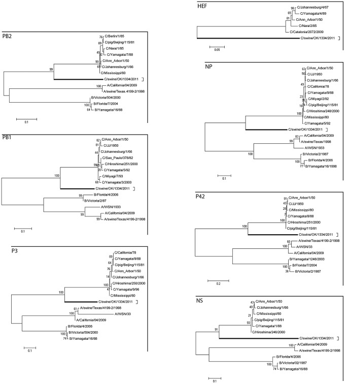
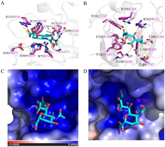
Of the Orthomyxoviridae family of viruses, only influenza A viruses are thought to exist as multiple subtypes and has non-human maintenance hosts. In April 2011, nasal swabs were collected for virus isolation from pigs exhibiting influenza-like illness. Subsequent electron microscopic, biochemical, and genetic studies identified an orthomyxovirus with seven RNA segments exhibiting approximately 50% overall amino acid identity to human influenza C virus. Based on its genetic organizational similarities to influenza C viruses this virus has been provisionally designated C/Oklahoma/1334/2011 (C/OK). Phylogenetic analysis of the predicted viral proteins found that the divergence between C/OK and human influenza C viruses was similar to that observed between influenza A and B viruses. No cross reactivity was observed between C/OK and human influenza C viruses using hemagglutination inhibition (HI) assays. Additionally, screening of pig and human serum samples found that 9.5% and 1.3%, respectively, of individuals had measurable HI antibody titers to C/OK virus. C/OK virus was able to infect both ferrets and pigs and transmit to naive animals by direct contact. Cell culture studies showed that C/OK virus displayed a broader cellular tropism than a human influenza C virus. The observed difference in cellular tropism was further supported by structural analysis showing that hemagglutinin esterase (HE) proteins between two viruses have conserved enzymatic but divergent receptor-binding sites. These results suggest that C/OK virus represents a new subtype of influenza C viruses that currently circulates in pigs that has not been recognized previously. The presence of multiple subtypes of co-circulating influenza C viruses raises the possibility of reassortment and antigenic shift as mechanisms of influenza C virus evolution.
Conflict of interest statement
The authors have read the journal's policy and have the following conflicts: BMH, EAC and RRS are employed by Newport Laboratories, a company that produces swine influenza virus vaccines. This does not alter the authors' adherence to all the PLoS Journal policies on sharing data and materials.
Figures


References
-
- Palese P, Shaw ML (2007) Orthomyxoviridae: The viruses and their replication. In: Knipe DM, Howley PM, editors. Fields Virology, Fifth Edition. Philadelphia: Lippincott Williams & Wilkins. pp. 1647–1690.
-
- Matsuzaki Y, Katsushima N, Nagai Y, Shoji M, Itagaki T, et al. (2006) Clinical features of influenza C virus infection in children. J Infect Dis 193: 1229–1235. - PubMed
-
- Antόn A, Marcos MA, Codoner FM, de Molina P, Martinez A, et al. (2011) Influenza C virus surveillance during the first influenza A (H1N1) 2009 pandemic wave in Catalonia, Spain. Diagn Microbiol Infect Dis 69: 419–427. - PubMed
Publication types
MeSH terms
Substances
Associated data
- Actions
- Actions
- Actions
- Actions
- Actions
- Actions
- Actions
Grants and funding
LinkOut - more resources
Full Text Sources
Other Literature Sources
Miscellaneous
