. 2013;9(2):e1003275.
doi: 10.1371/journal.pgen.1003275. Epub 2013 Feb 7.

Control of multicellular development by the physically interacting deneddylases DEN1/DenA and COP9 signalosome

Affiliations

Control of multicellular development by the physically interacting deneddylases DEN1/DenA and COP9 signalosome

Martin Christmann et al. PLoS Genet. 2013.

Abstract

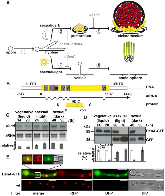
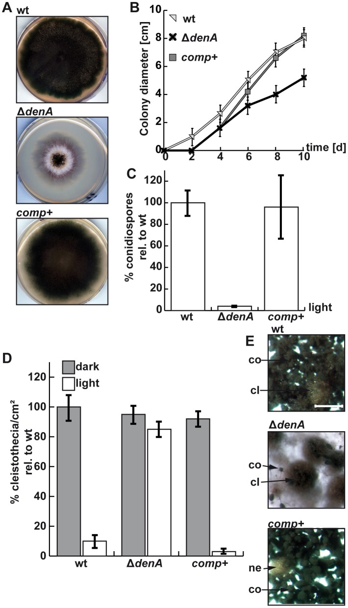
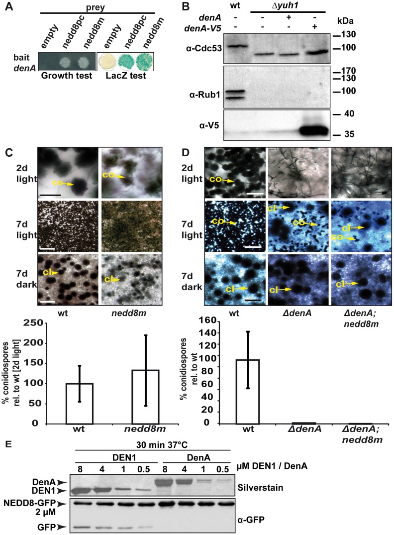
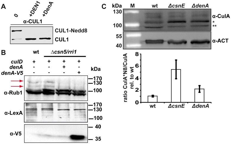
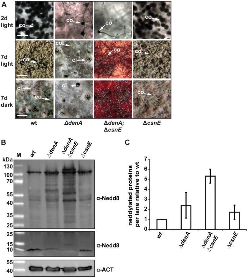
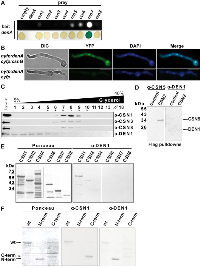
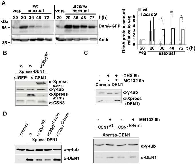
Deneddylases remove the ubiquitin-like protein Nedd8 from modified proteins. An increased deneddylase activity has been associated with various human cancers. In contrast, we show here that a mutant strain of the model fungus Aspergillus nidulans deficient in two deneddylases is viable but can only grow as a filament and is highly impaired for multicellular development. The DEN1/DenA and the COP9 signalosome (CSN) deneddylases physically interact in A. nidulans as well as in human cells, and CSN targets DEN1/DenA for protein degradation. Fungal development responds to light and requires both deneddylases for an appropriate light reaction. In contrast to CSN, which is necessary for sexual development, DEN1/DenA is required for asexual development. The CSN-DEN1/DenA interaction that affects DEN1/DenA protein levels presumably balances cellular deneddylase activity. A deneddylase disequilibrium impairs multicellular development and suggests that control of deneddylase activity is important for multicellular development.


Conflict of interest statement

The authors have declared that no competing interests exist.

Figures

Figure 1
Figure 1. Expression and localization of the DEN1 homolog DenA (AN10456) during development of the fungus Aspergillus nidulans.
(A) Life cycle of A. nidulans. 16 to 20 hours after spore germination vegetative filaments (hyphae) reach developmental competence . In darkness specialized filaments and globular, multi-nuclear cells (Hülle cells; yellow) embed the evolving developmental structure (primordium), which maturates to the sexual fruit body (cleistothecium) within 7 days. In the presence of light an aerial filament with an apical vesicle forms the asexual developmental structure (conidiophore) and releases the uninucleate conidiospores (green) into the air. Arrest points of the described deneddylase mutants are indicated in light grey. Sexual development is blocked (solid grey line) at the stage of primordia in ΔcsnE and even earlier in the double deletion strain (ΔcsnEdenA). Asexual development is drastically reduced (dashed grey line) in ΔdenA as well as in ΔcsnEdenA. (B) denA gene, transcript (RACE analysis) and protein. Positions within the coding sequence are given relative to the A of the start codon. The histidine (H), aspartate (D) and cysteine (C) typical for the active site of an Ulp1 family protease are indicated at their relative positions. (C) Northern hybridization analysis of denA mRNA at indicated stages of development. denA mRNA levels of repeated experiments were normalized to ribosomal RNA (rRNA) and presented as x-fold difference relative to 20 hours vegetative growth (graph). (D) Analysis of DenA protein abundance at indicated stages of development (vegetative, asexual, and sexual). GFP was fused C-terminally to DenA (DenA-GFP). Expression was driven by the native denA promoter. Strong accumulation of the released GFP tag (GFP) indicated that high amounts of very unstable fusion protein were produced. The proportion of DenA-GFP fusion protein and the remaining GFP-tag was analyzed quantitative and applied as a measure for DenA protein stability. Relative pixel density values are presented as percent of total GFP signal per lane shared between DenA-GFP and the remaining GFP-tag (n = 2). (E) Localization of C-terminal DenA-GFP in A. nidulans. The protein resides in the cytoplasm and accumulates in nuclei and around the septum (scale bar = 10 µm). The septal region is enlarged (white square; scale bar = 2 µm). Nuclei are marked by red fluorescence from ectopically integrated mRFP::H2A, membranes are visualized with FM4-64, as well visible through the RFP-filter.
Figure 2
Figure 2. denA function is required for A. nidulans development.
(A) Growth of the A. nidulans denA deletion strain. denA wild type (wt), denA deletion (ΔdenA) and the complemented deletion strain (comp+) were grown on agar plates containing selective minimal medium for 8 days in the presence of light and aeration. (B) Colony diameter of point inoculated colonies monitored over time (d). Plates were incubated at 30°C. (C) Deletion of denA causes a 25 fold reduction of spores compared to wild type. Percentage of conidia produced under asexual conditions (30°C, 48 hours, light and normal aeration) compared to wild type (wt). (D) The A. nidulans denA deletion strain is impaired in light response of development. Formation of sexual fruit bodies (cleistothecia) of wild type (wt), denA deletion (ΔdenA) and the complemented deletion strain (comp+) in light and dark were compared. (E) Agar surface pictures of the corresponding A. nidulans strains taken at the 7th day of growth during illumination (asexual conditions) (cl = cleistothecia; co = conidiophores; ne = nest; scale bar = 250 µm).
Figure 3
Figure 3. Fungal DenA developmental functions are independent of Nedd8 processing activity.
(A) Yeast-2-hybrid interaction between A. nidulans DenA and the precursor (nedd8pc) or the mature form (nedd8m) of Nedd8. (B) Western analysis with α-Cdc53 and α-Rub1/Nedd8 to visualize yeast cullin neddylation. Deletion of yuh1 prevents cleavage of the Rub1 precursor, therefore neddylated protein species can neither be recognized with α-Cdc53 nor with α-Rub1/Nedd8. DenA expression driven by the inducible GAL1 promoter was applied to test the processing ability of the protease towards the Rub1 precursor. Expression of DenA was visualized with α-V5. DenA expression was not sufficient to restore Rub1 processing in a yuh1 deficient S. cerevisiae strain . (C) An A. nidulans strain expressing a mature nedd8 construct (nedd8m) instead of normal nedd8 (wt) displayed a wild type like phenotype. Asexual structures (co) displayed on agar surface after 2 days of growth in the presence of light (scale bar = 20 µm) and after 7 days (scale bar = 500 µm). Sexual structures (cl) were only formed in the dark, but not in the light after 7 days incubation at 37°C (scale bar = 500 µm). Quantification of asexual spores from both strains after 2 days of incubation at 37°C in light. (D) The ΔdenA/nedd8m strain (mature Nedd8) and the ΔdenA (precursor Nedd8) are unresponsive to light-dependent inhibition of sexual development (compare rows 2 and 3; scale bar = 50 µm, first row; scale bar = 225 µm, second and third row) and impaired in asexual development (conidiation). (E) Human DEN1 cleaved human Nedd8 C-terminally fused with GFP while A. nidulans DenA did not. Nedd8-GFP substrate was combined with decreasing amounts of recombinant DEN1 or DenA, respectively (8–0.5 µM). The reaction mixture was incubated for 30 min at 37°C, immediately denatured, separated by SDS-PAGE and subjected to western blot analysis. α-GFP was applied to monitor cleavage of the Nedd8-GFP substrate. Samples were separated by additional SDS-PAGE and silver stained to prove for the presence of the respective deneddylase.
Figure 4
Figure 4. DenA deneddylase activity.
(A) Recombinant human DEN1 and fungal DenA deneddylate a human CUL1-Nedd8 substrate in vitro. SDS-PAGE and subsequent western analysis show cleavage of the substrate (∼60 kDa) producing the C-terminal CUL1 (∼50 kDa) as outlined in experimental procedures. (B) Deneddylation test in a heterologous yeast system. A. nidulans DenA can remove Rub1 from CulD in heterologous expression experiments in S. cerevisiae. DenA was expressed as native protein or C-terminally fused with a V5/His6 epitope tag. Both variants were driven by the inducible GAL1 promoter. CulD, N-terminal-fused with the LexA activation domain, was expressed under control of the constitutive ADH promoter. A. nidulans proteins were expressed in S. cerevisiae wild type and Δcsn5 background. Western analysis with antibodies against Rub1 (α-Rub1), the LexA epitope (α-LexA) and the V5 epitope (α-V5) were performed. Detection with α-Rub1 generated two additional signals upon culD expression, representing LexA-CulD and a second CulD pool where LexA was unspecifically cleaved off. Both signals disappeared upon co-expression of DenA indicating deneddylation activity (red arrows). The slower migrating signal of α-LexA western experiments corresponded to Rub1 modified LexA-CulD. This signal was absent when DenA was co-expressed. Detection of the V5 tag was applied to monitor DenA expression. The neddylated yeast cullin migrating at around 100 kDa was not affected by DenA activity. (C) Deneddylation of fungal CulA by CSN and DenA. Whole cell lysates of A. nidulans wild type, ΔcsnE and ΔdenA were probed with α-CulA. The ratio of neddylated CulA (CulA*N8; ∼106 kDa*) to non-neddylated CulA (∼96 kDa**) was calculated from three independent experiments. Membranes were reprobed with α-Actin (α-ACT) for normalization.
Figure 5
Figure 5. Double knock-out of the deneddylase encoding genes denA and csnE abolished fungal development.
(A) ΔdenA and ΔdenA/ΔcsnE strains were impaired in conidiophore formation while ΔcsnE and wild type (wt) form similar amounts of asexual structures. Even after seven days of asexual development only marginal numbers of conidiophores are formed in ΔdenA or ΔdenAcsnE strain. Sexual development after 7 days in the dark occurred for ΔdenA but was abolished in ΔdenA/ΔcsnE. Accumulated red color within the hyphae reflects impaired secondary metabolism. Initiation of sexual development is independent of light in both single deletion strains (7 days, light). ΔcsnE showed nest (yellowish filaments) formation in light and darkness, but could not progress further in sexual fruit body formation. The red color typical for csnE deletion is covered by the layer of hyphae and conidiophores in the pictures displayed here. The ΔdenA/ΔcsnE was impaired in sexual development and secondary metabolism (scale bar = 50 µm, first row; scale bar = 200 µm, second and third row). (B) Deneddylation deficient A. nidulans mutants accumulate neddylated proteins. Western analysis of A. nidulans wild type (wt), ΔdenA, ΔcsnE single deletion strains and ΔdenA/ΔcsnE double knock-out strain. Crude extracts were probed with A. nidulans α-Nedd8. Gels with identical samples were run for different times to allow proper separation of high migrating signals (higher panel) of neddylated proteins and to preserve the signal of free Nedd8 (lower panel). An increase of neddylated proteins correlates with a reduction of free Nedd8. (C) Semi-quantitive analysis of the Nedd8 signal intensity within each lane for ΔdenA, ΔcsnE single deletion strains and ΔdenA/ΔcsnE double knock-out strain relative to wild type. Signals were normalized by reprobing the membranes with α-Actin (α-ACT). Three independently repeated experiments were included into the quantification.
Figure 6
Figure 6. DEN1/DenA deneddylase interacts with the COP9 signalosome (CSN) in fungal and human cells.
(A) A. nidulans DenA-CSN interactions in yeast-2-hybrid growth and β-galactosidase activity test. (B) DenA and CSN7/CsnG interaction is enriched in the nucleus. BiFC studies in A. nidulans for DenA (nYFP::denA) and CSN7/CsnG (cYFP::csnG) interaction. The control strain expressed only the DenA fusion protein (nYFP::denA) whereas cYFP was expressed without a fused protein. Nuclei were visualized by DAPI staining (scale bar = 10 µm). (C) Human DEN1-CSN interaction. Density gradient centrifugation of lysate from HeLa cells and subsequent western experiments with fractions of different density. Endogenous DEN1 was detected by the α-DEN1 antibody and the position of the CSN was estimated by α-CSN1, α-CSN3 and α-CSN8 antibodies. The density gradient was calibrated with purified CSN about 400 kDa, ▴) and with purified 20S proteasome (about 700 kDa, Δ). (D) Flag-pull downs from lysates of Flag-CSN2 B8 cells. Proteins specifically eluted with the Flag peptide were analyzed by western experiments. CSN5 and DEN1 were detected by specific antibodies. Control pull downs were performed with lysates from B8 cells. (E) Far-western experiments with recombinant subunits CSN1, CSN2, CSN4, CSN6, CSN7 and CSN8. Membranes were incubated with 1 µg/ml DEN1 protein, washed and probed with the α-DEN1 antibody. (F) Recombinant full-length CSN1 (wt), CSN1(1–221) (N-term) and CSN1(222–527) (C-term) were analyzed by far-western blot. Membranes were incubated with DEN1 as in (E) and probed with the α-CSN1 or the α-DEN1 antibody.
Figure 7
Figure 7. CSN targets DEN1/DenA for degradation in fungal and human cells.
(A) Quantitative analysis of repeated western blot experiments displayed the differences in DenA abundance in fungal wild type and ΔcsnG cells. DenA levels of the different asexual developmental time points are shown relative to the vegetative (veg.) control for each strain. Anti-Actin was applied as loading control (statistics: 2-way ANOVA; n = 3; *p<0,05, **p<0,01). (B) Xpress-DEN1 was overexpressed in siGFP and siCSN1 human cells and steady state Xpress-DEN1 levels were estimated by western analysis with the α-Xpress antibody. Xpress-CSN1 was overexpressed in siCSN1 cells and DEN1 and CSN8 were probed with appropriate antibodies. (C) Xpress-DEN1 was overexpressed in siGFP human cells and the proteasome inhibitor MG132 was added 6 h before cell lysis at a final concentration of 10 µM. Cyclohexamide (CHX) was added in a final concentration of 10 µg/ml (D) Xpress-DEN1 was co-expressed in HeLa cells together with Xpress-CSN1wt, Xpress-CSN1(1–221) or Xpress-CSN1(222–527) in the absence or in the presence of MG132 (right panel), which was added 6 h before cell lysis. Cells were lyzed 24 h after co-transfection and lysates were analyzed by western blot using the α-DEN1 antibody (0 = only Xpress-DEN1).

Similar articles

Cited by

References

    1. Kerscher O, Felberbaum R, Hochstrasser M (2006) Modification of Proteins by Ubiquitin and Ubiquitin-Like Proteins. Annual Review of Cell and Developmental Biology - PubMed
    1. Hershko A, Ciechanover A (1998) The ubiquitin system. Annu Rev Biochem 67: 425–479. - PubMed
    1. Petroski MD, Deshaies RJ (2005) Function and regulation of cullin-RING ubiquitin ligases. Nat Rev Mol Cell Biol 6: 9–20. - PubMed
    1. von Zeska Kress MR, Harting R, Bayram O, Christmann M, Irmer H, et al... (2012) The COP9 signalosome prevents the accumulation of cullin SCF ubiquitin E3 RING ligases during fungal development. Mol Microbiol, accepted for publication. - PubMed
    1. Morimoto M, Nishida T, Nagayama Y, Yasuda H (2003) Nedd8-modification of Cul1 is promoted by Roc1 as a Nedd8-E3 ligase and regulates its stability. Biochemical and Biophysical Research Communications 301: 392–398. - PubMed

Publication types

MeSH terms