. 2013;9(2):e1003285.
doi: 10.1371/journal.pgen.1003285. Epub 2013 Feb 7.

Drosophila Yemanuclein and HIRA cooperate for de novo assembly of H3.3-containing nucleosomes in the male pronucleus

Affiliations

Drosophila Yemanuclein and HIRA cooperate for de novo assembly of H3.3-containing nucleosomes in the male pronucleus

Guillermo A Orsi et al. PLoS Genet. 2013.

Abstract

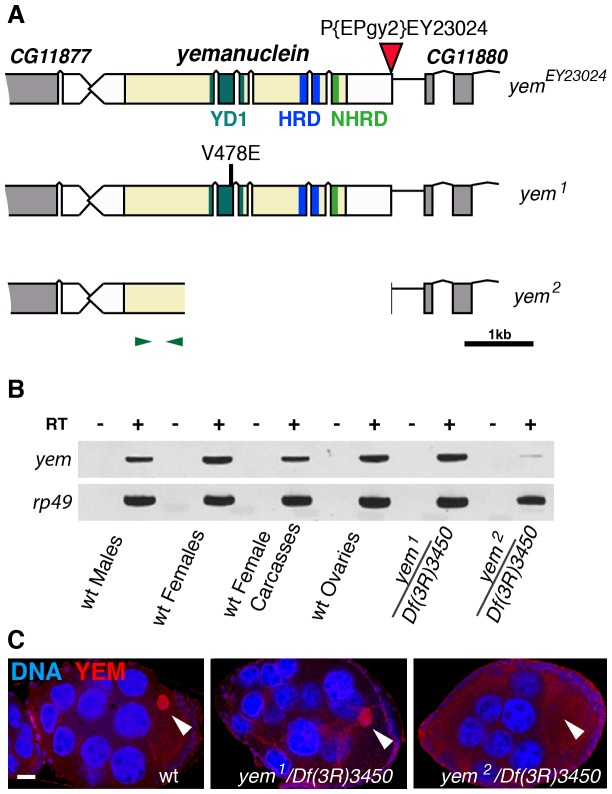
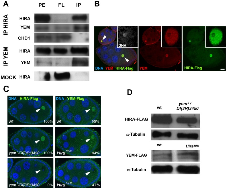
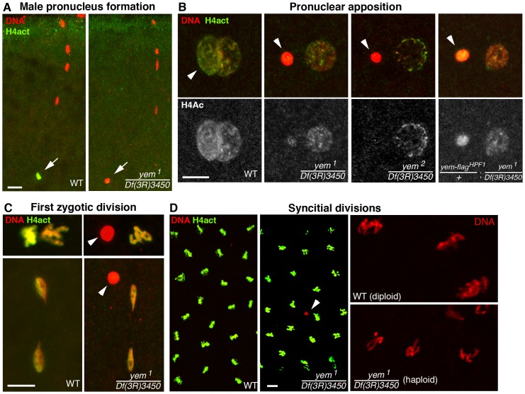
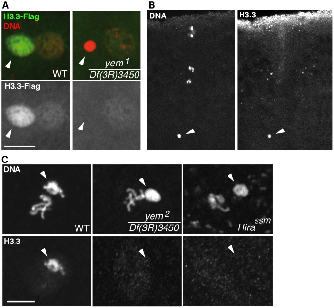
The differentiation of post-meiotic spermatids in animals is characterized by a unique reorganization of their nuclear architecture and chromatin composition. In many species, the formation of sperm nuclei involves the massive replacement of nucleosomes with protamines, followed by a phase of extreme nuclear compaction. At fertilization, the reconstitution of a nucleosome-based paternal chromatin after the removal of protamines requires the deposition of maternally provided histones before the first round of DNA replication. This process exclusively uses the histone H3 variant H3.3 and constitutes a unique case of genome-wide replication-independent (RI) de novo chromatin assembly. We had previously shown that the histone H3.3 chaperone HIRA plays a central role for paternal chromatin assembly in Drosophila. Although several conserved HIRA-interacting proteins have been identified from yeast to human, their conservation in Drosophila, as well as their actual implication in this highly peculiar RI nucleosome assembly process, is an open question. Here, we show that Yemanuclein (YEM), the Drosophila member of the Hpc2/Ubinuclein family, is essential for histone deposition in the male pronucleus. yem loss of function alleles affect male pronucleus formation in a way remarkably similar to Hira mutants and abolish RI paternal chromatin assembly. In addition, we demonstrate that HIRA and YEM proteins interact and are mutually dependent for their targeting to the decondensing male pronucleus. Finally, we show that the alternative ATRX/XNP-dependent H3.3 deposition pathway is not involved in paternal chromatin assembly, thus underlining the specific implication of the HIRA/YEM complex for this essential step of zygote formation.


Conflict of interest statement

The authors have declared that no competing interests exist.

Figures

Figure 1
Figure 1. Mutations affecting the yem gene.
(A) Schematic representation of the yem gene and mutant alleles. yem1 is a point mutation (V478E) and yem2 is a deletion that was generated by mobilizing the P-element insertion P{EPgy2}EY23024 (red triangle). Coding sequence is in yellow and untranslated regions are in white. The YD1 , HRD/HUN , and NHRD domains of YEM are indicated, as well as the position of primers used for RT-PCR analysis (green arrowheads). (B) RT-PCR analysis of yem expression in the indicated tissues and genotypes. RT-PCR amplification used the primer pair shown in (A). (C) Confocal images of wild-type or yem mutant egg chambers stained for DNA (blue) and with anti-YEM AS2 antibody (red). YEM protein accumulates in the germinal vesicle (arrowhead) in wild-type and yem1/Df(3R)3450 oocytes. In yem2/Df(3R)3450 mutant oocytes, only background staining is detected. Bar: 20 µm.
Figure 2
Figure 2. YEM and HIRA are interdependent for their localization in the germinal vesicle.
(A) HIRA and YEM interact in vivo. Ovary extracts were prepared from Hira-flag transgenic flies and immunoprecipitated with either YEM AS2 polyclonal antibody or anti-Flag monoclonal antibody in the conditions described in the Materials and Methods section. The mock immunoprecipitation was performed with a rabbit pre-immune serum. The protein extracts (PE), the flowthrough (FL) and the immunoprecipitated (IP) fractions were submitted to Western blot analysis. HIRA and YEM were revealed respectively with anti-Flag (1∶1000) and AS2 (1∶100) antibodies. Rabbit polyclonal antibody against CHD1 was used at a 1∶250 dilution. Note that YEM and HIRA were found in the same immune complex whereas CHD1 was recovered in the FL fraction. In the mock experiment, HIRA was recovered in the FL fraction, which assesses the specificity of the immunoprecipitation reactions. (B) YEM and HIRA colocalize in the GV (arrowheads) throughout oogenesis. Wild-type ovaries dissected from Hira-Flag transgenic females were stained with anti-Flag (green), anti-YEM antibodies (red) and DAPI (blue). Bar: 20 µm. (C) YEM and HIRA proteins are interdependent for their localization in the GV. Ovaries from wild-type (wt), yem1/Df(3R)3450 or yem2/Df(3R)3450 females bearing one copy of the Hira-Flag transgene were stained to visualize DNA (blue) and Flag (green). HIRA accumulation in the GV (arrowheads) is not affected in yem1/Df(3R)3450 but is abolished in yem2/Df(3R)3450 ovaries. Conversely, YEM-Flag GV localization was not significantly affected in Hirassm ovaries, but appeared highly variable in HiraHR1 ovaries. Percentages indicate the number of egg chambers with positive staining in the GV, equal or superior to the background fluorescence. At least 60 egg chambers were observed for each experiment. (D) Western-blot analysis of HIRA-Flag (left) and YEM-Flag (right) from protein extracts of ovaries of the indicated genotypes. α-Tubulin was used as a loading control.
Figure 3
Figure 3. YEM is essential for chromatin assembly in the male pronucleus at fertilization.
(A) Confocal images of fertilized eggs in telophase of meiosis II stained for DNA (red) and acetylated histone H4 (H4act, green). In wild-type eggs (WT), the male pronucleus (arrow) is brightly and specifically stained with anti-H4act antibodies. In yem mutant eggs, H4act incorporation in the male nucleus is very weak or absent. (B) Eggs at the pronuclear apposition stage. In yem1 and yem2 mutant eggs, the male pronucleus (arrowheads) fails to decondense (compare with wild-type) and contains very low or undetectable levels of H4act. Note that paternal chromatin assembly is partially restored in eggs laid by weakly fertile yem-flagHPF1/+; yem1/Df(3R)3450 females (see also Table 1). (C) Cycle 1 embryos in metaphase (top) and telophase (bottom), stained as in (A) and (B). In yem mutant eggs, the male nucleus (arrowheads) is excluded from the first zygotic division. (D) yem1/Df(3R)3450 females produce gynogenetic haploid embryos. Left: the male nucleus (red) is still detected (arrowhead) among the haploid cleavage nuclei containing chromosomes of maternal origin. Right: close-up of cleavage nuclei in metaphase from wild-type (diploid) and yem (haploid) embryos.
Figure 4
Figure 4. yem is required for H3.3 deposition in the male nucleus.
(A) At pronuclear apposition in wild-type eggs, the male nucleus (arrowhead) contains high levels of maternally expressed H3.3-Flag whereas the female pronucleus incorporates low levels of H3.3-Flag, presumably during S phase. In yem1/Df(3R)3450 mutant eggs, only the weak incorporation of H3.3-Flag in the female pronucleus is detected. (B) The monoclonal anti-H3.3 antibody specifically stains the male nucleus (arrowhead) at fertilization in wild-type eggs. Maternal chromosomes are in anaphase of the second meiotic division. (C) H3.3 is still detected in paternal chromosomes (arrowheads) during the first zygotic cycle in wild-type eggs but not in the male nucleus on cycle 1 yem or Hira mutant embryos. Bars: 10 µm.
Figure 5
Figure 5. HIRA and YEM are interdependent for their recruitment to the male pronucleus.
(A) YEM localizes to the decondensing male nucleus. Confocal images of eggs from yem-flagHPF3 yem2 homozygous females (upper panels) with the male nucleus (arrows) magnified in lower panels. YEM-Flag (green) is detected throughout the decondensing male nucleus but also accumulates in a small number of nuclear foci (arrowheads). Note that YEM-Flag is no longer detected at pronuclear apposition (right panels). Bar: 10 µm. (B) Hira mutations affect the general distribution of YEM-Flag in the male nucleus. Eggs from females of the indicated genotype were stained with anti-Flag antibodies and imaged as in (A). For each male nucleus, the presence of YEM-Flag (whole nucleus and/or foci) was evaluated and each category is represented as a percentage of the total number (n) of observed pronuclei. (C) YEM-Flag foci in the male nucleus do not colocalize with telomeres or centromeres. Upper panels: Nuclear foci of maternally expressed YEM-Flag (red arrow) do not localize to the cluster of telomeres marked with the paternal telomere marker GFP-K81 (green arrow). Lower panels: YEM-Flag nuclear foci (red arrows) do not colocalize with paternal centromeres (green arrows) in the male nucleus. (D) Confocal sections of male pronuclei in eggs laid by females of the indicated genotype (HIRA-Flag is shown in green, DNA in red) (n>20). In wild-type eggs, HIRA-Flag accumulates in the decondensing male nucleus. In eggs from yem mutant females however, HIRA-Flag is not detected.
Figure 6
Figure 6. XNP is not involved in paternal chromatin assembly.
(A) The dATRX/XNP protein (green) is expressed in the female germline and accumulates in the germinal vesicle (arrow) of the oocyte, here in a stage 10 egg chamber. (B) At fertilization (left panel), XNP is not observed in the male nucleus (inset) whereas it is detected in the nuclei of blastoderm embryo. (C) Confocal images of eggs or embryos from xnp mutant females stained for anti-H4act (green) and DNA (red). Left: in eggs from xnp mutant females, assembly of paternal chromatin seems to occur normally, as revealed by the strong incorporation of acetylated H4 in the male nucleus (inset). Middle: a cycle 1 embryo in prometaphase with the still separated sets of maternal and paternal chromosomes. Note the stronger anti-H4act staining of paternal chromosomes (arrowhead) relative to maternal chromosomes, as in wild-type embryos (see Figure 3C). Right: normal cleavage divisions in a syncytial embryo.

Similar articles

Cited by

References

    1. Polo SE, Almouzni G (2006) Chromatin assembly: a basic recipe with various flavours. Curr Opin Genet Dev 16: 104–111. - PubMed
    1. Ahmad K, Henikoff S (2002) Histone H3 variants specify modes of chromatin assembly. Proc Natl Acad Sci U S A 99 Suppl 4: 16477–16484. - PMC - PubMed
    1. Talbert PB, Ahmad K, Almouzni G, Ausio J, Berger F, et al. (2012) A unified phylogeny-based nomenclature for histone variants. Epigenetics Chromatin 5: 7. - PMC - PubMed
    1. Corpet A, Almouzni G (2009) Making copies of chromatin: the challenge of nucleosomal organization and epigenetic information. Trends Cell Biol 19: 29–41. - PubMed
    1. Elsaesser SJ, Goldberg AD, Allis CD (2010) New functions for an old variant: no substitute for histone H3.3. Curr Opin Genet Dev 20: 110–117. - PMC - PubMed

Publication types

MeSH terms

LinkOut - more resources