. 2013;8(2):e55289.
doi: 10.1371/journal.pone.0055289. Epub 2013 Feb 8.

Aberrant glycogen synthase kinase 3β is involved in pancreatic cancer cell invasion and resistance to therapy

Affiliations

Aberrant glycogen synthase kinase 3β is involved in pancreatic cancer cell invasion and resistance to therapy

Ayako Kitano et al. PLoS One. 2013.

Abstract

Background and purpose: The major obstacles to treatment of pancreatic cancer are the highly invasive capacity and resistance to chemo- and radiotherapy. Glycogen synthase kinase 3β (GSK3β) regulates multiple cellular pathways and is implicated in various diseases including cancer. Here we investigate a pathological role for GSK3β in the invasive and treatment resistant phenotype of pancreatic cancer.

Methods: Pancreatic cancer cells were examined for GSK3β expression, phosphorylation and activity using Western blotting and in vitro kinase assay. The effects of GSK3β inhibition on cancer cell survival, proliferation, invasive ability and susceptibility to gemcitabine and radiation were examined following treatment with a pharmacological inhibitor or by RNA interference. Effects of GSK3β inhibition on cancer cell xenografts were also examined.

Results: Pancreatic cancer cells showed higher expression and activity of GSK3β than non-neoplastic cells, which were associated with changes in its differential phosphorylation. Inhibition of GSK3β significantly reduced the proliferation and survival of cancer cells, sensitized them to gemcitabine and ionizing radiation, and attenuated their migration and invasion. These effects were associated with decreases in cyclin D1 expression and Rb phosphorylation. Inhibition of GSK3β also altered the subcellular localization of Rac1 and F-actin and the cellular microarchitecture, including lamellipodia. Coincident with these changes were the reduced secretion of matrix metalloproteinase-2 (MMP-2) and decreased phosphorylation of focal adhesion kinase (FAK). The effects of GSK3β inhibition on tumor invasion, susceptibility to gemcitabine, MMP-2 expression and FAK phosphorylation were observed in tumor xenografts.

Conclusion: The targeting of GSK3β represents an effective strategy to overcome the dual challenges of invasiveness and treatment resistance in pancreatic cancer.


Conflict of interest statement

Competing Interests: The authors have declared that no competing interests exist.

Figures

Figure 1
Figure 1. Expression, phosphorylation and activity of GSK3β in pancreatic cancer cells and primary pancreatic cancers.
(A) Protein extract from each cell line was analyzed by Western immunoblotting for the expression of GSK3β and its phosphorylation (p-GSK3βS9, p-GSK3βY216). β-actin expression was monitored as a loading control. (B) GSK3β activity was detected by NRIKA in the respective cells. As described in Materials and Methods, GSK3β activity is demonstrated by the presence of p-β-cateninS33/37/T41 in the test reaction (T) and by its absence in the negative control reaction (NC). The amount of immunoprecipitated GSK3β and the presence of substrate (β-catenin) in the kinase reaction were monitored by immunoblotting. (C, D) Serial paraffin sections of a primary pancreatic cancer and its adjacent non-neoplastic tissue (patient No. 5 in Table S1) were immunostained for GSK3β and p-GSK3βY216 (C), and for MMP-2, FAK, p-FAKY397 and p-FAKY861 (D). The scale bar in each panel indicates 100-µm in length.
Figure 2
Figure 2. Effects of GSK3β inhibition on the survival and proliferation of pancreatic cancer cells.
(A) Relative numbers of viable cells at the designated time points were measured by WST-8 assay for the respective cells in the presence of DMSO or AR-A014418 at the indicated concentrations. (B) Relative numbers of viable cells were measured for the respective cells after transfection of non-specific (NS) or GSK3β-specific siRNA. (C, D) The relative number of proliferating cells was determined by measuring the amount of BrdU incorporation. Proliferating cells were scored at 48 hrs after treatment with DMSO or AR-A014418 (10 µM, 20 µM) (C), or after transfection with non-specific (NS) or GSK3β-specific siRNA (D). Values shown in (A–D) are the means ± SD of five separate experiments. *p<0.05, statistically significant difference between cells treated with DMSO or AR-A014418 and between cells treated with non-specific and GSK3β-specific siRNA.
Figure 3
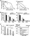
Figure 3. Combined effect of gemcitabine or ionizong radiation and GSK3β inhibitor against cancer cells and xenografts.
(A) The influence of AR-A014418 on the effect of gemcitabine was analyzed using the isobologram by plotting the IC50 of the combination therapy (Fig. S2, Table S4). (B) The combined effect of ionizing radiation and AR-A014418 was tested in PANC-1 and MIA PaCa-2 cells by colony formation assay. *p<0.05, statistically significant difference between cells treated with DMSO or AR-A014418. (C) The combined effect of gemcitabine and AR-A014418 was tested in PANC-1 xenografts. Athymic mice with PANC-1 xenograft were assigned to four groups for treatment with intraperitoneal injection (twice a week) of DMSO (control; 8 mice), gemcitabine (GEM; 20 mg/kg body weight; 9 mice) and AR-A014418 (AR; 2 mg/kg body weight; 8 mice), alone or in combination (GEM+AR; 9 mice). At the time after treatment for 10 weeks, tumor volume (cm3) was calculated using the formula 0.5×S2×L, where S is the smallest tumor diameter (cm) and L is the largest (cm) , . The mean tumor volume was compared between the 4 groups. *p<0.05, statistically significant difference between data.
Figure 4
Figure 4. Changes in expression and phosphorylation of the proteins in cancer cells following GSK3β inhibition.
(A) Immunoblotting analysis compares the expression of Rb, CDK4, CDK6 and cyclin D1, and the phosphorylation of Rb at S780 and S807/811 residues (p-RbS780, p-RbS807/811) between cells treated with DMSO (DM) or 10 µM AR-A014418 (AR) for 24 hrs. (B) Changes in levels of p-RbS780 and p-RbS807/811 and expression of Rb and cyclin D1 were examined in MIA PaCa-2 cells at the indicated time points after treatment with 10 µM AR-A014418. (C) Expression of Rb, cyclin D1, GSK3α and GSK3β proteins and levels of Rb phosphorylation (p-RbS780) were examined and compared between the same pancreatic cancer cells transfected with non-specific siRNA (NS) or GSK3β-specific siRNA (S) (10 nM each). (A–C) β-actin expression was monitored as a loading control.
Figure 5
Figure 5. Effects of GSK3β inhibition on the migration and invasion of pancreatic cancer cells.
(A) Upper panels show the time course for PANC-1 cell migration in a wound-healing assay in the presence of DMSO or AR-A014418 (AR). The lower panel shows the relative widths of wounds measured as a percentage of the initial gap at time zero. *p<0.05, statistically significant difference between cells treated with DMSO or AR-A014418. (B) Migrating cells through uncoated transwell and invading cells through matrigel-coated transwell were scored for PANC-1 and MIA PaCa-2 cells treated with DMSO or AR-A014418 (AR) for 22 hrs. Representative photomicroscopic findings in each assay are shown below the columns. *p<0.05, statistically significant difference between cells treated with DMSO or AR-A014418.
Figure 6
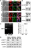
Figure 6. Changes in the invasive phenotype of pancreatic cancer cells following GSK3β inhibition.
(A) Phase-contrast microscopic findings (left panels), expression and subcellular localization of F-actin and Rac-1 (middle panels) and their merged images (right panels) in cancer cells along the wound edge (dashed line) were observed in the wound-healing assay in the presence of DMSO or AR-A014418 (AR). Arrows indicate lamellipodia. (B) Changes in the levels of active (Rac1-GTP) and total Rac1 examined by pull-down assay and Western blotting between the cancer cells treated with DMSO (DM) or 10 µM AR-A014418 (AR) for 24 hrs. (C) Changes in the secretion and mRNA expression of MMP-2 examined by gelatin zymography (left panel) and qRT-PCR (right panel) between PANC-1 cells treated with DMSO (DM) or AR-A014418 (AR) for 24 hrs. Values for the relative levels of mRNA expression are shown as means ± SDs of four separate experiments. *p<0.05, statistically significant difference between cells treated with DMSO or AR-A014418. (D) Changes in the expression of FAK and its phosphorylation (p-FAKY397, p-FAKY861) examined by Western blotting in cancer cells following GSK3β inhibition. The cells in confluent monolayer were wounded multiple times and cultured in the presence of DMSO (DM) or 10 µM AR-A014418 (AR) for 24 hrs. β-actin expression was monitored as a loading control.
Figure 7
Figure 7. Changes in the invasive phenotype of pancreatic cancer cells in xenografts following GSK3β inhibition.
(A) The left two panels showed represntative histological findings of the deeper part of PANC-1 xenografts in rodents after treatment with intraperitoneal injection (twice a week) of DMSO and AR-A014418, respectively, as shown in Fig. 3C. The serial sections of these tumors were immunostained for MMP-2, FAK, p-FAKY397 and p-FAKY861. Closed triangles in the lower panels delineate an interface between the xenograft and host stromal tissue. (B) These tumors were immunostained for GSK3β, p-GSK3βY216 and β-catenin. (A, B) The scale bar in each panel indicates 100-µm in length.

Similar articles

Cited by

References

    1. Siegel R, Naishadham D, Jemal A (2012) Cancer statistics, 2012. CA Cancer J Clin 62: 10–29. - PubMed
    1. Bardeesy N, DePinho RA (2002) Pancreatic cancer biology and genetics. Nat Rev Cancer 2: 897–909. - PubMed
    1. Li D, Xie K, Wolff R, Abbruzzese JL (2004) Pancreatic cancer. Lancet 363: 1049–1057. - PubMed
    1. Hidalgo M (2010) Pancreatic cancer. N Engl J Med 362: 1605–1617. - PubMed
    1. Furukawa T (2008) Molecular targeting therapy for pancreatic cancer: Current knowledge and perspectives from bench to bedside. J Gastroenterol 43: 905–911. - PubMed

Publication types

MeSH terms