. 2013;8(2):e55578.
doi: 10.1371/journal.pone.0055578. Epub 2013 Feb 7.

A new model of development of the mammalian ovary and follicles

Affiliations

A new model of development of the mammalian ovary and follicles

Katja Hummitzsch et al. PLoS One. 2013.

Abstract

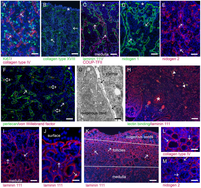
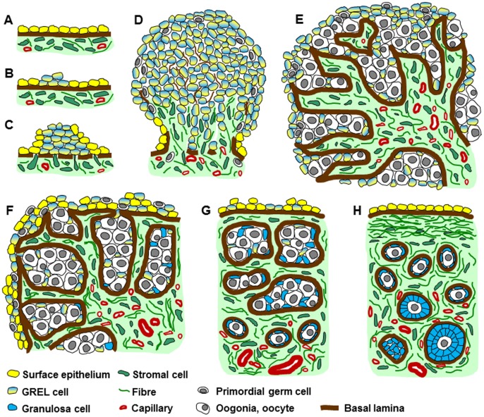
Ovarian follicular granulosa cells surround and nurture oocytes, and produce sex steroid hormones. It is believed that during development the ovarian surface epithelial cells penetrate into the ovary and develop into granulosa cells when associating with oogonia to form follicles. Using bovine fetal ovaries (n = 80) we identified a novel cell type, termed GREL for Gonadal Ridge Epithelial-Like. Using 26 markers for GREL and other cells and extracellular matrix we conducted immunohistochemistry and electron microscopy and chronologically tracked all somatic cell types during development. Before 70 days of gestation the gonadal ridge/ovarian primordium is formed by proliferation of GREL cells at the surface epithelium of the mesonephros. Primordial germ cells (PGCs) migrate into the ovarian primordium. After 70 days, stroma from the underlying mesonephros begins to penetrate the primordium, partitioning the developing ovary into irregularly-shaped ovigerous cords composed of GREL cells and PGCs/oogonia. Importantly we identified that the cords are always separated from the stroma by a basal lamina. Around 130 days of gestation the stroma expands laterally below the outermost layers of GREL cells forming a sub-epithelial basal lamina and establishing an epithelial-stromal interface. It is at this stage that a mature surface epithelium develops from the GREL cells on the surface of the ovary primordium. Expansion of the stroma continues to partition the ovigerous cords into smaller groups of cells eventually forming follicles containing an oogonium/oocyte surrounded by GREL cells, which become granulosa cells, all enclosed by a basal lamina. Thus in contrast to the prevailing theory, the ovarian surface epithelial cells do not penetrate into the ovary to form the granulosa cells of follicles, instead ovarian surface epithelial cells and granulosa cells have a common precursor, the GREL cell.


Conflict of interest statement

Competing Interests: The authors have declared that no competing interests exist.

Figures

Figure 1
Figure 1. The early-stage ovary.
At this stage the ovary was composed of mostly GREL cells, expressing cytokeratin (CK) 19 (A, green), interspersed with OCT3/4 positive PGCs (B, red, arrowheads). The ovary did not have a defined surface epithelium except at its base where it arose and protruded from the mesonephros and where it was strongly stained for cytokeratin 19 (A, green, open arrows) and was under laid by a basal lamina containing components of laminin 111 (A, red and B, green, arrows), collagen type IV (C, red), perlecan (D, red) and nidogens 1 (E, red) and 2 (F, red). Basal lamina material was interspersed throughout the ovary. Nidogen 2 and collagen type IV were more strongly expressed than components of laminin 111, nidogen 1 and perlecan, even in the cytoplasm of some cell clusters. Nuclei were counterstained with DAPI (blue). Gestational age: 63 days. Bars: A–D, F = 50 µm; E = 25 µm.
Figure 2
Figure 2. The ovarian cortex is rich in ovigerous cords separated from the penetrating stroma by a basal lamina.
The basal lamina (marked with arrows) contains collagens type IV (A, red) and type XVIII (B, green), components of laminin 111 (C, green and H, red), nidogens 1 (D, green) and 2 (E, red) and perlecan (F, green). The electron micrograph shows that the basal lamina was located adjacent to the surface of the ovigerous cords (G, arrows). Cells in the ovigerous cords and stroma were proliferating (A, Ki67 positive, green) and cords were still open at the surface (B–F, indicated with star). Stromal cells in the cortex and medulla expressed COUP-TFII (C, red nuclear staining). Stromal areas between ovigerous cords contained capillaries (F, open arrow, endothelial cells stained for von Willebrand factor, red) and their sub-endothelial basal laminas were composed of, amongst other things, perlecan (F, green) and components of laminin 111 (H, red). The medulla contained arterioles (H, open triangle, green lectin binding to endothelial cells surrounded by extensive basal lamina of smooth muscle cells identified by staining of components of laminin 111), capillaries (filled triangle) and mesonephric-derived tubules or rete ovarii close to the hilus (H, star). Later in development, increasing amounts of stroma led to a breakdown of large ovigerous cords into smaller cords (I, basal laminas stained for components of laminin 111, red) and the development of a basal lamina below layers of GREL cells on the ovarian surface (J, basal laminas stained for components of laminin 111,red). Primordial follicles firstly developed in the inner cortex/medulla region, whereas the outer cortex still contained ovigerous cords [k, basal laminas stained for components of laminin 111 (red)]. In addition to components of laminin 111 (K), the follicular basal lamina contained collagen type IV (L, red) and nidogen 2 (M, red). Nuclei were counterstained with DAPI (blue). Gestational ages: 73 (G), 86 (H), 93 (A), 98 (B–D,F), 120 (E), 127 (I), 144 (J), 148 (K,L) and 171 days (M). Bars: H, K, M = 100 µm; A–F, I, L = 50 µm; J = 25 µm; G = 1 µm.
Figure 3
Figure 3. Gradation of germ cell maturation.
After migrating into the early-stage ovary, PGCs proliferated as oogonia and together with the GREL cells constituted the ovigerous cords (A, methylene blue-stained semi-thin section, GREL cells marked with filled triangle and capillaries with asterisk). The least mature oogonia were located towards the outer cortex, undergoing mitosis, whilst oogonia in the inner cortex differentiated into oocytes. This zonal differentiation was mirrored in the expression of germ cell markers. Germ cells close to the surface expressed OCT3/4 (B, green; C and E, red), whilst deeper in the ovary they had commenced expressing cytoplasmic DAZL (B and D, red) and later VASA (E, green, open arrows; F, green) with concomitantly reduced OCT3/4 expression. Later in development germ cells in the outer cortex differentiated and expressed VASA (G, green and basal lamina stained for collagen type IV α1). Follicular oocytes expressed VASA (H, green) and OCT3/4 again (H, red). Morphological differences between germ cells and GREL cells were evident; germ cells were large and round with a round nucleus whereas GREL cells were smaller, elongated with an elongated nucleus and a larger nuclear/cytoplasmic ratio. GREL cells were located in the periphery of the ovigerous cords close to the surrounding basal lamina. Occasionally apoptotic germ cells were seen (A, open triangle). Basal laminas (arrows) are indicated by positive staining for nidogen 2 (C, green), perlecan (B and D, green), and collagen type IV α1 (F, red). Nuclei were counterstained with DAPI (blue). Gestational ages: 82 (B), 98 (E), 120 (C,D,F), 132 (G), 134 (A) and 171 days (H). Bars: G, H = 100 µm; B–E = 50 µm; A, F = 25 µm.
Figure 4
Figure 4. GREL cells in developing follicles.
Ovigerous cords were composed of germ cells (A, marked with OCT3/4, green) and GREL cells surrounded by a basal lamina (marked with arrows) composed of, amongst other components, perlecan (B,D,G and H, green) and nidogens 1 (E, red) and 2 (F, red). GREL cells expressed nuclear FOXL2 (A–C, red) and SF1 (D, red), cytoplasmic cytokeratin 18 (E, green) and cytokeratin 19 (F, green) and the desmosomal proteins plakophilin-2 (G, red) and desmoglein-2 (H, red). Cytokeratin 19 as well as plakophilin-2 and desmoglein-2 were more strongly expressed in the cells close to the surface of the ovary. FOXL2 expressing GREL cells (C, red) appeared to be non-proliferating, as there was no co-localization with Ki67-positive cells (C, green). Stromal cells expressed FOXL2 more weakly (A–C, red). The granulosa cells in primordial follicles and activated growing follicles expressed FOXL2 (I, red), plakophilin-2 (J, red), desmoglein-2 (K, red), cytokeratin 18 (L, green), cytokeratin 19 (M, green) and SF1 (N, red). The follicular basal lamina was composed of perlecan (I and N, green), nidogens 1 (L, red) and 2 (M, red), components of laminin 111 (O, red) and collagens type IV and type XVIII (not shown). Follicular oocytes again expressed OCT3/4 (J and K, green). Stromal tissue surrounding follicles contained short fibres of nidogens 1 (L, red) and 2 (M, red) and thick fibres of versican (O, green). Nuclei were counterstained with DAPI (blue). Gestational ages: 86 (E,F), 90 (A–D’,G,H), 171(I–K,M,N), 181 (L) and 193 days (O), respectively. Bars: A–H, J, K = 50 µm; I, L–O = 25 µm.
Figure 5
Figure 5. Identification of stroma.
The ovigerous cords were separated from the stroma by a basal lamina (marked with arrows), containing amongst other components, collagen type XVIII (C, green), collagen type IV (D, red), perlecan (F, red), components of laminin 111 (G, red) and nidogen 2 (I, red). Stromal cells proliferated and synthesised matrix fibrils containing fibrillin 3 (A, green), collagen type I (C, red), fibrillin 1 (D, green; co-localised yellow with collagen type IV), decorin (E, green) and fibronectin (H, green). Additionally small aggregates of perlecan (F, red) and nidogen 2 (I, red) were observed in the stroma. Versican was localised mainly to the stroma and to a lesser amount to the cytoplasm of ovigerous cord cells in the outer cortex (G, green). Stromal cells expressed nuclear COUP-TFII (A and B, red); but also FOXL2 weakly (B, green, white-yellowish co-localized with COUP-TFII, closed triangle). Conversely FOXL2 was highly expressed in the GREL cells (B, green, open triangle). The tunica albuginea was composed of fibrillin 1 (M, green), collagen type IV (M, red, star) and decorin (R, green) with less laminin (L, green, star and R, red) and perlecan (O, red, star). The stroma of the medulla and cortex contained fibres strongly stained for fibrillin 1 (M, green), collagen type I (N, red), fibronectin (P, green) and decorin (R, green). During development, fibrillin 3 expression declined, showing only remaining fibres around some cell clusters (J, green, open triangle). Versican was restricted to the tunica albuginea and the outer cortex (Q, green) and not expressed in the medulla (S, green). In general the medullar stroma appeared less cellular than the cortical stroma (S-V). Decorin (T, green) and fibrillin 1 (U, green) were localised to the medullar stroma, whereas components of laminin 111 (L and S, Q, R and V, red), collagen type IV (M and U, red), collagen type XVIII (N, green) and perlecan (O, red) were restricted to the basal laminas. Stromal cells in the medulla continued to express COUP-TFII (J and K, red, nuclear) and FOXL2 weakly (K, green, co-localization with COUP-TFII appears red-yellowish), which was mainly expressed in granulosa cells. Nuclei were counterstained with DAPI (blue). Gestational ages: 86 (E), 98 (A–D,F–H), 120 (I), 171 (J–O,U), 183 (P), 190 (T), 229 (Q,R), 236 (S) and 268 days (V). Bars: A–H, J–M, O–V = 50 µm; I, N = 25 µm.
Figure 6
Figure 6. Surface epithelium formation.
Early-stage ovaries did not have a specialised surface epithelium under laid by a basal lamina, except at their base where they connected to the mesonephros. However, GREL cells in the most outer layers were connected to each other by adherens junctions (A, electron micrograph, open arrows). During development stromal cells migrated closer to the surface (B, methylene blue-stained semi-thin section, arrows) and expanded laterally over the top of the ovigerous cords until the whole ovarian surface had some layers of GREL cells under laid by a basal lamina and stroma (C, methylene-blue stained semi-thin section, arrows). These GREL cells differentiated into surface epithelium and expressed cytokeratin 18 (D, green) and cytokeratin 19 (E, green), the desmosomal proteins plakophilin-2 (F, red) and desmoglein-2 (G, red) in their cell membranes, and SF1 (H, red) and FOXL2 (I, red). Nidogens 1 (D, red) and 2 (E, red), and perlecan (H and I, green) were localized to the basal lamina (marked with arrows) underlying the ovarian surface epithelium. Nuclei were counterstained with DAPI (blue). Gestational ages: 73 (A), 148 (B), 171 (C), 190 (D), 229 (G,H) and 241 days (E,F). Bars: D–I = 50 µm; B = 25 µm; C = 10 µm; A = 1 µm.
Figure 7
Figure 7. Schematic diagram of ovarian development.
(A) The development of the ovary commences at the mesonephric surface epithelium (yellow cells) in the location of the future gonadal ridge. (B) Some mesonephric surface epithelial cells change phenotype into GREL cells (yellow-blue cells). (C) The GREL cells proliferate and the basal lamina underlying the mesonephric surface epithelium breaks down allowing stromal cells (green) to penetrate into the gonadal ridge. (D) GREL cells continue to proliferate and PGCs (grey) migrate into the ridge between the GREL cells. Mesonephric stroma including vasculature (red) continues to penetrate and expand in the ovary. (E) Oogonia proliferate and stroma penetrates further towards the ovarian surface enclosing oogonia and GREL cells into ovigerous cords. The cords are surrounded by a basal lamina at their interface with stroma, but are open to the ovarian surface. Stromal areas including those between the ovigerous cords contain capillaries. (F) A compartmentalization into cortex and medulla becomes obvious. The cortex is characterised by alternating areas of ovigerous cords and stroma, whereas the medulla is formed by stromal cells, vasculature and tubules originating from the mesonephros (rete ovarii). Once stroma penetrates below the cells on the surface it spreads laterally. The GREL cells at the surface are then aligned by a basal lamina at their interface with the stroma and begin to differentiate into typical ovarian surface epithelium (yellow cells). Some germ cells at the surface are also compartmentalized to the surface as stroma expands below it. (G) Ovigerous cords are partitioned into smaller cords and eventually into follicles. These contain GREL cells that form granulosa cells (blue cells) and oogonia that form oocytes. The first primordial follicles appear in the inner cortex-medulla region, surrounded by a basal lamina. A now fully intact basal lamina underlies multiple layers of surface epithelial cells. (H) At the final stage the surface epithelium becomes mostly single-layered and a tunica albuginea, densely packed with fibres, develops from the stroma below the surface epithelial basal lamina. Some primordial follicles become activated and commence development into primary and preantral follicles.

Similar articles

Cited by

References

    1. Norman RJ, Dewailly D, Legro RS, Hickey TE (2007) Polycystic ovary syndrome. Lancet 370: 685–697. - PubMed
    1. Hatzirodos N, Bayne RA, Irving-Rodgers HF, Hummitzsch K, Sabatier L, et al. (2011) Linkage of regulators of TGF-beta activity in the fetal ovary to polycystic ovary syndrome. FASEB J 25: 2256–2265. - PMC - PubMed
    1. Coulam CB, Adamson SC, Annegers JF (1986) Incidence of premature ovarian failure. Obstet Gynecol 67: 604–606. - PubMed
    1. Wilhelm D, Palmer S, Koopman P (2007) Sex determination and gonadal development in mammals. Physiol Rev 87: 1–28. - PubMed
    1. Maheshwari A, Fowler PA (2008) Primordial follicular assembly in humans–revisited. Zygote 16: 285–296. - PubMed

Publication types