

Extraocular muscle characteristics related to myasthenia gravis susceptibility
- PMID: 23409007
- PMCID: PMC3568149
- DOI: 10.1371/journal.pone.0055611
Extraocular muscle characteristics related to myasthenia gravis susceptibility
Abstract
Background: The pathogenesis of extraocular muscle (EOM) weakness in myasthenia gravis might involve a mechanism specific to the EOM. The aim of this study was to investigate characteristics of the EOM related to its susceptibility to myasthenia gravis.
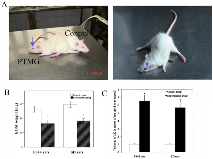
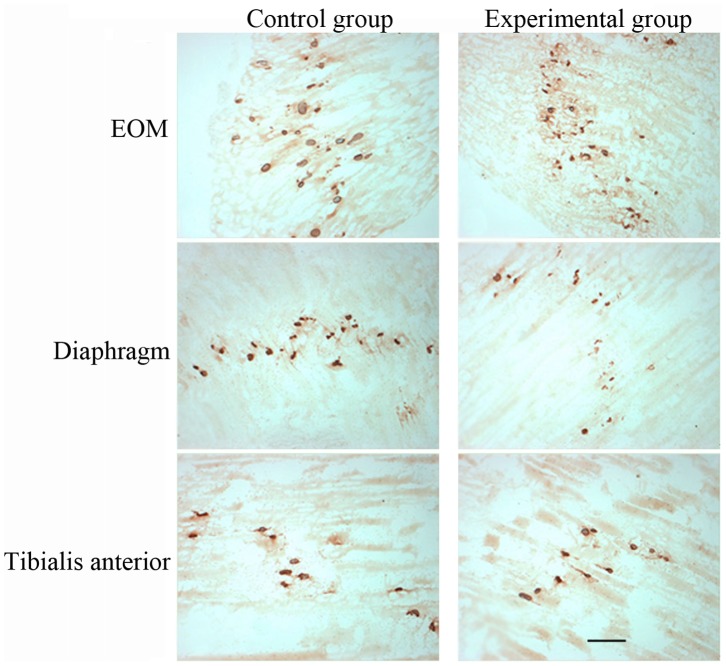
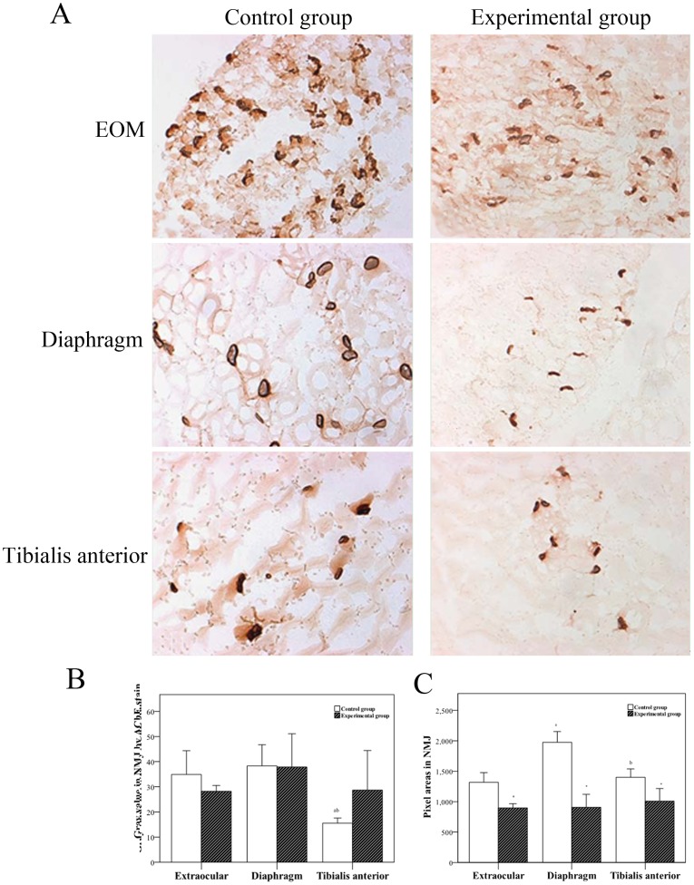
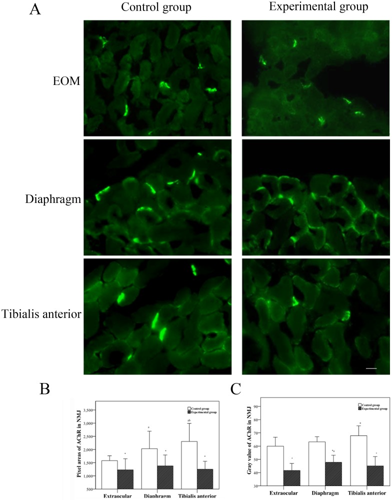
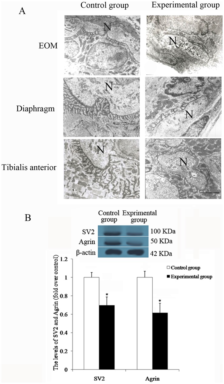
Methods: Female F344 rats and female Sprague-Dawley rats were assigned to experimental and control groups. The experimental group received injection with Ringer solution containing monoclonal antibody against the acetylcholine receptor (AChR), mAb35 (0.25 mg/kg), to induce experimental autoimmune myasthenia gravis, and the control group received injection with Ringer solution alone. Three muscles were analyzed: EOM, diaphragm, and tibialis anterior. Tissues were examined by light microscopy, fluorescence histochemistry, and transmission electron microscopy. Western blot analysis was used to assess marker expression and ELISA analysis was used to quantify creatine kinase levels. Microarray assay was conducted to detect differentially expressed genes.
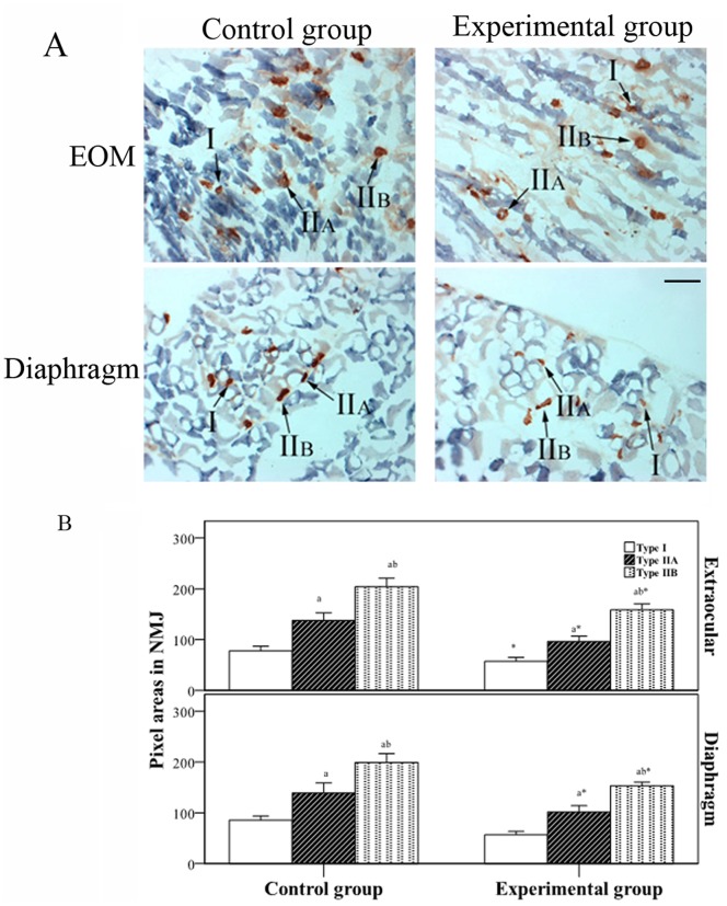
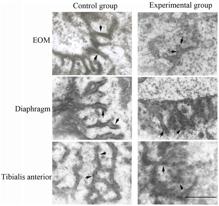
Results: In the experimental group, the EOM showed a simpler neuromuscular junction (NMJ) structure compared to the other muscles; the NMJ had fewer synaptic folds, showed a lesser amount of AChR, and the endplate was wider compared to the other muscles. Results of microarray assay showed differential expression of 54 genes in the EOM between the experimental and control groups.
Conclusion: Various EOM characteristics appear to be related to the increased susceptibility of the EOM and the mechanism of EOM weakness in myasthenia gravis.
Conflict of interest statement
Figures
Similar articles
-
RNA expression analysis of passive transfer myasthenia supports extraocular muscle as a unique immunological environment.Invest Ophthalmol Vis Sci. 2014 Jun 10;55(7):4348-59. doi: 10.1167/iovs.14-14422. Invest Ophthalmol Vis Sci. 2014. PMID: 24917137 Free PMC article.
-
Factors contributing to failure of neuromuscular transmission in myasthenia gravis and the special case of the extraocular muscles.Ann N Y Acad Sci. 2011 Sep;1233:26-33. doi: 10.1111/j.1749-6632.2011.06123.x. Ann N Y Acad Sci. 2011. PMID: 21950972
-
Reduction of miniature end-plate potential amplitude in extraocular and limb muscles in an animal model of myasthenia gravis.Exp Neurol. 1983 Apr;80(1):258-62. doi: 10.1016/0014-4886(83)90022-5. Exp Neurol. 1983. PMID: 6832272
-
Neuromuscular transmission failure in myasthenia gravis: decrement of safety factor and susceptibility of extraocular muscles.Ann N Y Acad Sci. 2012 Dec;1275(1):129-35. doi: 10.1111/j.1749-6632.2012.06841.x. Ann N Y Acad Sci. 2012. PMID: 23278588 Free PMC article. Review.
-
Pathophysiology of myasthenia gravis.Semin Neurol. 2004 Mar;24(1):21-30. doi: 10.1055/s-2004-829585. Semin Neurol. 2004. PMID: 15229789 Review.
Cited by
-
Ophthalmologic manifestations in myasthenia gravis: presentation and prognosis.Acta Neurol Belg. 2021 Oct;121(5):1131-1140. doi: 10.1007/s13760-020-01556-3. Epub 2021 Jan 4. Acta Neurol Belg. 2021. PMID: 33439450
-
Controversies in Ocular Myasthenia Gravis.Front Neurol. 2020 Nov 30;11:605902. doi: 10.3389/fneur.2020.605902. eCollection 2020. Front Neurol. 2020. PMID: 33329368 Free PMC article. Review.
-
Ocular Weakness in Myasthenia Gravis: Changes in Affected Muscles are a Distinct Clinical Feature.J Neuromuscul Dis. 2019;6(3):369-376. doi: 10.3233/JND-190407. J Neuromuscul Dis. 2019. PMID: 31424417 Free PMC article.
-
Investigation of the differential susceptibility of extraocular muscles in patients diagnosed with ocular myasthenia gravis based on the computerized diplopia test and the Ocular Motor Nerve Palsy Scale.Front Neurol. 2024 May 30;15:1353248. doi: 10.3389/fneur.2024.1353248. eCollection 2024. Front Neurol. 2024. PMID: 38872815 Free PMC article.
-
Immunopathogenesis in Myasthenia Gravis and Neuromyelitis Optica.Front Immunol. 2017 Dec 12;8:1785. doi: 10.3389/fimmu.2017.01785. eCollection 2017. Front Immunol. 2017. PMID: 29312313 Free PMC article. Review.
References
-
- Hughes BW, De Casillas MLM, Kaminski HJ (2004) Pathophysiology of myasthenia gravis. Semin Neurol 24: 21–30. - PubMed
-
- Kaminski HJ, Ruff RL (1997) Ocular muscle involvement by myasthenia gravis. Ann Neurol 41: 419–420. - PubMed
-
- Kaminski HJ, Li Z, Richmonds C, Ruff RL, Kushner L (2003) Susceptibility of ocular tissues to autoimmune diseases. Ann N Y Acad Sci 998: 362–374. - PubMed
-
- Kaminski HJ, Kusner LL, Block CH (1996) Expression of acetylcholine receptor isoforms at extraocular muscle endplates. Invest Ophthalmol Vis Sci 37: 345–351. - PubMed
-
- Wang ZY, Diethelm-Okita B, Okita DK, Kaminski HJ, Howard JF, et al. (2000) T cell recognition of muscle acetylcholine receptor in ocular myasthenia gravis. J of Neuroimmunol 108: 29–39. - PubMed
Publication types
MeSH terms
LinkOut - more resources
Full Text Sources
Other Literature Sources
Medical
