. 2013;8(2):e55784.
doi: 10.1371/journal.pone.0055784. Epub 2013 Feb 7.

Genetic and pharmacological modifications of thrombin formation in apolipoprotein e-deficient mice determine atherosclerosis severity and atherothrombosis onset in a neutrophil-dependent manner

Affiliations

Genetic and pharmacological modifications of thrombin formation in apolipoprotein e-deficient mice determine atherosclerosis severity and atherothrombosis onset in a neutrophil-dependent manner

Julian I Borissoff et al. PLoS One. 2013.

Abstract

Background: Variations in the blood coagulation activity, determined genetically or by medication, may alter atherosclerotic plaque progression, by influencing pleiotropic effects of coagulation proteases. Published experimental studies have yielded contradictory findings on the role of hypercoagulability in atherogenesis. We therefore sought to address this matter by extensively investigating the in vivo significance of genetic alterations and pharmacologic inhibition of thrombin formation for the onset and progression of atherosclerosis, and plaque phenotype determination.

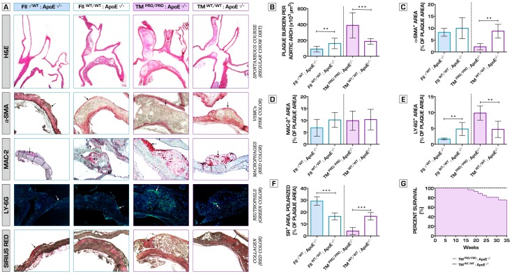
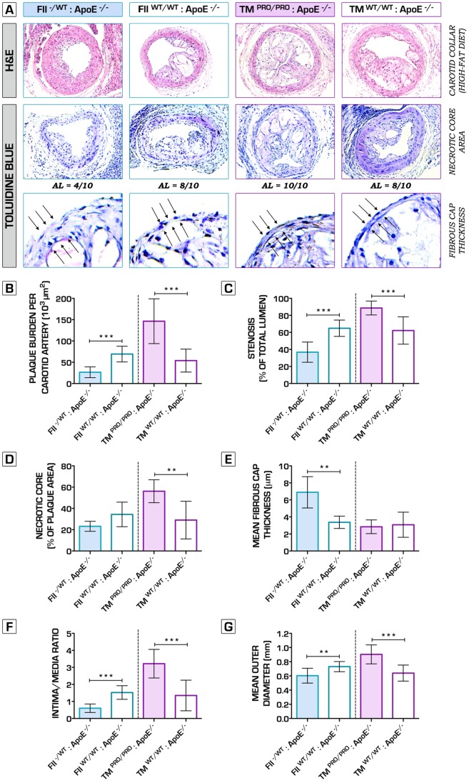
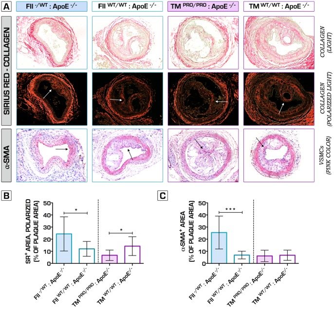
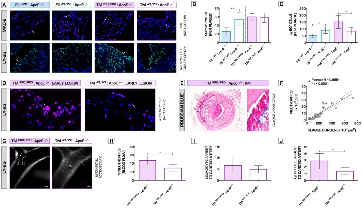
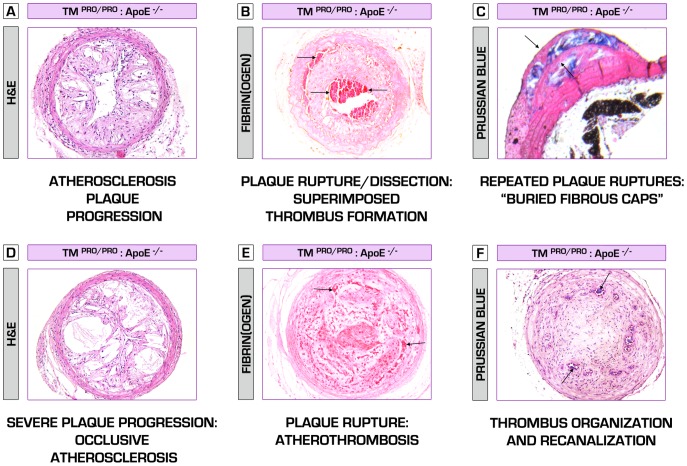
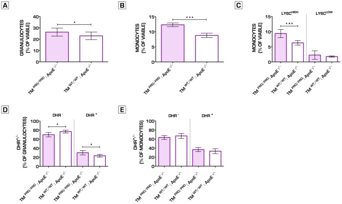
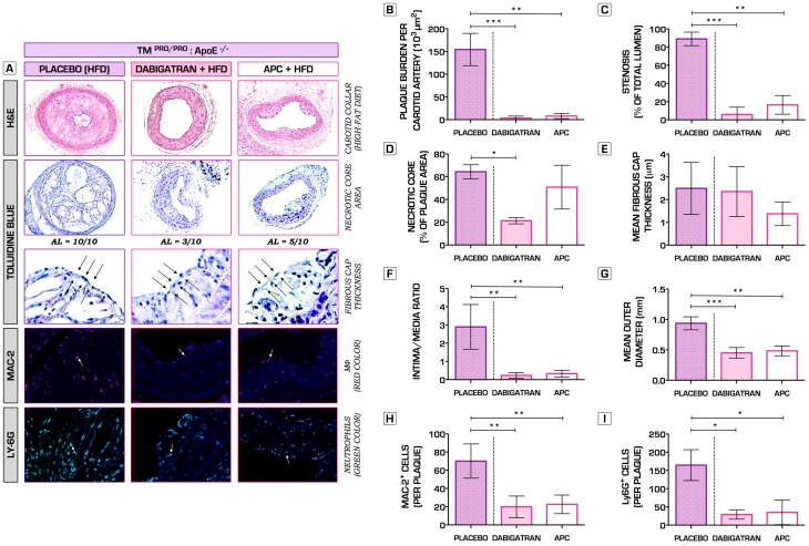
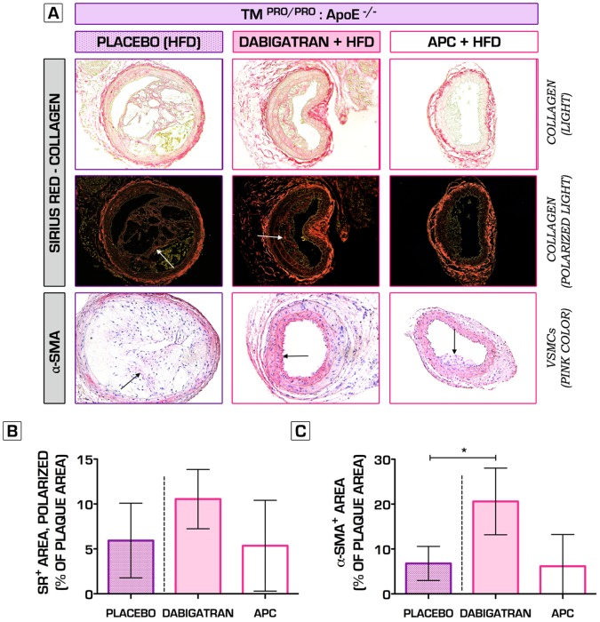
Methodology/principal findings: We generated transgenic atherosclerosis-prone mice with diminished coagulant or hypercoagulable phenotype and employed two distinct models of atherosclerosis. Gene-targeted 50% reduction in prothrombin (FII(-/WT):ApoE(-/-)) was remarkably effective in limiting disease compared to control ApoE(-/-) mice, associated with significant qualitative benefits, including diminished leukocyte infiltration, altered collagen and vascular smooth muscle cell content. Genetically-imposed hypercoagulability in TM(Pro/Pro):ApoE(-/-) mice resulted in severe atherosclerosis, plaque vulnerability and spontaneous atherothrombosis. Hypercoagulability was associated with a pronounced neutrophilia, neutrophil hyper-reactivity, markedly increased oxidative stress, neutrophil intraplaque infiltration and apoptosis. Administration of either the synthetic specific thrombin inhibitor Dabigatran etexilate, or recombinant activated protein C (APC), counteracted the pro-inflammatory and pro-atherogenic phenotype of pro-thrombotic TM(Pro/Pro):ApoE(-/-) mice.

Conclusions/significance: We provide new evidence highlighting the importance of neutrophils in the coagulation-inflammation interplay during atherogenesis. Our findings reveal that thrombin-mediated proteolysis is an unexpectedly powerful determinant of atherosclerosis in multiple distinct settings. These studies suggest that selective anticoagulants employed to prevent thrombotic events may also be remarkably effective in clinically impeding the onset and progression of cardiovascular disease.


Conflict of interest statement

Competing Interests: The authors have the following interests. Boehringer Ingelheim GmbH provided Dabigatran etexilate-supplemented and placebo diets for this study and is the employer of Joanne van Ryn. Dr. ten Cate has received speaker fees from Bayer, Boehringer Ingelheim, GlaxoSmithKline and Leo Pharma. There are no further patents, products in development or marketed products to declare. This does not alter the authors‚ adherence to all the PLOS ONE policies on sharing data and materials.

Figures

Figure 1
Figure 1. The effects of variations in coagulation potential on atherogenesis in a spontaneous atherosclerosis model at 35 weeks on a regular chow diet.
(A) Top row represents images of the aortic arch and its main branches, stained with hematoxylin and eosin (H&E), used to analyze the extent of atherosclerotic plaque burden. To determine plaque phenotype characteristics, sections were stained against α-smooth muscle actin (vascular smooth muscle cell content – second row), MAC-2+ (macrophage infiltration – third row), Ly-6G (neutrophil recruitment – fourth row) and with Sirius red (collagen – bottom row). (B) Hypocoagulability in FII−/+:ApoE−/− significantly attenuated atherosclerosis plaque development (90.6±35.1*103 µm2 total plaque burden) when compared to normal ApoE−/− mice (160.6±65.9*103 µm2)(n = 10 per group, p = 0.0084). Total plaque area in TMPro/Pro:ApoE−/− mice was established 389.1±158.4*103 vs. 187.0±35.1*103 µm2 in the corresponding control ApoE−/− group (n = 10 per group, p = 0.0010). (C) TMPro/Pro:ApoE−/− mice atherosclerotic plaques demonstrated a significant decrease in intimal vascular smooth muscle cell content (2.2±1.3% of plaque area) compared to ApoE−/− mice (8.7±2.9% of plaque area)(n = 10 per group, p = 0.0016). Recruitment of macrophages within the lesions did not differ between all experimental groups (D). Neutrophil infiltration was significantly diminished in the lesions of hypocoagulable FII−/+:ApoE−/− mice (n = 10 per group, p = 0.0092 vs. ApoE−/− mice), and substantially increased in the TMPro/Pro:ApoE−/− intima (n = 10 per group, p = 0.0094 vs. ApoE−/− mice) (E). A similar trend was observed with regard to collagen deposition within the atherosclerotic plaques. In FII−/+:ApoE−/− mice, 29.3±3.6% of the plaque area stained collagen-positive (n = 10 per group, p = 0.0002 vs. ApoE−/− mice). In contrast, Sirius red staining showed only 4.1±3.0% positivity for collagen in the TMPro/Pro:ApoE−/− lesions (n = 10 per group, p = 0.0002 vs. ApoE−/− mice) (F). By 35 weeks (established duration of the experiment), we recorded the following fatal events: 6 of 16 TMPro/Pro:ApoE−/−, 1 of 11 FII−/+:ApoE−/− and 0 of 20 ApoE−/− control mice. Dead mice were not included from the study analyses. The exact cause of death remained unclear. Kaplan-Meier analysis of the survival data comparing TMPro/Pro:ApoE−/− vs. ApoE−/− mice, as determined by the Gehan-Breslow-Wilcoxon test, indicated that hypercoagulability is linked to significantly higher spontaneous mortality rates (p = 0.0165) (G). No significant difference was found between FII−/+:ApoE−/− and ApoE−/− control mice (p = 0.3173) (data not shown).*p<0.05; **p<0.01; ***p<0.001. Error bars represent mean ± SD. Arrows indicate examples of positive staining. Abbreviations: H&E – hematoxylin and eosin; α-SMA - α-smooth muscle actin; SR – Sirius red.
Figure 2
Figure 2. Morphometrical analysis of periadventitial cuff-induced atherosclerosis in mice with genetically imposed alterations in blood coagulation potential.
(A) Representative hematoxylin and eosin (H&E)-stained sections of carotid arteries of FII−/+:ApoE−/−, TMPro/Pro:ApoE−/− and control ApoE−/− mice (top row). Necrotic core areas of the atherosclerotic lesions were identified and quantified by using toluidine blue (TB) staining (second and third row). (B, C) Whereas hypocoagulable mice were significantly protected against plaque progression (26.5±12.6*103 in FII−/+:ApoE−/− vs. 69.2±18.4*103 µm2 in ApoE−/− control mice, n = 10 per group, p<0.0001), pro-thrombotic mice developed severe and occlusive atherosclerotic burden (146.4±52.7*103 in TMPro/Pro:ApoE−/− vs. 53.9±27.0*103 µm2 in ApoE−/− control mice, n = 10 per group, p = 0.0001). The degree of stenosis in TMPro/Pro:ApoE−/− reached an average of 88.6±8.1% (vs. 62.2±16.1% in ApoE−/− mice, n = 10 per group, p = 0.0002), whereas it was substantially lower in FII−/+:ApoE−/− mice (36.8±11.9% vs. 64.9±9.6% in ApoE−/− mice, n = 10 per group, p<0.0001). (A, D) Pearson's chi-squared test (χ 2) detected a significant difference in the number of advanced atherosclerotic lesions (presence of fibrous cap atheromata [54]) formed between FII−/+:ApoE−/− (4 out of 10) and TMPro/Pro:ApoE−/− mice (10 out of 10) (n = 10 per group, p = 0.0108). In fact, the necrotic area within the lesions of the hypercoagulable mice was significantly increased: 56.2±10.8% of the total plaque area, as compared to 29.0±17.7% in the control ApoE−/− group (n = 10 per group, p = 0.0024). (E) Hypocoagulable mice showed more stable advanced lesions, as indicated by the significantly thicker fibrous caps in comparison to ApoE−/− mice (n = 10 per group, p = 0.0081). (F) Intima/media ratio was significantly increased in TMPro/Pro:ApoE−/− mice, whereas profoundly decreased in FII−/+:ApoE−/− mice. Of note, the average outer diameter of the common carotid artery is 0.36 mm , thus suggesting that TMPro/Pro:ApoE−/− atherosclerotic plaques undergo a dramatic outward remodeling as indicated in panel (G). *p<0.05; **p<0.01; ***p<0.001. Error bars represent mean ± SD. Arrows indicate examples of positive staining/fibrous cap thickness. Abbreviations: H&E – hematoxylin and eosin; AL – advanced atherosclerotic lesion.
Figure 3
Figure 3. The role of hypo- and hypercoagulability in plaque fibrosis.
Picrosirius red-stained sections assessed by light (A, top row) and polarized light (A, second row), indicate a significant decrease in the levels of collagen in TMPro/Pro:ApoE−/− carotid atherosclerotic plaques (6.7±4.3% vs. 14.3±7.8% of total plaque area in ApoE−/− mice, n = 10 per group, p = 0.0193) (B). Hypocoagulable FII−/+:ApoE−/− mice lesions showed a pro-fibrotic appearance, testified by increased collagen deposition (24.4±14.1% vs. 12.0±6.1% of total plaque area in ApoE−/− mice, n = 10 per group, p = 0.0435) and α-smooth muscle actin content (25.5±13.6% vs. 6.9±3.2% of total plaque area in ApoE−/− mice, n = 10 per group, p = 0.0003) (B, C). *p<0.05; **p<0.01; ***p<0.001. Error bars represent mean ± SD. Arrows indicate examples of positive staining. Abbreviations: SR – (Picro)sirius red; α-SMA - α-smooth muscle actin.
Figure 4
Figure 4. Hypercoagulability promotes neutrophil intraplaque recruitment and severe plaque phenotype.
(A) Representative sections of atherosclerotic lesions formed in the carotid arteries of TMPro/Pro:ApoE−/− and FII−/+:ApoE−/− mice (incl. control ApoE−/− mice), stained for the presence of macrophages (MAC-2, red color, top row) and neutrophils (Ly-6G, green color, bottom row). Arrows show examples of positive cells. DNA is counterstained in blue. Macrophage and neutrophil infiltration were expressed as the absolute number of Mac-2+ and Ly-6G+ cells per plaque. (A, B) Hypocoagulability in FII−/+:ApoE−/− mice promoted an anti-inflammatory plaque profile, indicated by a significant decrease in macrophage infiltration compared to control ApoE−/− mice: 26±9 vs. 54±15 cells per plaque (n = 10 per group, p = 0.0067). No difference in macrophage content was observed between TMPro/Pro:ApoE−/− and ApoE−/− mice atherosclerotic plaques: 60±8 vs. 58±11 cells per plaque (n = 10 per group, p = 0.7479). (A, C) Neutrophil accumulation was significantly increased in TMPro/Pro:ApoE−/− lesions (151±48 vs. 83±28 cells per plaque in ApoE−/− mice, respectively; n = 10 per group; p = 0.0260). Interestingly, the opposite trend was observed in mice with genetically imposed hypoprothrombinemia (51±12 vs. 90±24 cells per plaque in ApoE−/− mice, respectively; n = 10 per group; p = 0.0127). (D) Representative sections of early atherosclerotic lesions in external carotid artery of a TMPro/Pro:ApoE−/− mouse (left), abundantly infiltrated by Ly-6G+ cells (Ly-6G, red color), suggesting that hypercoagulability triggers lesion formation in a neutrophil-dependent manner. ApoE−/− control mice are shown on the right hand side. (E) The panel represents an atherosclerotic dissection with superimposed thrombus formation in a TMPro/Pro:ApoE−/− mouse at 6 weeks after carotid collar placement. Using Perl’s Prussian blue stain (blue color), we detected free ferric ions deposited within the sites of plaque dissection, indicating the areas of intraplaque hemorrhage. Whereas the carotid lesions in 5 out of 10 TMPro/Pro:ApoE−/− mice were associated with either rupture, dissection or intraplaque hemorrhage, none of the control ApoE−/− mice plaques had any signs of severe plaque vulnerability (Pearson's chi-squared test (χ 2), n = 10 per group, p = 0.0325). Statistical analysis including all experimental groups indicated that the number of circulating neutrophils in peripheral blood was strongly correlated to the extent of atherosclerotic plaque burden (F). Using intravital microscopy, we confirmed that the relative percentage of circulating neutrophils in TMPro/Pro:ApoE−/− mice was found significantly higher than ApoE−/− control mice (n = 6 per group, p = 0.0107). Whereas there were no differences found in the general leukocyte rolling and arrest between TMPro/Pro:ApoE−/− and ApoE−/− mice after 6 weeks on a high-fat diet (Rhodamine-labeled leukocytes) (n = 6 per group, p = 0.2886), Ly-6G+ neutrophils in TMPro/Pro:ApoE−/− mice were significantly more adherent to atherosclerotic lesions in the common carotid artery than in ApoE−/− control mice (n = 6 per group, p = 0.0139). Bar represents 100 µm. (G,H,I, J). *p<0.05; **p<0.01; ***p<0.001. Error bars represent mean ± SD. Arrows indicate examples of positive staining. DNA was counterstained with Hoechst-33342 (blue). Abbreviations: MФ – Macrophages; IPH – intraplaque hemorrhage; TAT – thrombin-antithrombin complex.
Figure 5
Figure 5. Hypercoagulable TMPro/Pro:ApoE−/− mice – a new mouse model of atherosclerotic plaque vulnerability.
Here we present a new hypercoagulable atherosclerosis model, which closely mimics the composition and events leading to plaque destabilization, as normally observed in human atherothrombosis. In a series of sections, demonstrating carotid atherosclerotic plaques, obtained from TMPro/Pro:ApoE−/− mice at 6 weeks after collar placement on high-fat diet regimen, we show multiple signs of plaque vulnerability. (A) A non-occlusive but rapidly progressing atherosclerotic lesion, characterized by abundant infiltration of leukocytes. (B) TMPro/Pro:ApoE−/− mice plaques tend to rupture and dissect (upper arrow) even during the non-occlusive phase, accompanied by “silent” intraluminal thrombosis (lower arrows). Despite the detrimental pathologic characteristics of those lesions, these data confirm the hypothesis that arterial thrombosis might exist long before a fatal event takes place. This is further consolidated by the presence of so called “buried fibrous caps” (indicated by the arrows) in TMPro/Pro:ApoE−/− mice plaques , considered a marker of healed plaque ruptures, and also observed in human atherosclerosis. Blue color denotes a massive intraplaque hemorrhage (iron ions deposition) (C). Hypercoagulability induces a severe inflammatory and pro-necrotic intraplaque environment, leading to the formation of enormous necrotic core, thin fibrous caps, further plaque destabilization (D) and atherothrombosis (occlusive intraluminal thrombosis/abundant fibrin(ogen) deposition (indicated by the arrows)) (E). Thrombi undergo fibrotic organization involving vascular smooth muscle cells and fibroblasts ingrowth, and are then partially recanalized by newly formed vessels (arrows, blue color – iron deposition/presence of erythrocytes)(F).
Figure 6
Figure 6. Hypercoagulability induces oxidative stress in granulocytes within the bone marrow compartment.
Granulocytes and monocytes cell fractions in the bone marrow were significantly increased in TMPro/Pro:ApoE−/− as compared to ApoE−/− control mice after 8 weeks on a regular chow diet (Granulocytes: 26.3±3.6% vs. 22.9±3.4%; n = 12 per group, p = 0.0292)(Monocytes: 12.3±0.6% vs. 8.8±0.7%; n = 12 per group, p<0.0001) (A, B). The significant increase in monocytes can be explained by the higher relative numbers of Ly6CHIGH monocyte cells in TMPro/Pro:ApoE−/− mice bone marrow (Ly6CHIGH cells: 9.4±1.3% vs. 6.3±0.8%; n = 12 per group, p = 0.0002) (C). Using DHR123 FACS analysis, we analyzed the amount of oxidative burst activity in granulocytes and monocytes in the bone marrow after PMA stimulation. The monocytes did not show any differences in DHR signal and thus ROS activity, whereas in the granulocytes of the TMPro/Pro:ApoE−/− mice, a significant increase was observed in the DHR signal when compared to ApoE−/− mice, indicating enhanced oxidative stress upon PMA stimulation in the TMPro/Pro:ApoE−/− granulocytes present in the bone marrow (D, E). *p<0.05; **p<0.01; ***p<0.001. Error bars represent mean ± SD. Abbreviations: DHR123– Dihydrorhodamine 123; ROS – Reactive Oxygen Species; PMA - Phorbol 12-Myristate 13-Acetate.
Figure 7
Figure 7. Inhibition of thrombin activity by administration of direct thrombin inhibitor Dabigatran etexilate or recombinant murine APC substantially attenuates leukocyte recruitment and prevents against severe atherosclerosis progression and atherothrombosis.
(A) Representative hematoxylin and eosin (H&E)-stained sections of atherosclerotic lesions formed in carotid arteries of TMPro/Pro:ApoE−/− mice, which were assigned to different intervention arms (oral Dabigatran etexilate - 7.5 mg DE/gram chow; i.p. administered bolus doses of recombinant murine APC - 2.5 mg/kg/per every 5 days; or placebo) for a total of 6 weeks after cuff placement around the common carotid arteries (top row). Toluidine blue (TB) stainings were used to quantify the size of necrotic core areas (second and third row). Whereas placebo treated TMPro/Pro:ApoE−/− mice all developed advanced lesions (identified by the presence of necrotic core and fibrous cap formation), Dabigatran etexilate- (3 out of 10, Pearson's chi-squared test (χ 2), n = 10 per group, p = 0.0031 vs. placebo) and rAPC-treated mice (5 out of 10, Pearson's chi-squared test (χ 2), n = 10 per group, p = 0.0325 vs. placebo) had significantly reduced atheromata formed. A total of 5 out 10 animals in the placebo group showed signs of severe plaque vulnerability, whereas none were observed in the intervention arms. Atherosclerotic plaques were further analyzed for the presence of macrophages (MAC-2, red color, fourth row) and neutrophils (Ly-6G, green color, bottom row). Arrows show examples of positive cells. Macrophage and neutrophil infiltration were expressed as the absolute number of Mac-2+ and Ly-6G+ cells per plaque. (B) Administration of either Dabigatran etexilate or rAPC rescued the phenotype and pronouncedly reduced atherosclerotic plaque burden (Placebo: 154.3±35.5*103 µm2; Dabigatran Etexilate: 3.3±4.4*103 µm2, p<0.001; rAPC: 7.9±5.5*103 µm2, p<0.01; n = 10 per group). (C, F, G) These findings were further consolidated by a significant decrease in the degree of stenosis (with ∼80%), intima/media ratio and outward remodeling within the treatment arms of the study (n = 10 per group). (D, E) Except for a significant reduction of the necrotic core area in the Dabigatran etexilate-treated mice as compared to placebo group (n = 10 per group, p<0.05), no other effects were observed with regard to necrotic core formation or fibrous cap thickness. Of note, only mice having advanced lesions were included in these analyses (Dabigatran etexilate: n = 3; rAPC: n = 5). (H, I) In addition, TMPro/Pro:ApoE−/− mice treated with direct thrombin inhibitor or natural anticoagulant rAPC developed an anti-inflammatory stable plaque phenotype, associated with substantially reduced levels of macrophage and neutrophil recruitment. *p<0.05; **p<0.01; ***p<0.001. Error bars represent mean ± SD. Arrows indicate examples of positive staining/fibrous cap thickness. DNA was counterstained with Hoechst-33342 (blue). Abbreviations: HFD – high-fat diet; AL – advanced atherosclerotic lesion; MФ- macrophage; rAPC – recombinant murine activated protein C.
Figure 8
Figure 8. The effects of direct and indirect inhibition of thrombin activity on plaque fibrosis.
Picrosirius red-stained sections assessed by light (A, top row) and polarized light (A, second row), indicate no significant changes in collagen content in TMPro/Pro:ApoE−/− mice after 6-week treatment with either Dabigatran etexilate or mouse rAPC, as compared to placebo (B). Administration of oral Dabigatran etexilate led to a significant increase in α-smooth muscle actin intraplaque content in TMPro/Pro:ApoE−/− mice vs. placebo treatment (20.6±7.4% vs. 6.8±3.8% of total plaque area, n = 10 per group, p<0.05) (A, C). rAPC therapy did not have an effect (6.2±7.1% of total plaque area, n = 10 per group, p>0.05) (A, C). *p<0.05; **p<0.01; ***p<0.001. Error bars represent mean ± SD. Arrows indicate examples of positive staining. Abbreviations: SR – (Picro)sirius red; α-SMA - α-smooth muscle actin; HFD – high-fat diet; VSMC – vascular smooth muscle cells; rAPC – recombinant mouse activated protein C.

Similar articles

Cited by

References

    1. Degen JL, Bugge TH, Goguen JD (2007) Fibrin and fibrinolysis in infection and host defense. Journal of thrombosis and haemostasis : JTH 5 Suppl 124–31. - PubMed
    1. Delvaeye M, Conway EM (2009) Coagulation and innate immune responses: can we view them separately? Blood 114: 2367–2374. - PubMed
    1. Massberg S, Grahl L, von Bruehl ML, Manukyan D, Pfeiler S, et al. (2010) Reciprocal coupling of coagulation and innate immunity via neutrophil serine proteases. Nature medicine 16: 887–896. - PubMed
    1. Ross R (1999) Atherosclerosis - an inflammatory disease. N Engl J Med 340: 115–126. - PubMed
    1. Libby P (2002) Inflammation in atherosclerosis. Nature 420: 868–874. - PubMed

Publication types

MeSH terms