. 2013;8(2):e55887.
doi: 10.1371/journal.pone.0055887. Epub 2013 Feb 11.

Urinary vitamin D binding protein: a potential novel marker of renal interstitial inflammation and fibrosis

Affiliations

Urinary vitamin D binding protein: a potential novel marker of renal interstitial inflammation and fibrosis

Katarina Mirković et al. PLoS One. 2013.

Abstract

Non-invasive tubulointerstitial damage markers may allow better titration and monitoring of renoprotective therapy. We investigated the value of urinary vitamin D binding protein excretion (uVDBP) as a tubulointerstitial inflammation and fibrosis marker in adriamycin rats, and tested whether uVDBP parallels renal damage and responds to therapy intensification in humans. In adriamycin (ADR) rats, uVDBP was strongly elevated vs controls (CON) already 6 wks after nephrosis induction (ADR: 727±674 [mean±SD] vs CON: 9±12 µg/d, p<0.01), i.e. before onset of pre-fibrotic and inflammatory tubulointerstitial damage, and at all following 6-wk time points until end of follow up at 30 wks (ADR: 1403±1026 vs CON: 206±132 µg/d, p<0.01). In multivariate regression analysis, uVDBP was associated with tubulointerstitial macrophage accumulation (standardized beta = 0.47, p = 0.01) and collagen III expression (standardized beta = 0.44, p = 0.02) independently of albuminuria. In humans, uVDBP was increased in 100 microalbuminuric subjects (44±93 µg/d) and in 47 CKD patients with overt proteinuria (9.2±13.0 mg/d) compared to 100 normoalbuminuric subjects (12±12 µg/d, p<0.001). In CKD patients, uVDBP responded to intensification of renoprotective therapy (ACEi+liberal sodium: 9.2±13.0 mg/d vs dual RAAS blockade+low sodium: 2747±4013, p<0.001), but remained still >100-fold increased during maximal therapy vs normoalbuminurics (p<0.001), consistent with persisting tubulointerstitial damage. UVDBP was associated with tubular and inflammatory damage markers KIM-1 (standardized beta = 0.52, p<0.001), beta-2-microglobuline (st.beta = 0.45, p<0.001), cystatin C (st.beta = 0.40, p<0.001), MCP-1 (st.beta = 0.31, p<0.001) and NGAL (st.beta = 0.20, p = 0.005), independently of albuminuria. UVDBP may be a novel urinary biomarker of tubulointerstitial damage. Prospectively designed studies are required to validate our findings and confirm its relevance in the clinical setting.


Conflict of interest statement

Competing Interests: The authors have declared that no competing interests exist.

Figures

Figure 1
Figure 1. Urinary vitamin D binding protein (uVDBP) and albuminuria levels, and structural renal changes over time in adriamycin and control rats. A.
Graphs illustrating the development of albuminuria (orange) and uVDBP (purple) over time in the unilateral adriamycin nephropathy model and in sham-operated control animals. B. Development of tubulointerstitial damage as reflected by interstitial α-smooth muscle actin (blue bars) and collagen III staining (green bars), and interstitial macrophage accumulation (red bars) in adriamycin rats (dark bars) and controls (light bars).
Figure 2
Figure 2. Representative photographs of renal damage markers in adriamycin-exposed and control kidneys.
Interstitial α-SMA increased progressively in proteinuric kidneys during follow up (A, B). α-SMA was located around dilated tubules (arrows) and in blood vessels (arrowheads). In control kidneys at week 30, only very limited expression of interstitial α-SMA was present (C). Interstitial macrophage influx increased in ADR animals (D, E). ED-1 positive cells were found mostly around damaged tubules (arrows) and sporadically present in the interstitium of control kidneys (F). Immunostaining of collagen III was found in ADR animals (G, H; arrows), and rarely in controls (I).
Figure 3
Figure 3. Urinary vitamin D binding protein (uVDBP) levels in patients with normoalbuminuria, microalbuminuria, and overt proteinuria.
Box-whisker plots indicating the median, interquartile range, and range of 24-h uVDBP per patient category: general population with normoalbuminuria, general population with microalbuminuria, chronic kidney disease with overt proteinuria treated with ACE inhibitor and liberal sodium diet, and chronic kidney disease with overt proteinuria treated with ACE inhibitor+ARB+low sodium diet. UVDBP increased with albuminuria (first three groups, p<0.001) and responded to intensification of anti-proteinuric treatment (two last groups, p<0.001).
Figure 4
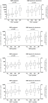
Figure 4. Urinary vitamin D binding protein (uVDBP) and established renal tubular damage markers in patients with renal damage.
Box-whisker plots illustrating the relation between uVDBP and established renal tubular damage markers kidney injury molecule-1 (KIM-1) (A), beta-2-microglobulin (B2M) (B), Cystatin C (CysC) (C) and monocyte chemoattractant protein-1 (MCP-1) (D) in normo- and microalbuminuric subjects (both n = 100). The uVDBP is presented per quartile of the established renal damage marker studied. In all panels (A-D), the left four box whisker plots represent the unadjusted VDBP excretion, and the right four box whisker plots represent the uVDBP adjusted for albuminuria. All biomarkers are positively associated with uVDBP, also when VDBP was adjusted for albuminuria (p<0.001, Kruskal Wallis).

Similar articles

Cited by

References

    1. Liu Y (2011) Cellular and molecular mechanisms of renal fibrosis. Nat Rev Nephrol 7: 684–696. - PMC - PubMed
    1. Remuzzi G, Benigni A, Remuzzi A (2006) Mechanisms of progression and regression of renal lesions of chronic nephropathies and diabetes. J Clin Invest 116: 288–296. - PMC - PubMed
    1. Perico N, Cattaneo D, Remuzzi G (2009) Kidney injury molecule 1: In search of biomarkers of chronic tubulointerstitial damage and disease progression. Am J Kidney Dis 53: 1–4. - PubMed
    1. Verboven C, Rabijns A, De Maeyer M, Van Baelen H, Bouillon R, et al. (2002) A structural basis for the unique binding features of the human vitamin D-binding protein. Nat Struct Biol 9: 131–136. - PubMed
    1. Nykjaer A, Dragun D, Walther D, Vorum H, Jacobsen C, et al. (1999) An endocytic pathway essential for renal uptake and activation of the steroid 25-(OH) vitamin D3. Cell 96: 507–515. - PubMed

Publication types