. 2013;8(2):e55904.
doi: 10.1371/journal.pone.0055904. Epub 2013 Feb 11.

Differential roles of Hath1, MUC2 and P27Kip1 in relation with gamma-secretase inhibition in human colonic carcinomas: a translational study

Affiliations

Differential roles of Hath1, MUC2 and P27Kip1 in relation with gamma-secretase inhibition in human colonic carcinomas: a translational study

Frédérique Souazé et al. PLoS One. 2013.

Abstract

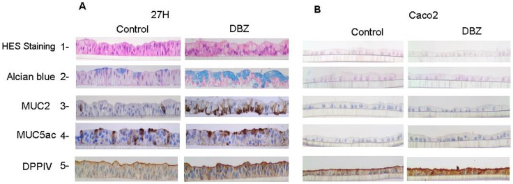
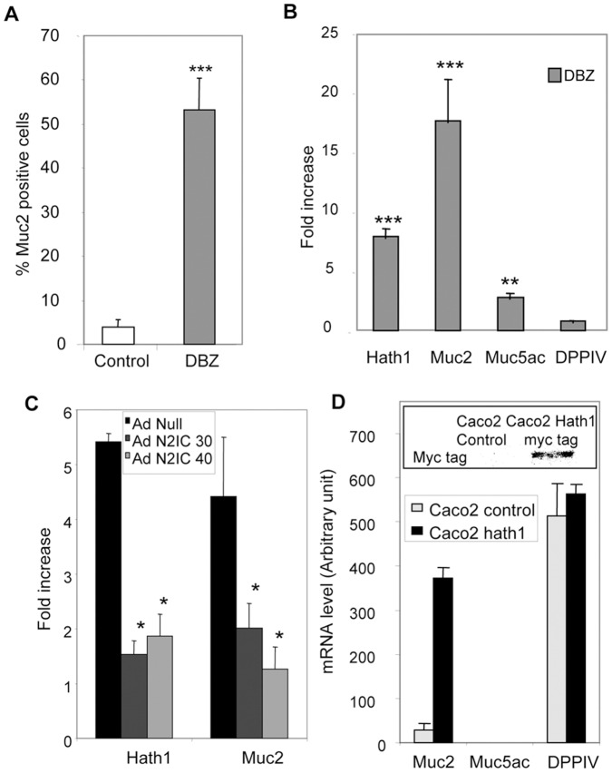
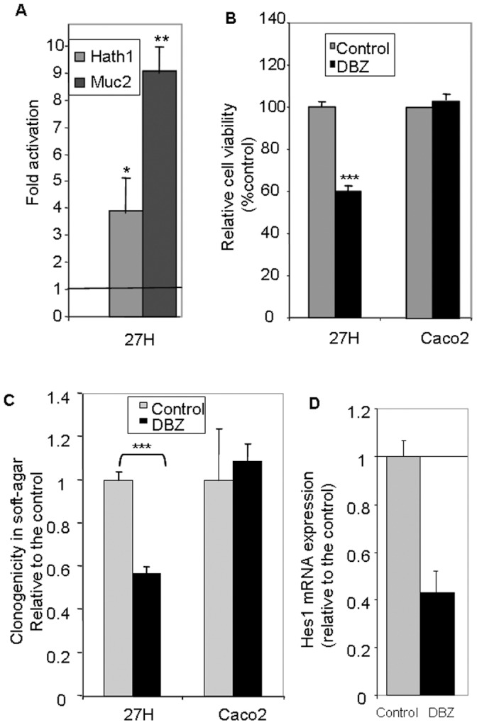
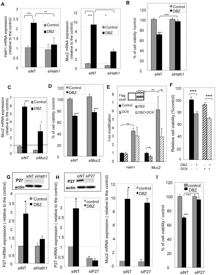
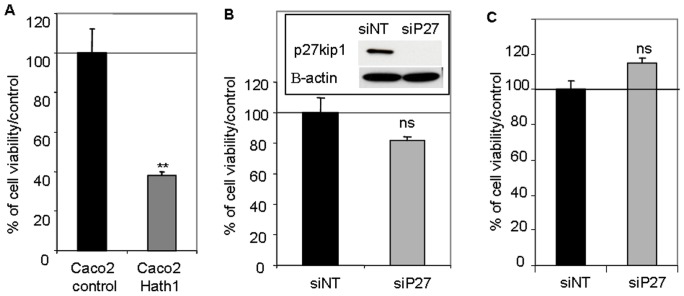
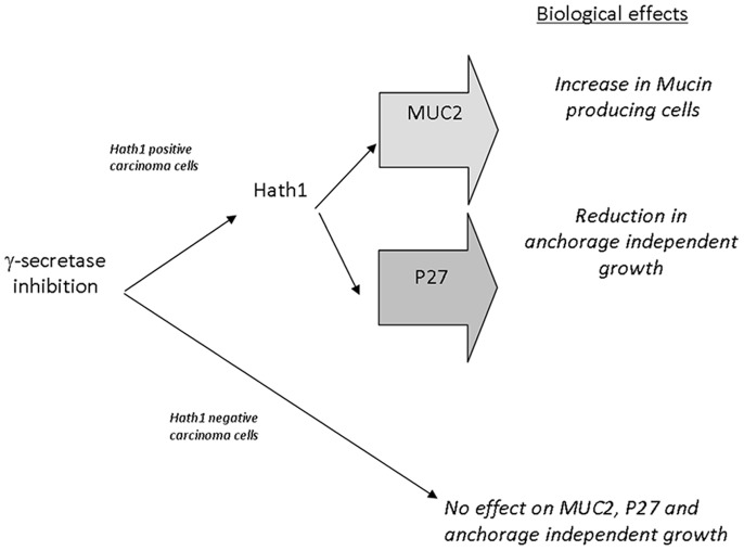
Hath1, a bHLH transcription factor negatively regulated by the γ-secretase-dependent Notch pathway, is required for intestinal secretory cell differentiation. Our aim was fourfold: 1) determine whether Hath1 is able to alter the phenotype of colon cancer cells that are committed to a differentiated phenotype, 2) determine whether the Hath1-dependent alteration of differentiation is coupled to a restriction of anchorage-dependent growth, 3) decipher the respective roles of three putative tumor suppressor genes Hath1, MUC2 and P27kip1 in this coupling and, 4) examine how our findings translate to primary tumors. Human colon carcinoma cell lines that differentiate along a mucin secreting (MUC2/MUC5AC) and/or enterocytic (DPPIV) lineages were maintained on inserts with or without a γ-secretase inhibitor (DBZ). Then the cells were detached and their ability to survive/proliferate in the absence of substratum was assessed. γ-secretase inhibition led to a Hath1-mediated preferential induction of MUC2 over MUC5AC, without DPPIV modification, in association with a decrease in anchorage-independent growth. While P27kip1 silencing relieved the cells from the Hath1-induced decrease of anchorage-independent growth, MUC2 silencing did not modify this parameter. Hath1 ectopic expression in the Hath1 negative enterocytic Caco2 cells led to a decreased anchorage-independent growth in a P27kip1-independent manner. In cultured primary human colon carcinomas, Hath1 was up-regulated in 7 out of 10 tumors upon DBZ treatment. Parallel MUC2 up-regulation occurred in 4 (4/7) and P27kip1 in only 2 (2/7) tumors. Interestingly, the response patterns of primary tumors to DBZ fitted with the hierarchical model of divergent signalling derived from our findings on cell lines.


Conflict of interest statement

Competing Interests: The authors have declared that no competing interests exist.

Figures

Figure 1
Figure 1. Morphological and immunohistochemical patterns of differentiation of filter-grown HT29-Cl.27H and Caco2 cells upon γ-secretase inhibition.
HT29-Cl.27H (A) or Caco2 (B) cells were seeded at high density on filters and treated or not (control, DMSO 0.01%) with DBZ (0.46 µM) for 21 days (see Material and Methods). Then the filters were fixed, embedded in paraffin and sectioned perpendicularly to the culture plane. The slides were stained with HES and Alcian Blue. MUC2, MUC5AC, and DPPIV expression were detected by immunohistochemistry as described in material and methods.
Figure 2
Figure 2. Effect of γ-secretase inhibition on the percentage of MUC2 positive cells and the expression of differentiation-associated genes.
A- Percentage of MUC2 positive cells in control and DBZ-treated cells maintained on filters was determined by immunohistochemistry as described in material and methods. Mean ± SEM of 3 filters after counting at least 200 cells/filter (***, p<0.001 DBZ vs control). B- RT-PCR detection of Hath1, MUC2, MUC5AC, DPPIV mRNA in HT29-Cl.27H filter cultures treated with DBZ for 21 days. The results of real-time quantitative PCR are expressed relative to the expression level of control cultures after normalization to actin gene expression. Mean relative expression to the control DMSO; Mean ± SEM of 4 experiments; ***, P<0.001; **, P<0.01 (DBZ vs control). C- RT-PCR detection of Hath1 and MUC2 mRNA in DBZ-treated HT29-Cl.27H cells infected by AdN2ICD Adenovirus (30 or 40 moi/cell); Mean ± SEM of 3 experiments; *, P<0.05. D- RT-PCR detection of MUC2, MUC5AC and DPP-IV mRNA in polyclonal populations of Caco2 cells stably transfected by Hath1 expression vector. (For comparison purpose the Cts for MUC2 PCR are: Caco2-cmv Ct = 32; Caco2-Hath1 Ct = 26.5 and Ht29-Cl.27H Ct = 26). Insert: c-myc tag was detected by immunoblot on polyclonal populations of Caco2 cells stably transfected by empty vector or Hath1 expression vector.
Figure 3
Figure 3. Effect of γ-secretase inhibition on cellular viability in anchorage-independent culture conditions in relation with Hath1 and MUC2 expression.
A and B- Filter-grown HT29-Cl.27H or Caco2 cells, treated or not (control) with DBZ for 21 days, were resuspended and plated on polyHEMA-coated wells (nonadherent conditions) for 72 hours culture in 10% FCS containing medium without DBZ. A- RT-PCR detection of Hath1 and MUC2 mRNA: mean expression relative to control DMSO after normalization to actin gene expression; Mean ± SEM of 3 experiments; *, P<0.05; **, p<0.01. B- Relative cell viability versus control determined by trypan blue dye exclusion cell count. C- Filter-grown HT29-Cl.27H or Caco2 cells, pre-treated or not (control) with DBZ for 21 days were detached and seeded at 10, 000 cells per 60 mm dish in 0.35% agarose. The cells were maintained in culture for additional 15 days without supplementation of DBZ. Clusters of minimum 10 cells were counted. Mean relative expression to the control DMSO; Mean ± SEM of 3 experiments (3–4 dishes per experiment); ***, p<0.001. D- RT-PCR detection of Hes1 in Caco2 cells, treated or not (control) with DBZ for 21 days on filters: mean expression relative to control DMSO after normalization to actin gene expression; Mean ± SEM of 3 experiments.
Figure 4
Figure 4. Effect of Hath1, MUC2 or P27Kip1 gene silencing on anchorage independent-growth.
A,B, C,D,G,H,I-Filter-grown HT29-Cl.27H, treated or not (control) with DBZ for 21 days, were resuspended and plated on polyHEMA-coated wells (nonadherent conditions) for 72 hours culture in the presence of the indicated siRNA target smart pool (NT, non target; Hath1; MUC2 or P27Kip1). A- RT-PCR detection of Hath1 (left) and Muc-2 (right) mRNA in HT29-Cl.27H cells transfected by siRNA (NT, or Hath1): mean expression relative to siNT DMSO after normalization to actin gene expression; Mean ± SEM of 3 experiments; *, P<0.05; **, p<0.01; ***, p<0.001. B- Relative cell viability determined by Trypan blue dye exclusion cell count. Mean ± SEM of 3 experiments; ***, p<0.001. C- RT-PCR detection of MUC2 mRNA in HT29-Cl.27H cells transfected by siRNA (NT and MUC2): mean expression relative to siNT DMSO after normalization to actin gene expression; Mean ± SEM of 3 experiments; *, P<0.05; ***, p<0.001. D- Relative cell viability determined by Trypan blue dye exclusion cell count. Mean ± SEM of 3 experiments; ***, p<0.001. E- Filter-grown HT29-Cl.16E sox9 cells, treated or not (control) with DBZ for 21 days, were resuspended and plated on polyHEMA-coated wells (nonadherent conditions) for 72 hours culture in 10% FCS containing medium with or without addition of doxycycline. RT-PCR detection of Hath1 and MUC2 mRNA: relative expression to control DMSO after normalization to actin gene expression; Mean ± SEM of 3 experiments; **, P<0.01; *, P<0.05. Insert: Flag-Sox9 was detected by immunoblot on cellular extracts after 72-hour doxycycline induction. F- Relative cell viability determined by Trypan blue dye exclusion cell count (in the same culture conditions as Fig. 4E). Mean ± SEM of 3 experiments; ***, p<0.001. G- and H- RT-PCR detection of P27Kip1 mRNA (G and H left) and MUC2 mRNA (H right) in HT29-Cl.27H cells transfected by siRNA (G -NT and Hath1, H- NT and P27): mean expression relative to siNT DMSO after normalization to actin gene expression; Mean ± SEM of 3 experiments; ***, p<0.001. Insert: P27Kip1 and β-actin were detected by immunoblot on DBZ pre-treated cells transfected by siRNA (G -NT and Hath1, H left- NT and P27). I- Relative cell viability determined by Trypan blue dye exclusion cell count of HT29-Cl.27H cells transfected by siRNA NT or P27; Mean ± SEM of 3 experiments; ***, p<0.001.
Figure 5
Figure 5. Effect of Hath1 ectopic expression and P27Kip1 silencing on anchorage-independent cell growth of Caco2 cells.
A- Polyclonal populations of Caco2 cells stably transfected by empty vector (Caco2 control) or Hath1 expression vector (Caco2 Hath1) were plated on polyHEMA-coated wells (nonadherent conditions) for 72 hours culture in 10% FCS containing medium. Relative cell viability determined by Trypan blue dye exclusion cell count. Mean ± SEM; **, p<0.01. B-C- Caco2 cells (B) or Hath1 over-expressing Caco2 cells (C) plated on polyHEMA-coated wells (nonadherent conditions) for 72 hours culture with siRNA target smart pool (NT or P27). Relative cell viability determined by Trypan blue dye exclusion cell count. (B) Insert: P27Kip1 and β-actin were detected by immunoblot on cells transfected by siRNA.
Figure 6
Figure 6. Proposed divergent signalling for gamma-secretase-dependent pathways of differentiation and anchorage-independent growth in colon cancer cells.
Biological effects induced by γ-secretase inhibition in colonic cancer cells were dependent on Hath1 expression. In Hath1 non-expressing cells, γ-secretase inhibition had no effect on MUC2, P27Kip1 and anchorage independent growth. In Hath1 positive cells, γ-secretase inhibition led to an increase of Hath1 expression leading to two separate effects on 1- increase in mucin producing cells with MUC2 expression and 2- reduction in anchorage independent growth mediated by the activation of the cyclin dependent kinase inhibitor P27Kip1.

Similar articles

Cited by

References

    1. Sikandar SS, Pate KT, Anderson S, Dizon D, Edwards RA, et al. (2010) NOTCH signaling is required for formation and self-renewal of tumor-initiating cells and for repression of secretory cell differentiation in colon cancer. Cancer Res 70: 1469–1478. - PMC - PubMed
    1. Fre S, Pallavi SK, Huyghe M, Lae M, Janssen KP, et al. (2009) Notch and Wnt signals cooperatively control cell proliferation and tumorigenesis in the intestine. Proc Natl Acad Sci U S A 106: 6309–6314. - PMC - PubMed
    1. Reedijk M, Odorcic S, Zhang H, Chetty R, Tennert C, et al. (2008) Activation of Notch signaling in human colon adenocarcinoma. Int J Oncol 33: 1223–1229. - PMC - PubMed
    1. Veenendaal LM, Kranenburg O, Smakman N, Klomp A, Borel Rinkes IH, et al. (2008) Differential Notch and TGFbeta signaling in primary colorectal tumors and their corresponding metastases. Cell Oncol 30: 1–11. - PMC - PubMed
    1. De Strooper B, Annaert W, Cupers P, Saftig P, Craessaerts K, et al. (1999) A presenilin-1-dependent gamma-secretase-like protease mediates release of Notch intracellular domain. Nature 398: 518–522. - PubMed

Publication types

MeSH terms

Substances