. 2013;8(2):e55945.
doi: 10.1371/journal.pone.0055945. Epub 2013 Feb 8.

Suppression of natural killer cells by sorafenib contributes to prometastatic effects in hepatocellular carcinoma

Affiliations

Suppression of natural killer cells by sorafenib contributes to prometastatic effects in hepatocellular carcinoma

Qiang-Bo Zhang et al. PLoS One. 2013.

Abstract

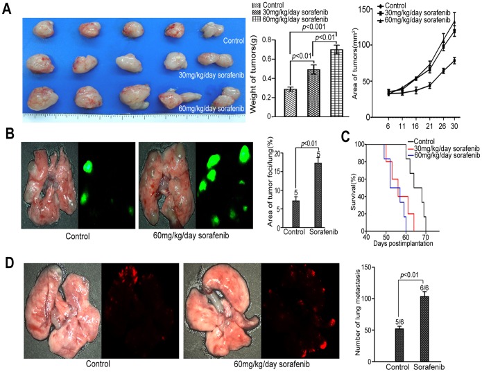
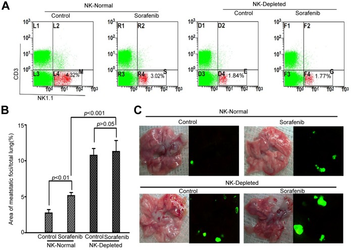
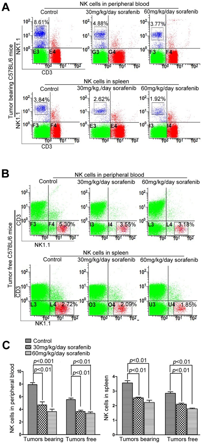
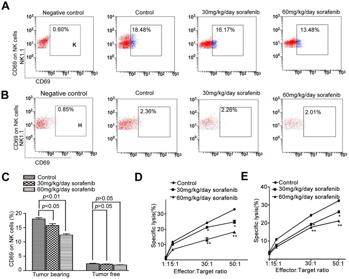
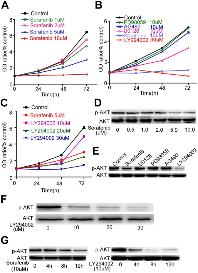
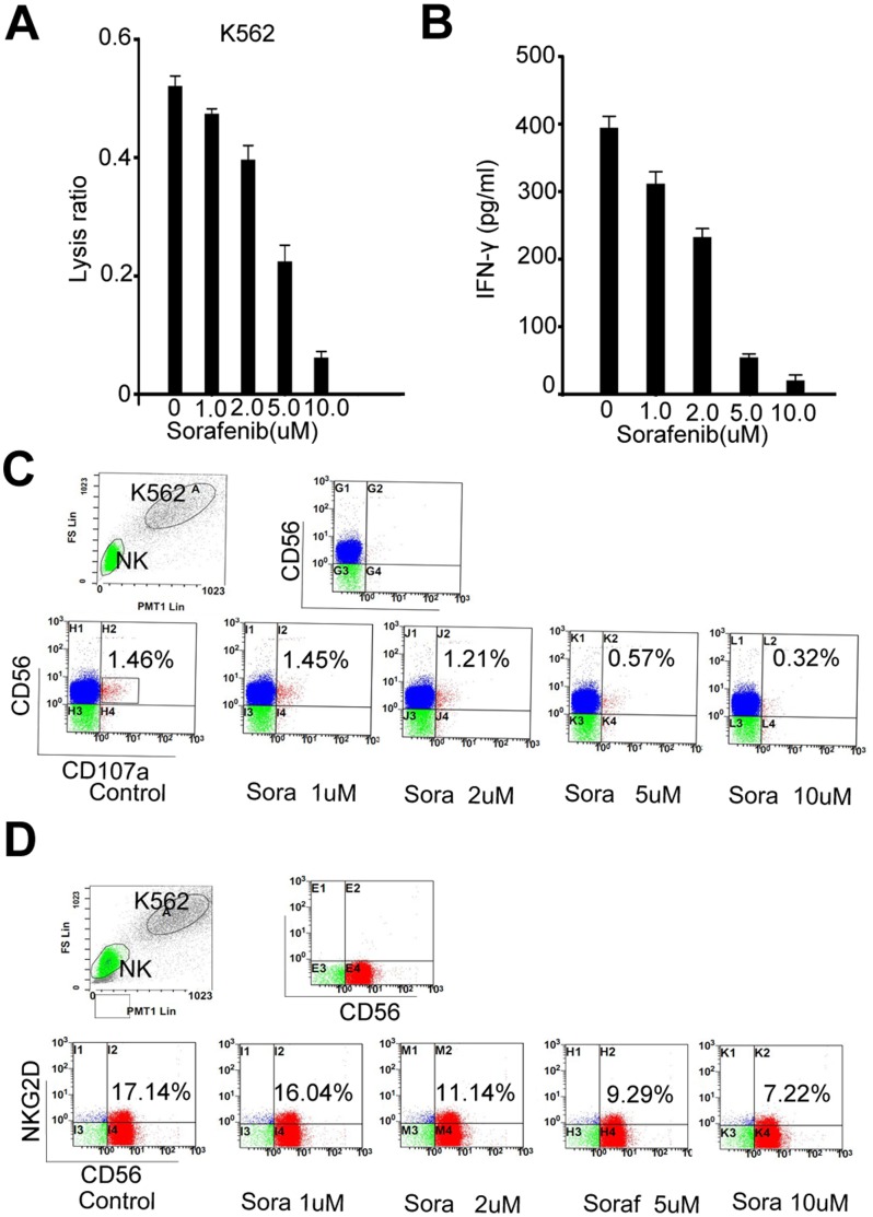
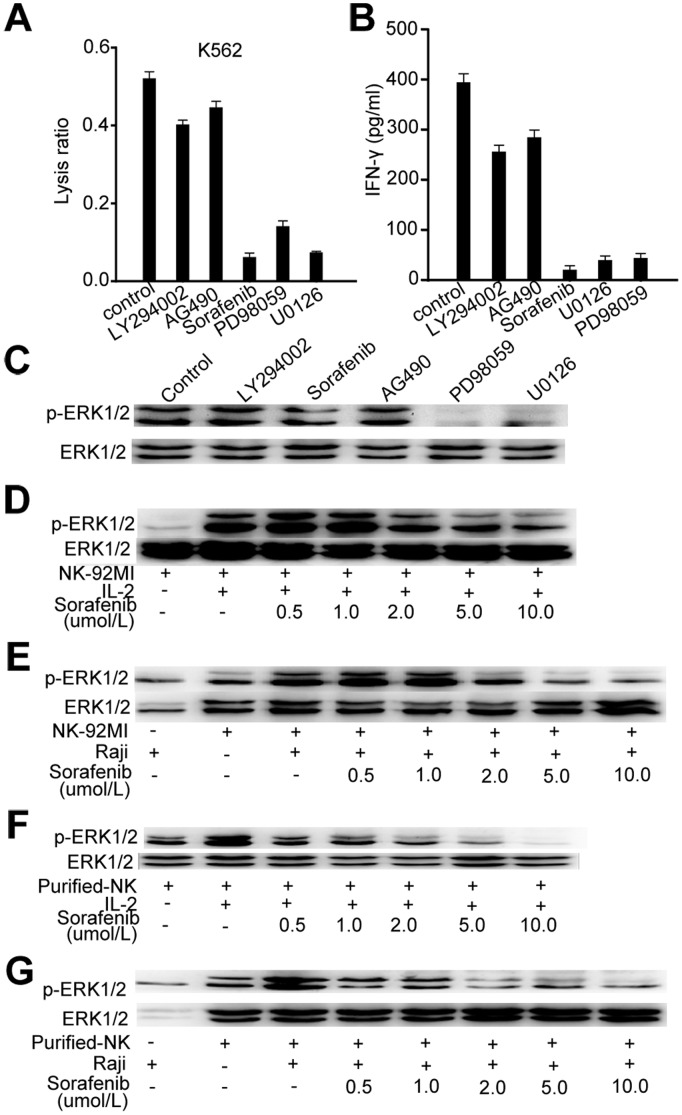
Sorafenib, a multi-tyrosine kinase inhibitor, is a standard treatment for advanced hepatocellular carcinoma (HCC). The present study was undertaken to determine whether the growth and metastasis of HCC were influenced in mice receiving sorafenib prior to implantation with tumors, and to investigate the in-vivo and in-vitro effect of sorafenib on natural killer (NK) cells. In sorafenib-pretreated BALB/c nu/nu mice and C57BL/6 mice, tumor growth was accelerated, mouse survival was decreased, and lung metastasis was increased. However, the depletion of NK1.1(+) cells in C57BL/6 mice eliminated sorafenib-mediated pro-metastatic effects. Sorafenib significantly reduced the number of NK cells and inhibited reactivity of NK cells against tumor cells, in both tumor-bearing and tumor-free C57BL/6 mice. Sorafenib down-regulated the stimulatory receptor CD69 in NK cells of tumor-bearing mice, but not in tumor-free mice, and inhibited proliferation of NK92-MI cells, which is associated with the blocking of the PI3K/AKT pathway, and inhibited cytotoxicity of NK cells in response to tumor targets, which was due to impaired ERK phosphorylation. These results suggest immunotherapeutic approaches activating NK cells may enhance the therapeutic efficacy of sorafenib in HCC patients.


Conflict of interest statement

Competing Interests: The authors have declared that no competing interests exist.

Figures

Figure 1
Figure 1. Sorafenib pretreatment accelerated tumor growth, promoted lung metastasis, and decreased overall survival in nude mice.
(A) In the HepG2 subcutaneous tumors model, sorafenib pretreatment accelerated the growth of ectopic tumors in nude mice. Tumor weight was 0.31±0.05 g, 0.51±0.09 g, and 0.73±0.09 g in the vehicle, 30 mg·kg−1·day−1 sorafenib, and 60 mg·kg−1·day−1 sorafenib groups, respectively (n = 5 for each group) (p<0.01, right panel). (B) In the HepG2-GFP experimental lung metastases model, sorafenib pretreatment enhanced the formation of lung metastatic foci, which was 7.19±2.16% in the vehicle group and 17.32±2.92% in the sorafenib group (n = 5 for each group) (p<0.01, right panel). (C) In the LM3-RFP orthotopic model of nude mice, sorafenib pretreatment increased the number of lung-metastatic foci, compared to the vehicle group (n = 6 for each group) (105.0±19.80 versus 59.7±16.8 per lung, p<0.01, right panel), but did not increase the rate of lung metastasis, which was 6 of 6 in the sorafenib group and 5 of 6 in controls. (D) Sorafenib significantly reduced median survival compared to vehicle in the nude mice inoculated HepG2 cells. Mice pretreated with 30 mg·kg−1·day−1 and 60 mg·kg−1·day−1 sorafenib survived 52 and 56 days, respectively, compared to 64 days for the controls (p<0.01).
Figure 2
Figure 2. Sorafenib pretreatment promoted lung metastasis only in mice with intact NK cells.
(A) At day 7, the ratio of NK cells in the spleens was determined by flow cytometry. (B) The metastatic index was measured by the ratio of the area of GFP-positive lung metastatic foci to total lung area. There was no significant difference between vehicle and sorafenib in NK cell–depleted groups (n = 5 for each group).
Figure 3
Figure 3. Sorafenib reduced the ratio of NK cells dose-dependently, in both tumor-free and tumor-bearing C57BL/6 mice.
Sorafenib treatment was started one week after tumor implantation and continued for two weeks. Then, NK cells were harvested for further measurement. (A, C) The ratio of NK cells was 7.92±0.61% in the controls and 4.70±0.98%, and 3.66±0.74% in the 30 mg·kg−1·day−1 sorafenib and 60 mg·kg−1·day−1 sorafenib groups (n = 5 for each group), respectively (p<0.001). For tumor-bearing mice, the ratio was 3.56±0.31% in controls and 2.54±0.99% and 2.23±0.30% in the 30 mg·kg−1·day−1 sorafenib and 60 mg·kg−1·day−1 sorafenib groups, respectively (p<0.01). (B, C) Sorafenib also reduced the ratio of NK cells dose-dependently in the peripheral blood and spleens of tumor-free mice (n = 5 for each group): 2.85±0.21% in controls and 2.11±0.15% and 1.78±0.11% in the 30 mg·kg−1·day−1 and 60 mg·kg−1·day−1 groups, respectively, in the spleens (p<0.001, right panel); and 5.54±0.49% in the controls and 3.66±0.43% and 3.37±0.45% in the 30 mg·kg−1·day−1 and 60 mg·kg−1·day−1 groups, respectively, in peripheral blood.
Figure 4
Figure 4. Sorafenib inhibited the activation and cytotoxicity reaction of NK cells in vivo.
(A, C) The ratio of CD69+ NK cells was 18.0±0.91% in controls, and 15.75±1.22% and 12.41±0.85% in the 30 and 60 mg·kg−1·day−1 groups, respectively (p<0.05) (n = 5 for each group). (B, C) NK cells were not activated in tumor-free mice, and sorafenib did not reduce the expression of CD69 (p>0.05). (D, E) Sorafenib substantially inhibited the cytolytic activity of isolated NK cells in response to YAC-1 cells from both tumor-bearing mice and blank mice (n = 5 for each group). *, 30 mg·kg−1·day−1 sorafenib group compared with control group; **, 60 mg·kg−1·day−1 sorafenib group compared with control group; *,**,P<0.05.
Figure 5
Figure 5. Sorafenib inhibited proliferation of NK-92MI cells mainly by blocking the PI3K/AKT pathway.
(A) Sorafenib inhibited proliferation of NK-92MI cells dose-dependently. (B) Addition of different specific inhibitors had different effects on the proliferation of NK-92MI cells at the indicated time. (C) Different concentrations of LY294002 had an inhibitory effect on proliferation of NK-92MI cells at the indicated time. (D, E) Cells were treated with different concentrations of Sorafenib and LY294002 for 6 h. (F) Cells were treated with different inhibitors (10 µM) for 6 h. (G) Cells were treated with sorafenib (10 µM) and LY294002 (10 µM) for different lengths of time.
Figure 6
Figure 6. Sorafenib impaired the reactivity of isolated human NK cells in vitro.
(A) Sorafenib reduced the lysis ratio of NK cells in response to K562 cells (E:T = 30∶1) in a dose-dependent manner. (B) Sorafenib decreased the production of IFN-γ in NK cells when cocultured with K562 at the ratio of 3∶1 for 12 h. (C) Sorafenib reduced the expression of CD107a in NK cells mixed with K562 cells at the ratio of 3∶1 for 2 h. Representative flow cytometry dot plots were showed. (D) The expression of NKG2D by NK cells was also reduced when they were mixed with K562 cells at the ratio of 3∶1 for 8 h. Representative flow cytometry dot plots were showed.
Figure 7
Figure 7. Sorafenib inhibited Raf/MEK/ERK signaling involved in NK activation.
(A) NK cells were cultured with K562 cells (E:T = 30∶1) in the presence of sorafenib (10 µM) and the indicated specific inhibitors (10 µM). (B) NK cells were cultured with K562 tumor cells in the presence of sorafenib (10 µM) and the indicated specific inhibitors (10 µM). After 24 h, IFN-γ levels in cell culture supernatants were determined by ELISA. (C) Isolated human NK cells were treated with sorafenib and the indicated specific inhibitors for 30 min and exposed to IL-2 for 10 min. Subsequently, cell lysates were analyzed for phosphorylated ERK (pERK1/2) and whole (panERK) ERK1/2 protein. (D, E) NK92-MI cells were treated with different concentrations of sorafenib for 30 min and exposed to IL-2 for 10 min or fixed Raji cells for 15 min. Subsequently, cell lysates were analyzed as described in C. (F, G) Isolated human NK cells were treated with different concentrations of sorafenib for 30 min and exposed to IL-2 for 10 min or fixed Raji cells for 15 min.

Similar articles

Cited by

References

    1. Jemal A, Bray F (2011) Center MM, Ferlay J, Ward E, et al (2011) Global cancer statistics. CA Cancer J Clin 61: 69–90. - PubMed
    1. Clavien PA, Petrowsky H, DeOliveira ML, Graf R (2007) Strategies for safer liver surgery and partial liver transplantation. N Engl J Med 356: 1545–1559. - PubMed
    1. Carr BI (2004) Hepatocellular carcinoma: current management and future trends. Gastroenterology 127: S218–224. - PubMed
    1. Johnson P, Billingham L (2009) Sorafenib for liver cancer: the horizon broadens. Lancet Oncol 10: 4–5. - PubMed
    1. Palmer DH (2008) Sorafenib in advanced hepatocellular carcinoma. N Engl J Med 359: 2498; author reply 2498–2499. - PubMed

Publication types

MeSH terms