. 2013;8(2):e55983.
doi: 10.1371/journal.pone.0055983. Epub 2013 Feb 7.

Identification of injury specific proteins in a cell culture model of traumatic brain injury

Affiliations

Identification of injury specific proteins in a cell culture model of traumatic brain injury

Camilla Lööv et al. PLoS One. 2013.

Abstract

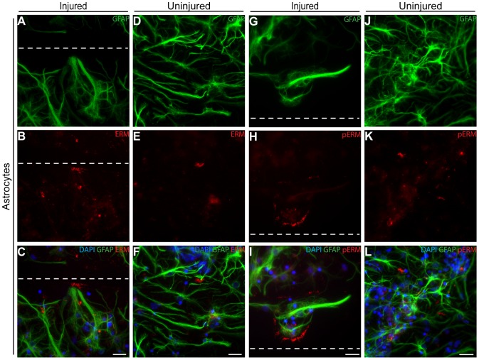
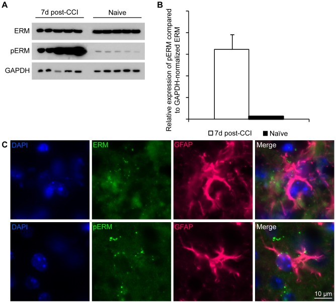
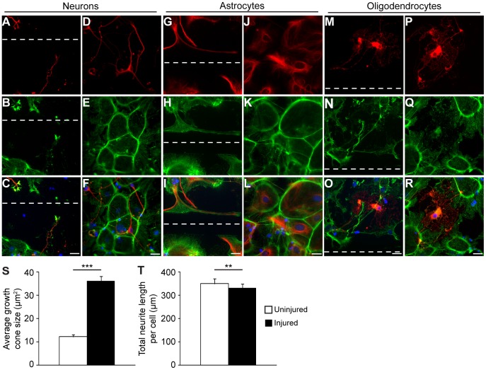
The complicated secondary molecular and cellular mechanisms following traumatic brain injury (TBI) are still not fully understood. In the present study, we have used mass spectrometry to identify injury specific proteins in an in vitro model of TBI. A standardized injury was induced by scalpel cuts through a mixed cell culture of astrocytes, oligodendrocytes and neurons. Twenty-four hours after the injury, cell culture medium and whole-cell fractions were collected for analysis. We found 53 medium proteins and 46 cell fraction proteins that were specifically expressed after injury and the known function of these proteins was elucidated by an extensive literature survey. By using time-lapse microscopy and immunostainings we could link a large proportion of the proteins to specific cellular processes that occur in response to trauma; including cell death, proliferation, lamellipodia formation, axonal regeneration, actin remodeling, migration and inflammation. A high percentage of the proteins uniquely expressed in the medium after injury were actin-related proteins, which normally are situated intracellularly. We show that two of these, ezrin and moesin, are expressed by astrocytes both in the cell culture model and in mouse brain subjected to experimental TBI. Interestingly, we found many inflammation-related proteins, despite the fact that cells were present in the culture. This study contributes with important knowledge about the cellular responses after trauma and identifies several potential cell-specific biomarkers.


Conflict of interest statement

Competing Interests: The authors have no competing interests.

Figures

Figure 1
Figure 1. Differences in regeneration of neurons, astrocytes and oligodendrocytes after injury.
(A) Phase-contrast of an injured culture. The scalpel cut create clear injury (dashed line) with surrounding cells that remain unharmed. (B–D) Displaying the individual cell types' appearance 24 h after injury. (B) The neurons have regenerated new axons towards and along the laceration, without breaching the boundary of the cut. (C) Astrocytes reach towards and along the injury, and like neurons, do not grow into the laceration. (D) Oligodendrocytes are more hesitant in their regeneration of the injury and unless in its direct vicinity, does not grow along the cut but rather avert it. (E–M) The actin patterns of the cells 24 h after injury implicates it as an important regeneratory protein in wound healing. (E–G) The outgrowing new growth cones along the injury are highly reactive for actin. (H–J) The astrocytes have extended a multitude of actin-positive lamellipodia towards and along the cut at 24 h after injury. (K–M) Twenty-four hours after injury, oligodendrocytes in direct proximity to the injury have some lamellipodia-like extensions towards the cut, although they appear more reluctant in covering the cut compared to both neurons and astrocytes. Scale bars equal 50 µm (A–D) or 10 µm (E–M) and dashed lines represent the injury.
Figure 2
Figure 2. Astrocytes are most likely the source of the ezrin and moesin found specifically in medium after injury.
(A–F) The ERM is expressed by astrocytes in a fingerlike pattern at 24 h post-injury (A–C) in comparison to the disorganized expression mostly seen in uninjured cultures (D–F). (G–L) Stainings with antibodies specifically against the active, phosphorylated form of ERM reveal the intracellular ERM expressed by astrocytes appear to be phosphorylated as it co-localizes with the expression patterns of ERM after injury (G–I) as well as the occasional patches of ERM seen in uninjured cultures (J–L). Scale bars equal 50 µm and dashed lines represent the injury.
Figure 3
Figure 3. Phosphorylation of ERM is greatly enhanced after TBI in mice.
(A) Western blot analysis of cell lysates from cerebral cortex of mice that had been subjected to TBI (n = 5, lanes to the left) and uninjured controls (n = 5, lanes to the right) show that, although ERM expression remain stable post-CCI, injury induce activation of ERM by phosphorylation. (B) There is a 25-fold increase in pERM after injury as compared to naïve animals. Bars represent the average relative expression of pERM as compared to GAPDH normalized ERM. Error bars represent SEM. (C) Similarly to the in vitro injury, ERM and pERM is expressed in lamellipodia-like extensions in astrocytes reaching towards the injury. Representative images of ERM and pERM expression seen in brain slices from mice 7 days post-CCI.
Figure 4
Figure 4. Injury results in intracellular reorganization of actin and changes the neurite characteristics.
After injury, the neurons start to generate new and bigger actin-positive growth cones, which stretch towards the cut (A–C) compared to the neurons in the uninjured culture, which have fewer and smaller growth cones (D–F). The astrocytes develop an abundance of actin-rich lamellipodia that stretch towards and along the injury (G–I). The uninjured astrocytes, on the other hand, express actin in a tile-like pattern (J–L), which is disrupted by the injury. Oligodendrocytic actin expression is diffuse and remains largely unchanged after injury (M–O) compared to uninjured controls (P–R). Scale bars equal 50 µm and dashed lines represent the cut. (S) There is a 3-fold increase in growth cone size after injury. The sizes of 30 growth cones in injured and uninjured cultures of three independent experiments were measured and the average area per growth cone was calculated. Error bars represent SEM. (T) Injury leads to shorter neurites. The total neurite lengths of 50 neurons in injured and uninjured cultures of three independent cultures were measured and an average of the total length of the neurites per cell was calculated. Error bars represent SEM.

Similar articles

Cited by

References

    1. Ingebrigtsen T, Romner B (2003) Biochemical serum markers for brain damage: a short review with emphasis on clinical utility in mild head injury. Restor Neurol Neurosci 21: 171–176. - PubMed
    1. Marklund N, Hillered L (2011) Animal modelling of traumatic brain injury in preclinical drug development: where do we go from here? Br J Pharmacol 164: 1207–1229. - PMC - PubMed
    1. Morrison B 3rd, Elkin BS, Dolle JP, Yarmush ML (2011) In vitro models of traumatic brain injury. Annu Rev Biomed Eng 13: 91–126. - PubMed
    1. Kumaria A, Tolias CM (2008) In vitro models of neurotrauma. Br J Neurosurg 22: 200–206. - PubMed
    1. Morrison B 3rd, Saatman KE, Meaney DF, McIntosh TK (1998) In vitro central nervous system models of mechanically induced trauma: a review. J Neurotrauma 15: 911–928. - PubMed

Publication types

MeSH terms