. 2013;8(2):e56192.
doi: 10.1371/journal.pone.0056192. Epub 2013 Feb 8.

Spatial distribution and receptor specificity of zebrafish Kit system--evidence for a Kit-mediated bi-directional communication system in the preovulatory ovarian follicle

Affiliations

Spatial distribution and receptor specificity of zebrafish Kit system--evidence for a Kit-mediated bi-directional communication system in the preovulatory ovarian follicle

Kai Yao et al. PLoS One. 2013.

Abstract

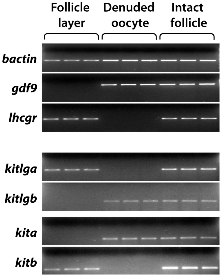
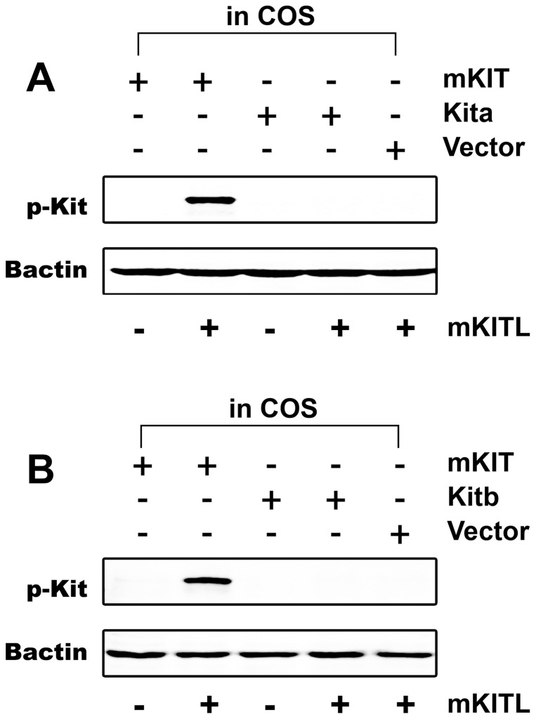
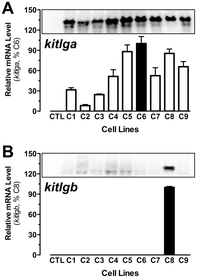
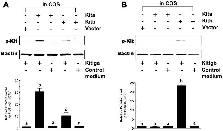
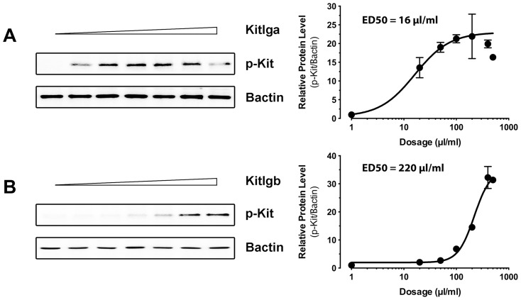
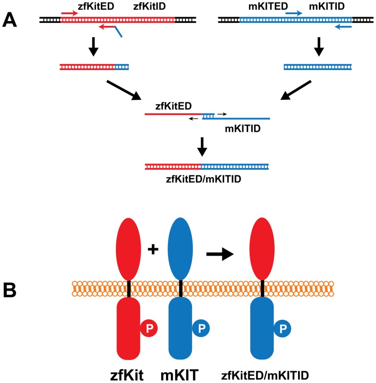
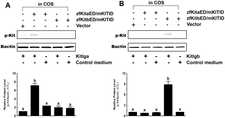
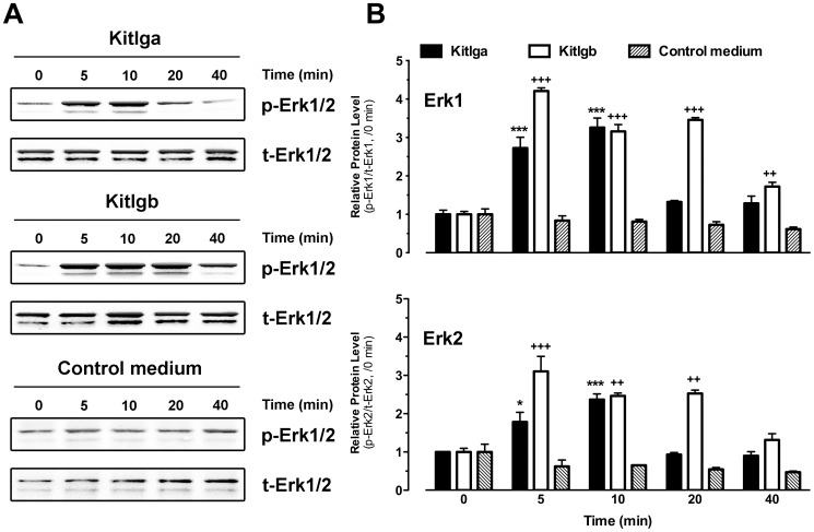
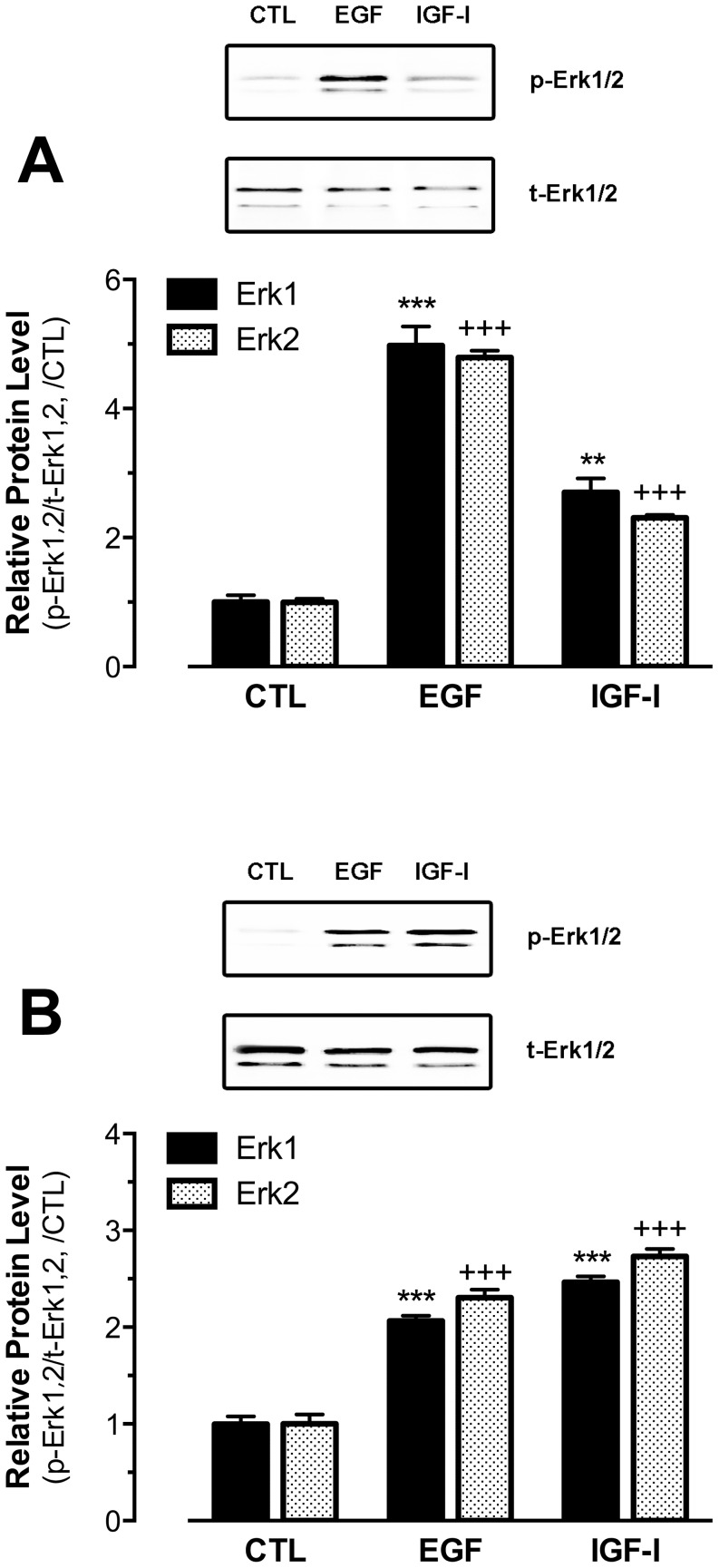
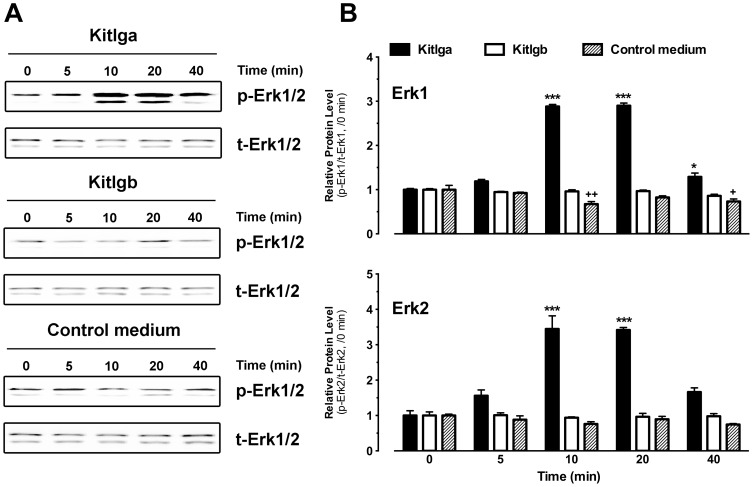
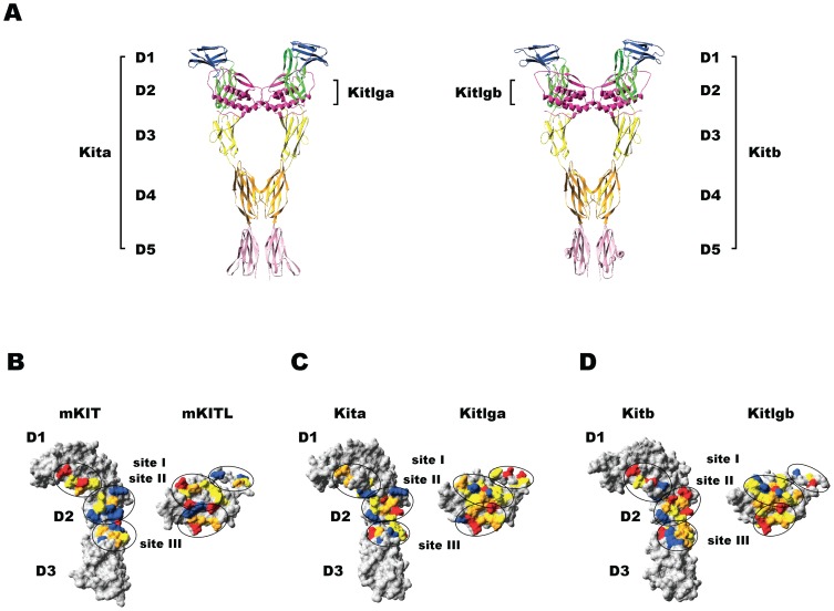
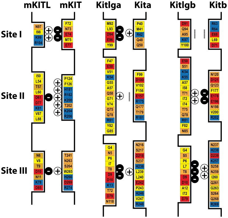
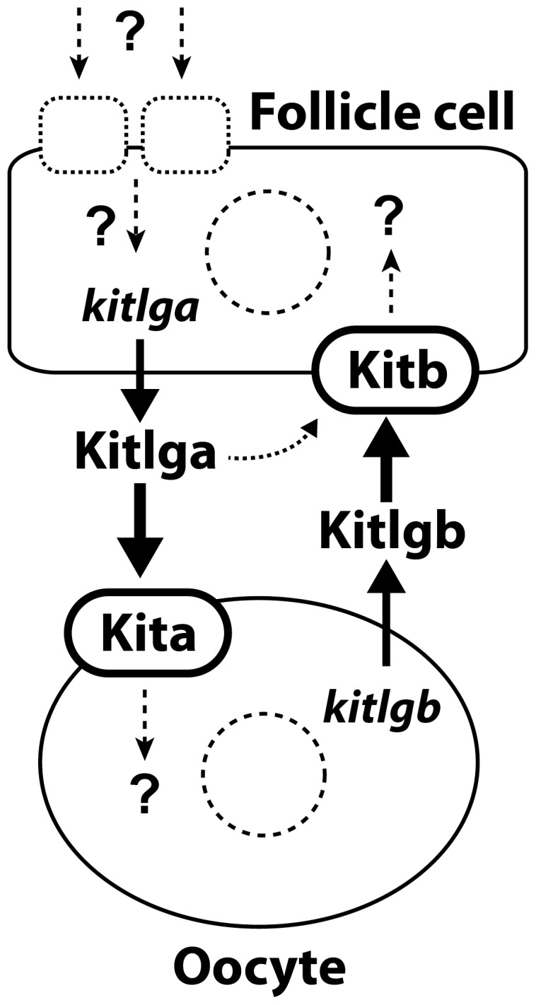
Consisting of Kit ligand and receptor Kit, the Kit system is involved in regulating many ovarian functions such as follicle activation, granulosa cell proliferation, and oocyte growth and maturation. In mammals, Kit ligand is derived from the granulosa cells and Kit receptor is expressed in the oocyte and theca cells. In the zebrafish, the Kit system contains two ligands (Kitlga and Kitlgb) and two receptors (Kita and Kitb). Interestingly, Kitlga and Kitb are localized in the somatic follicle cells, but Kitlgb and Kita are expressed in the oocyte. Using recombinant zebrafish Kitlga and Kitlgb, we demonstrated that Kitlga preferentially activated Kita whereas Kitlgb specifically activated Kitb by Western analysis for receptor phosphorylation. In support of this, Kitlgb triggered a stronger and longer MAPK phosphorylation in follicle cells than Kitlga, whereas Kitlga but not Kitlgb activated MAPK in the denuded oocytes, in agreement with the distribution of Kita and Kitb in the follicle and their specificity for Kitlga and Kitlgb. Further analysis of the interaction between Kit ligands and receptors by homology modeling showed that Kitlga-Kita and Kitlgb-Kitb both have more stable electrostatic interaction than Kitlgb-Kita or Kitlga-Kitb. A functional study of Kit involvement in final oocyte maturation showed that Kitlga and Kitlgb both suppressed the spontaneous maturation significantly; in contrast, Kitlgb but not Kitlga significantly promoted 17α, 20β-dihydroxy-4-pregnen-3-one (DHP) -induced oocyte maturation. Our results provided strong evidence for a Kit-mediated bi-directional communication system in the zebrafish ovarian follicle, which could be part of the complex interplay between the oocyte and the follicle cells in the development of follicles.


Conflict of interest statement

Competing Interests: The authors have declared that no competing interests exist.

Figures

Figure 1
Figure 1. Distribution of the Kit system within the zebrafish follicle.
The somatic follicle layer was separated from the oocyte followed by RNA extraction and semi-quantitative PCR detection in the two compartments (follicle layer and denuded oocyte). Each sample represents the total RNA pooled from 5 follicles. The housekeeping gene bactin was used as the internal control for all samples, whereas gdf9 and lhcgr were used as the markers for denuded oocytes and somatic follicle layers, respectively.
Figure 2
Figure 2. Species specificity of Kit activation by Kit ligand.
Plasmids expressing mouse KIT (mKIT), zebrafish Kita (A) or Kitb (B) were transfected into the COS-1 cells. After treatment with recombinant mouse KITL (mKITL) for 10 min, the phosphorylation of Kit was detected by an antibody recognizing the tyrosine phosphorylation site in the intracellular domain. Plasmid pCMV-Script (Vector) was used as a negative control. Bactin was used as the internal control for all samples. p-Kit, Kit phosphorylation; +, recombinant ligand proteins were added or cells were transfected with specific receptor plasmids; −, no recombinant ligands or transfection with receptor plasmids.
Figure 3
Figure 3. Characterization of recombinant CHO clones expressing zebrafish Kitlga and Kitlgb.
Nine clones (C1–9) for Kitlga (A) and one clone (C8) for Kitlgb (B) were chosen for analysis by real-time qPCR (bottom) and Northern blot hybridization (top). CTL, control CHO cells transfected with the vector pcDNA5/FRT.
Figure 4
Figure 4. Response of zebrafish Kita and Kitb to zebrafish Kitlga and Kitlgb.
The COS-1 cells expressing Kita and Kitb were treated with conditioned media containing recombinant Kitlga (A) and Kitlgb (B) for 10 min followed by Western blot analysis for Kit phosphorylation. The control medium was from the CHO cells carrying the control plasmid pcDNA5/FRT. The expression plasmid pCMV-Script (Vector) was used as the control in the COS-1 cells. The densitometric analysis of the Western signals is shown at the bottom of each graph. The data were normalized to Bactin and expressed as the fold change compared to the first group (mean ± SEM, n = 3). Different letters indicate statistical significance (P<0.05). p-Kit, Kit phosphorylation; +, recombinant ligand proteins were added or cells were transfected with specific receptor plasmids; −, no recombinant ligands or transfection with receptor plasmids.
Figure 5
Figure 5. Dose response activation of Kita and Kitb by Kit ligands.
The COS-1 cells expressing Kita and Kitb were treated with conditioned media containing recombinant Kitlga (A) and Kitlgb (B) for 10 min followed by Western blot analysis for Kit phosphorylation (left) and characterization of Kitlga and Kitlgb potency (right). The ED50 for each ligand was defined as 1 Unit. p-Kit, Kit phosphorylation.
Figure 6
Figure 6. Schematic illustration of plasmid construction for expressing chimeric receptors.
The mouse Kit receptor is shown in blue and zebrafish Kita or Kitb is in red. The extracellular domain of zebrafish Kita or Kitb (zfKitED) and intracellular domain of mouse KIT (mKITID) were amplified by PCR followed by extension of the PCR products on each other to generate a DNA fragment (A) coding for the fusion protein zfKitED/mKITID (B). “P” represents the phosphorylated tyrosine site in the intracellular domain that is recognized by the antibody.
Figure 7
Figure 7. Response of the chimeric zebrafish/mouse Kit receptors to zebrafish Kitlga and Kitlgb.
The COS-1 cells expressing zfKitaED/mKITID and zfKitbED/mKITID were treated with recombinant Kitlga (A) and Kitlgb (B) for 10 min followed by Western blot analysis for Kit phosphorylation. The control medium was from the CHO cells carrying the control plasmid pcDNA5/FRT. The expression plasmid pCMV-Script (Vector) was used as the control in the COS-1 cells. The densitometric analysis of the Western signals is shown at the bottom of each graph. The data were normalized to Bactin and expressed as the fold change compared to the first group (mean ± SEM, n = 3). Different letters indicate statistical significance (P<0.05). p-Kit, Kit phosphorylation; +, recombinant ligand proteins were added or cells were transfected with specific receptor plasmids; −, no recombinant ligands or transfection with receptor plasmids.
Figure 8
Figure 8. Effects of Kitlga and Kitlgb on MAPK phosphorylation in cultured follicle cells.
(A) Western blot analysis of MAPK (Erk1/2) phosphorylation in response to Kitlga (200 µl Kitlga conditioned medium+200 µl control medium per ml), Kitlgb (400 µl/ml Kitlgb conditioned medium) and control medium (400 µl per ml). (B) Densitometric quantification of Erk1 (upper) and Erk2 (lower) phosphorylation. The data were normalized to total Erk1 or Erk2 and expressed as the fold change compared to the first group at time 0 min (mean ± SEM, n = 3). */+ P<0.05; **/++ P<0.01; ***/+++ P<0.001. p-Erk1/2, phosphorylated Erk1/2; t-Erk1/2, total Erk1/2.
Figure 9
Figure 9. Effects of EGF and IGF-I on MAPK phosphorylation in immature intact follicles or ovulated mature oocytes.
The immature intact full-grown follicles (A) or ovulated mature oocytes (B) were treated with human EGF or IGF-I for 10 min followed by Western blot analysis for MAPK (Erk1/2) phosphorylation. The Western blot image is shown on the top of each graph and the densitometric quantification is at the bottom. The data were normalized to the total Erk1 or Erk2 and expressed as the fold change compared to the control (CTL) (mean ± SEM, n = 3). **/++ P<0.01; ***/+++ P<0.001. p-Erk1/2, phosphorylated Erk1/2; t-Erk1/2, total Erk1/2.
Figure 10
Figure 10. Effects of Kitlga and Kitlgb on MAPK phosphorylation in mature and ovulated oocytes.
(A) Western blot analysis of MAPK (Erk1/2) phosphorylation in response to Kitlga (200 µl Kitlga conditioned medium+200 µl control medium per ml), Kitlgb (400 µl/ml Kitlgb conditioned medium) and control medium (400 µl per ml). (B) Densitometric quantification of Erk1 (upper) and Erk2 (lower) phosphorylation. The data were normalized to total Erk1 or Erk2 and expressed as the fold change compared to the first group at time 0 min (mean ± SEM, n = 3). */+ P<0.05; ***/+++ P<0.001. p-Erk1/2, phosphorylated Erk1/2; t-Erk1/2, total Erk1/2.
Figure 11
Figure 11. Molecular modeling of the three-dimensional structures of zebrafish Kit ligands (Kitlga and Kitlgb) and receptors (Kita and Kitb).
(A) Ribbon model of the Kitlga-Kita (left) and Kitlgb-Kitb (right) complexes. The motif D1 is colored in blue, D2 in green, D3 in yellow, D4 in orange, and D5 in pink. Kit ligands are colored in magenta. (B–D) Surface models of the interphase of the mouse KITL-KIT (B), zebrafish Kitlga-Kita (C) and Kitlgb-Kitb (D) complexes. The Kit receptor part involving D1, D2 and D3 is shown on the left and the ligand part is on the right. The amino acids with negative charge, positive charge, polar and hydrophobic side chains are shown in red, blue, orange and yellow, respectively. The ligand-receptor binding sites (site I, site II, and site III) are circled.
Figure 12
Figure 12. Schematic illustration of the three ligand-receptor binding sites and the amino acids involved in the mouse and zebrafish.
The amino acids with negative charge, positive charge, polar and hydrophobic side chains are shown in red, blue, orange and yellow, respectively. The amount of net charges at each site is indicated by plus or minus signs, and the vertical dotted bar indicates neutral site.
Figure 13
Figure 13. Effects of recombinant zebrafish Kitlga and Kitlgb on oocyte maturation.
Full-grown immature follicles were pretreated for 6 h with Kitlga (200 µl Kitlga conditioned medium+200 µl control medium per ml), Kitlgb (400 µl/ml Kitlgb conditioned medium) and control medium (CTL, 400 µl per ml)), respectively, followed by treatment with DHP (5 ng/ml) for 9 h. Germinal vesicle breakdown (GVBD) was scored at different time points and expressed as the percentage (mean ± SEM, n = 4).
Figure 14
Figure 14. The hypothetical model for the existence of a Kit-mediated bi-directional communication network in the zebrafish ovarian follicle.
The follicle cell-derived Kitlga specifically targets its receptor Kita on the oocyte with a minor action on Kitb on the follicle cells as well, similar to the KITL-KIT system in mammals. The Kitlgb, however, is exclusively expressed in the oocyte and it acts on its specific receptor Kitb on the follicle cells, representing a potential paracrine signaling pathway for the oocyte to control the follicle cells. The expression of Kitlga in the follicle cells is likely subject to regulation by various endocrine and/or paracrine factors, therefore mediating the actions of these factors on the oocyte.

Similar articles

Cited by

References

    1. Copeland NG, Gilbert DJ, Cho BC, Donovan PJ, Jenkins NA, et al. (1990) Mast cell growth factor maps near the steel locus on mouse chromosome 10 and is deleted in a number of steel alleles. Cell 63: 175–183. - PubMed
    1. Huang E, Nocka K, Beier DR, Chu TY, Buck J, et al. (1990) The hematopoietic growth factor KL is encoded by the Sl locus and is the ligand of the c-kit receptor, the gene product of the W locus. Cell 63: 225–233. - PubMed
    1. Matsui Y, Zsebo KM, Hogan BL (1990) Embryonic expression of a haematopoietic growth factor encoded by the Sl locus and the ligand for c-kit. Nature 347: 667–669. - PubMed
    1. Yarden Y, Kuang WJ, Yang-Feng T, Coussens L, Munemitsu S, et al. (1987) Human proto-oncogene c-kit: A new cell surface receptor tyrosine kinase for an unidentified ligand. Embo J 6: 3341–3351. - PMC - PubMed
    1. Chabot B, Stephenson DA, Chapman VM, Besmer P, Bernstein A (1988) The proto-oncogene c-kit encoding a transmembrane tyrosine kinase receptor maps to the mouse W locus. Nature 335: 88–89. - PubMed

Publication types

MeSH terms

Substances

LinkOut - more resources