. 2013;8(2):e56273.
doi: 10.1371/journal.pone.0056273. Epub 2013 Feb 8.

Circulating angiopoietin-2 is a marker for early cardiovascular disease in children on chronic dialysis

Affiliations

Circulating angiopoietin-2 is a marker for early cardiovascular disease in children on chronic dialysis

Rukshana C Shroff et al. PLoS One. 2013.

Abstract

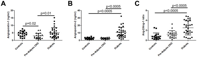
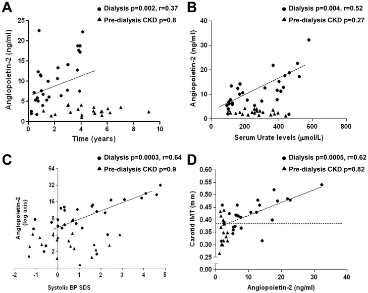
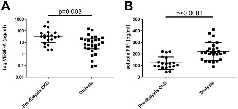
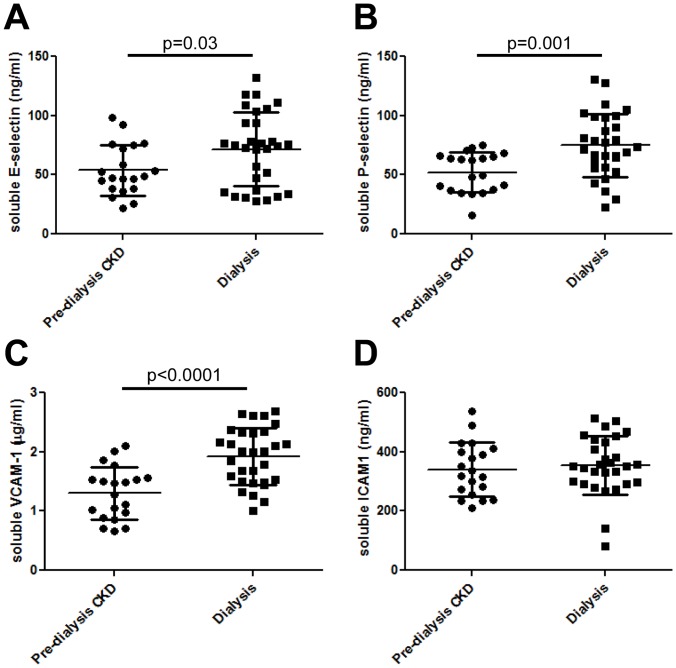
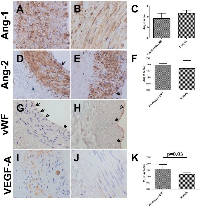
Cardiovascular disease (CVD) is increasingly recognised as a complication of childhood chronic kidney disease (CKD) even in the absence of diabetes and hypertension. We hypothesized that an alteration in angiopoietin-1 and -2, growth factors which regulate endothelial and vascular function could be involved. We report that the endothelial survival factor, angiopoietin-1 is low in children with pre-dialysis CKD whereas the pro-inflammatory angiopoietin-2 is elevated in children on dialysis. In dialysis patients, angiopoietin-2 positively correlated with time on dialysis, systolic blood pressure, and carotid artery intima media thickness. Elevated angiopoietin-2 levels in dialysis versus pre-dialysis CKD patients were also associated with an anti-angiogenic (high soluble VEGFR-1 and low VEGF-A) and pro-inflammatory (high urate, E-selectin, P-selectin and VCAM-1) milieu. Ang-2 was immunodetected in arterial biopsy samples whilst the expression of VEGF-A was significantly downregulated in dialysis patients. Serum urate correlated with angiopoietin-2 levels in dialysis patients and addition of uric acid was able to induce rapid release of angiopoietin-2 from cultured endothelial cells. Thus, angiopoietin-2 is a marker for cardiovascular disease in children on chronic dialysis and may act as an anti-angiogenic and pro-inflammatory effector in this context. The possibility that the release of angiopoietin-2 from endothelia is mediated by urate should be explored further.


Conflict of interest statement

Competing Interests: The authors have declared that no competing interests exist.

Figures

Figure 1
Figure 1. Circulating Ang levels in pre-dialysis CKD and dialysis patients.
Serum Ang-1 levels (A) were significantly lower in pre-dialysis CKD patients compared with healthy controls. In dialysis patients Ang-1 levels were similar to values found in healthy controls. Similar levels of both circulating Ang-2 (B) and Ang-2/Ang-1 (C) were found in healthy children and those with pre-dialysis CKD, but these were significantly increased in the dialysis group.
Figure 2
Figure 2. Correlation of Ang-2 levels with clinical and vascular parameters.
Ang-2 levels in dialysis individuals correlated positively with time on dialysis (A), serum urate levels (B), systolic blood pressure SDS (C) and cIMT (D). Independent variables are shown on the x-axis. Regression lines account for dialysis patients only. Dotted line in D indicates the value for cIMT in healthy age-matched controls. There was no correlation between Ang-2 and any clinical and vascular measures in pre-dialysis CKD patients.
Figure 3
Figure 3. Circulating levels of VEGF-A and sFlt-1 in pre-dialysis CKD and dialysis patients.
VEGF-A levels were significantly lower in individuals on dialysis compared with pre-dialysis CKD patients (A). In contrast, sFlt-1 were significantly higher in the dialysis patients (B)
Figure 4
Figure 4. Circulating levels of soluble cell adhesion molecules.
Compared with pre-dialysis CKD individuals, patients treated with dialysis had significantly elevated levels of soluble E-selectin (A), P-selectin (B) and VCAM-1 (C); there was no difference in ICAM-1 levels (D).
Figure 5
Figure 5. Immunolocalisation of vascular growth factors in arteries.
Ang-1 was detected in the media of vessels from both pre-dialysis CKD (A) and dialysis patients (B); no differences in staining intensity were observed between the two groups (C). Ang-2 was immunodetected in both the media and endothelia (arrows) in pre-dialysis CKD (D) and dialysis (E) vessels with similar intensity (F). The endothelial later was also positive for von Willebrand factor (arrows, G and H). VEGF-A immunostaining was prominent in the media of pre-dialysis CKD vessels (I), but was significantly decreased in dialysis patients (J and K). All fields taken with ×40 objective.
Figure 6
Figure 6. Effect of uric acid on Ang-2 secretion in HUVECs.
A) Both non-stimulated and uric acid stimulated HUVECs expressed the mRNA for the transporter Urat1 but not Oat1-4; they were also positive for Ang-1, Ang-2, Tie-1, Tie-2 and Tlr4. Sizes were determined using a 100 bp marker (m), positive (+ive) controls consisted of total kidney cDNA and negative controls were without cDNA addition. (B) Uric acid stimulation for 15 minutes, but not 72 hours (C) led to elevated Ang-2 secretion in the conditioned media of HUVEC cells. Within the cells, uric acid stimulation led to a decreased abundance of Ang-2 mRNA after 6 hours of stimulation (D). a = p<0.05 compared with controls, b = p<0.01 compared with controls, c = p<0.001 compared with controls, d = p<0.01 compared with HUVEC stimulated with 3 mg/dl uric acid, e = p<0.01 compared with HUVEC stimulated with 6 mg/dl uric acid, f = p<0.05 compared with HUVEC stimulated with 9 mg/dl uric acid.

Similar articles

Cited by

References

    1. Goodman WG, Goldin J, Kuizon BD, Yoon C, Gales B, et al. (2000) Coronary-artery calcification in young adults with end-stage renal disease who are undergoing dialysis. N Engl J Med 342: 1478–1483. - PubMed
    1. Covic A, Mardare N, Gusbeth-Tatomir P, Brumaru O, Gavrilovici C, et al. (2006) Increased arterial stiffness in children on haemodialysis. Nephrol Dial Transplant 21: 729–735. - PubMed
    1. Shroff RC, McNair R, Figg N, Skepper JN, Schurgers L, et al. (2008) Dialysis accelerates medial vascular calcification in part by triggering smooth muscle cell apoptosis. Circulation 118: 1748–1757. - PubMed
    1. Dursun I, Poyrazoglu HM, Gunduz Z, Ulger H, Yykylmaz A, et al. (2009) The relationship between endothelial microparticles and arterial stiffness and atherosclerosis in children with chronic kidney disease. Nephrol Dial Transplant 24: 2511–2518. - PubMed
    1. Mitsnefes MM, Barletta GM, Dresner IG, Chand DH, Geary D, et al. (2006) Severe cardiac hypertrophy and long-term dialysis: the Midwest Pediatric Nephrology Consortium study. Pediatr Nephrol 21: 1167–1170. - PubMed

Publication types

MeSH terms