. 2013;8(2):e56377.
doi: 10.1371/journal.pone.0056377. Epub 2013 Feb 11.

In vitro characterization of circulating endothelial progenitor cells isolated from patients with acute coronary syndrome

Affiliations

In vitro characterization of circulating endothelial progenitor cells isolated from patients with acute coronary syndrome

Diana Campioni et al. PLoS One. 2013.

Abstract

Background: The current understanding of the functional characteristics of circulating endothelial progenitor cells (EPC) is limited, especially in patients affected by cardiovascular diseases. In this study, we have analyzed the in vitro clonogenic capacity of circulating EPC, also known as endothelial colony-forming cells (ECFC), in patients with acute coronary syndrome (ACS), in comparison to the colony forming unit-endothelial-like cells (CFU-EC) of hematopoietic/monocytic origin.

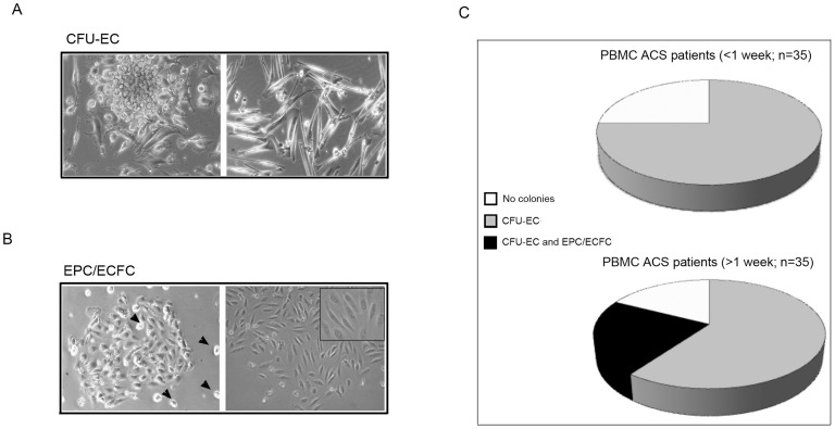
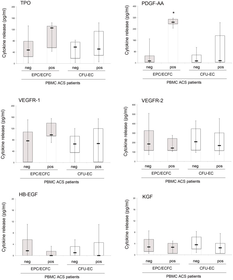
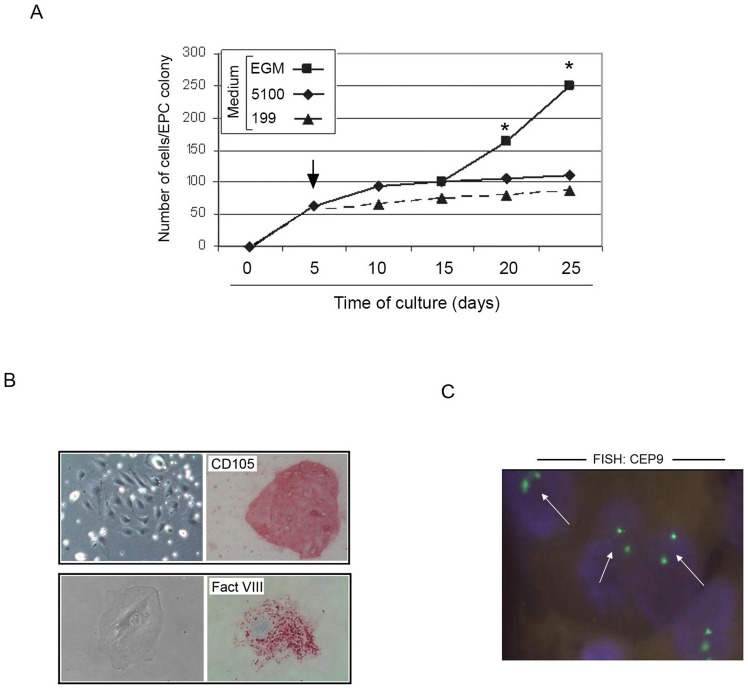
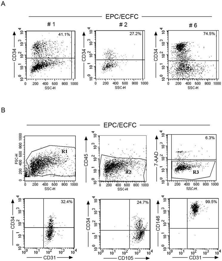
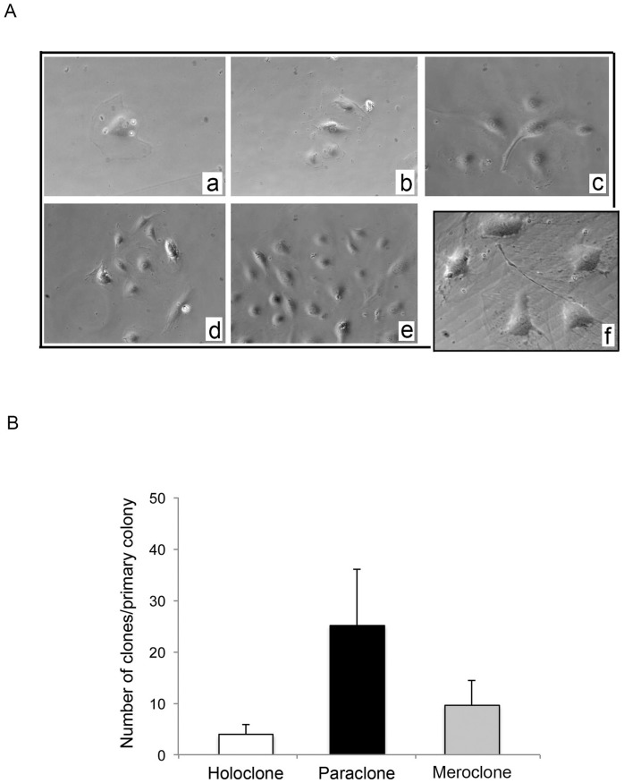
Methodology/principal findings: By culturing peripheral blood mononuclear cells (PBMC) of patients with ACS (n = 70), CFU-EC were frequently isolated (from 77% of ACS patients), while EPC/ECFC were obtained only in a small subset (13%) of PBMC samples, all harvested between 7-14 days after the acute cardiovascular event. Notably, ex-vivo generation of EPC/ECFC was correlated to a higher in vitro release of PDGF-AA by the corresponding ACS patient PBMC. By using specific endothelial culture media, EPC/ECFC displayed in vitro expansion capacity, allowing the phenotypic and functional characterization of the cells. Indeed, after expansion, EPC/ECFC exhibited a normal diploid chromosomal setting by FISH analysis and an immunophenotype characterized by: i) uniform positivity for the expression of CD105, CD31, CD146 and Factor VIII, i) variable expression of the CD34, CD106 and CD184 markers, and iii) negativity for CD45, CD90, CD117 and CD133. Of interest, in single-cell replanting assays EPC/ECFC exhibited clonogenic expansion capacity, forming secondary colonies characterized by variable proliferation capacities.

Conclusion/significance: Our data indicate that a careful characterization of true EPC is needed in order to design future studies in the clinical autologous setting of patients with ACS.


Conflict of interest statement

Competing Interests: The authors have declared that no competing interests exist.

Figures

Figure 1
Figure 1. Characterization of the clonogenic potential of PBMC derived from ACS patients.
PBMC samples obtained from ACS patients (n = 70) were seeded in collagen I coated wells for short-term primary colony assay in liquid culture medium. Cultures were monitored for 15 days for the presence of adherent colonies, scored on the basis of morphological features as: CFU-EC (A, left panel) or EPC/ECFC (B, left panel; arrowheads: hemopoietic mononucleated cells). In A, the right panel shows a monolayer of spindle-shaped endothelial-like monocytes. In B, the right panel shows a representative image of CFU-EC after in vitro expansion. Original magnification: 20X and 25X for the inset. In C, frequency of detection of CFU-EC and EPC/ECFC in PBMC of ACS patients, divided on the basis of the time of blood withdrawal after the hospital admission for the acute cardiovascular events.
Figure 2
Figure 2. Analysis of pro-angiogenic cytokines release by PBMC derived from ACS patients.
After 48 hour of culture, PBMC conditioned media were collected and analyzed for the release of angiogenic cytokines. Cytokine levels were analyzed in relation to the ability of the PBMC ACS patient samples to generate EPC/ECFC and/or CFU-EC colonies: EPC/ECFCneg vs EPC/ECFCpos gray box-plots) or CFU-ECneg vs CFU-ECpos (white box-plots). Horizontal bars are median, upper and lower edges of box are 75th and 25th percentiles, lines extending from box are 10th and 90th percentiles. Asterisk, p<0.01.
Figure 3
Figure 3. Identification of optimal culture conditions for the ex-vivo expansion of ACS PB-derived EPC/ECFC.
Primary EPC/ECFC colonies were generated by plating patient PBMC in M5100 medium, as detailed in the Method section. In A, after the colony identification (at day 5 after plating), medium was change (arrow) and replaced either with fresh M5100, or MEGM or M199 and the development of the colonies was monitored over the time. The growth kinetics of a representative experiment out of five independent experiments is shown. At each indicated time point, the mean cell number/ECFC was determined by two independent operators; standard deviations were below 10% and are not shown. Asterisk, p<0.05. In B, immunocytochemical analysis of in vitro expanded EPC/ECFC documenting positivity for CD105 antigen (original magnification: 20X) and for the specific endothelial marker Factor VIII (original magnification: 40X). In C, FISH analysis performed on in vitro expanded EPC/ECFC by using the centromeric enumeration probe CEP9 (white arrows) documenting a normal diploid chromosomal pattern (original magnification: 40X).
Figure 4
Figure 4. Immunophenotype of EPC/ECFC generated from the PBMC of ACS patients.
After ex-vivo expansion, primary EPC/ECFC colonies were trypsinized and assessed for their immuno-phenotype by multi-colors flow cytometry. In A, the variable expression of the CD34 antigene is documented by 3 independent examples of EPC/ECFC colonies. In B, 4-colors flow cytometric analysis of EPC/ECFC cells. A representative example of 7 independent experiments is shown.
Figure 5
Figure 5. Subcloning potential of EPC/ECFC generated from the PBMC of ACS patients.
After ex-vivo expansion, primary EPC/ECFC colonies were trypsinized and assessed for clonogenic potential capacity by single cells replating assay. In A, single cells derived from EPC/ECPF colonies were seeded in collagen I coated wells and monitored day by day (a: day 1; b: day 2; c: day 3; e–f: day 4; a–e: original magnification 25X; f: original magnification 40X). One representative experiment is shown. In B, secondary clones were classified on the basis of their proliferation properties. Data are mean±SD derived from six independent experiments.

References

    1. Asahara T, Murohara T, Sullivan A, Silver M, van der Zee R, et al. (1997) Isolation of putative progenitor endothelial cells for angiogenesis. Science 275: 964–967. - PubMed
    1. Isner JM, Asahara T (1999) Angiogenesis and vasculogenesis as therapeutic strategies for postnatal vascularization. J Clin Invest 103: 1231–1234. - PMC - PubMed
    1. Schatteman GC, Dunnwald M, Jiao C (2006) Biology of bone marrow derived endothelial precursors. Am J Physiol Heart Circ Physiol 292: 1–18. - PubMed
    1. Shi Q, Rafii S, Wu MHD, Wijelath ES, Cong Y, et al. (1998) Evidence for circulating bone marrow-derived endothelial cells. Blood 92: 362–367. - PubMed
    1. Rafii S (2000) Circulating endothelial precursors: mystery, reality and promise. J Clin Invest 105: 17–19. - PMC - PubMed

Publication types

MeSH terms