. 2013;8(2):e56484.
doi: 10.1371/journal.pone.0056484. Epub 2013 Feb 7.

Effect of delayed peripheral nerve repair on nerve regeneration, Schwann cell function and target muscle recovery

Affiliations

Effect of delayed peripheral nerve repair on nerve regeneration, Schwann cell function and target muscle recovery

Samuel Jonsson et al. PLoS One. 2013.

Abstract

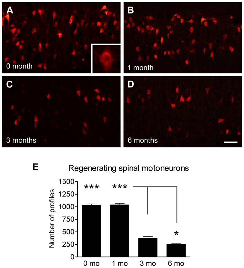
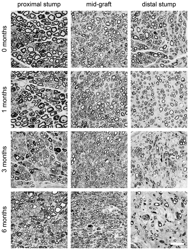
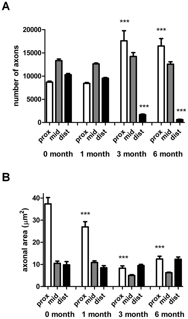
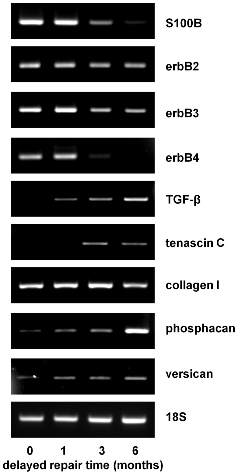
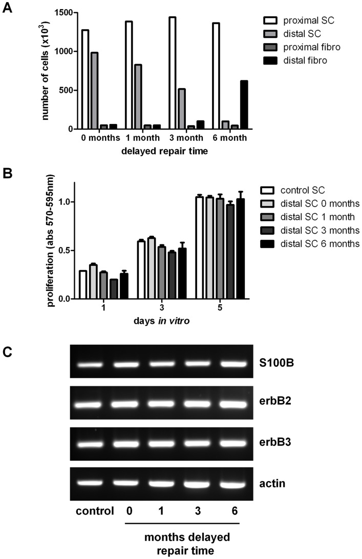
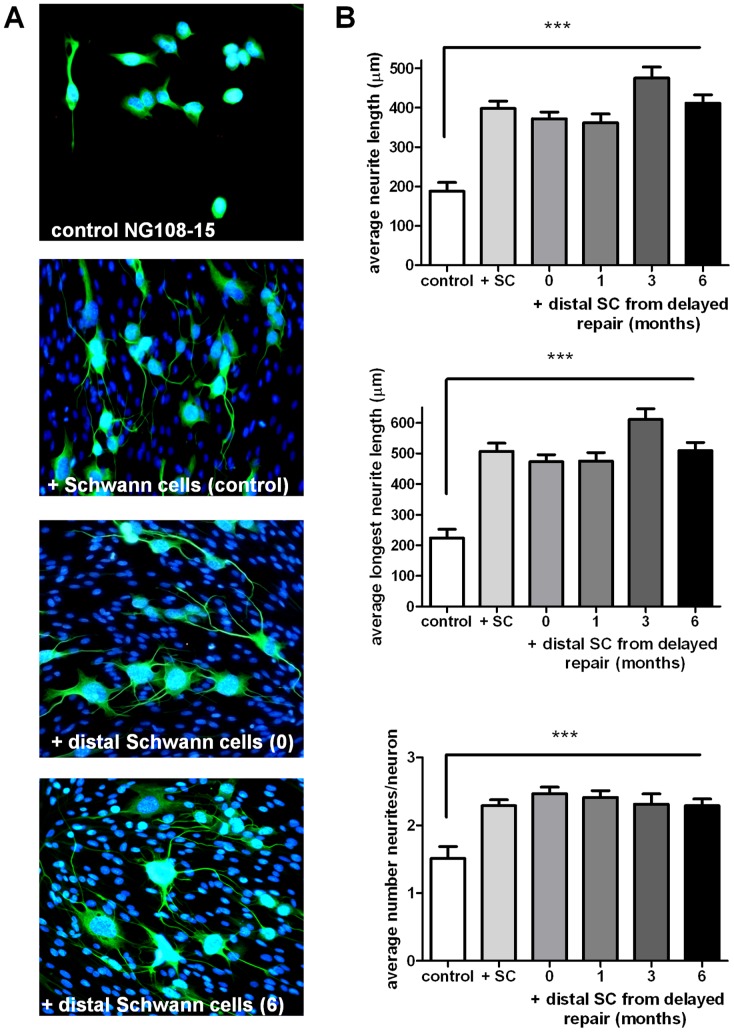
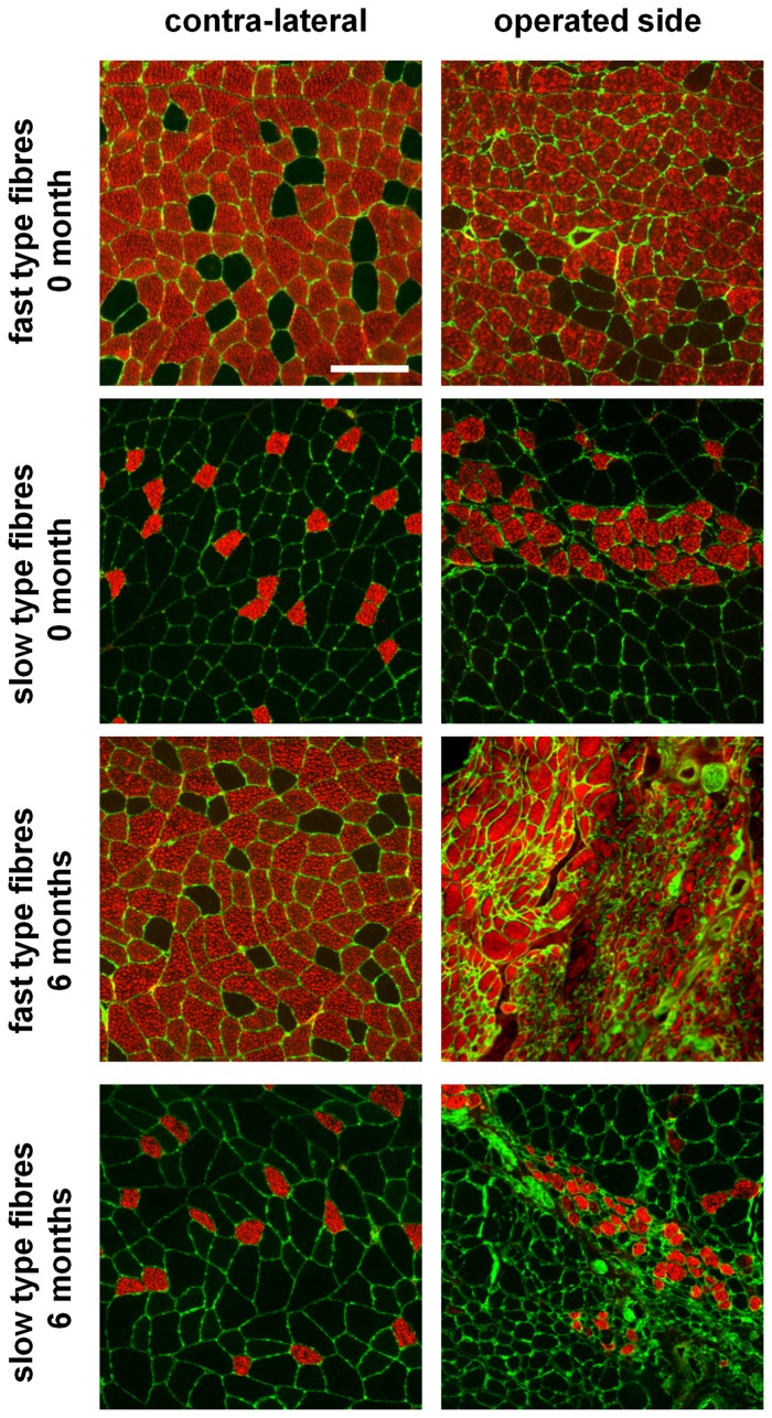
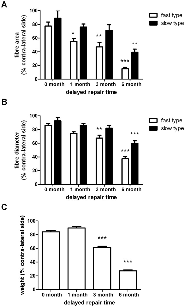
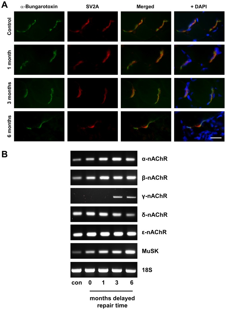
Despite advances in surgical techniques for peripheral nerve repair, functional restitution remains incomplete. The timing of surgery is one factor influencing the extent of recovery but it is not yet clearly defined how long a delay may be tolerated before repair becomes futile. In this study, rats underwent sciatic nerve transection before immediate (0) or 1, 3, or 6 months delayed repair with a nerve graft. Regeneration of spinal motoneurons, 13 weeks after nerve repair, was assessed using retrograde labeling. Nerve tissue was also collected from the proximal and distal stumps and from the nerve graft, together with the medial gastrocnemius (MG) muscles. A dramatic decline in the number of regenerating motoneurons and myelinated axons in the distal nerve stump was observed in the 3- and 6-months delayed groups. After 3 months delay, the axonal number in the proximal stump increased 2-3 folds, accompanied by a smaller axonal area. RT-PCR of distal nerve segments revealed a decline in Schwann cells (SC) markers, most notably in the 3 and 6 month delayed repair samples. There was also a progressive increase in fibrosis and proteoglycan scar markers in the distal nerve with increased delayed repair time. The yield of SC isolated from the distal nerve segments progressively fell with increased delay in repair time but cultured SC from all groups proliferated at similar rates. MG muscle at 3- and 6-months delay repair showed a significant decline in weight (61% and 27% compared with contra-lateral side). Muscle fiber atrophy and changes to neuromuscular junctions were observed with increased delayed repair time suggestive of progressively impaired reinnervation. This study demonstrates that one of the main limiting factors for nerve regeneration after delayed repair is the distal stump. The critical time point after which the outcome of regeneration becomes too poor appears to be 3-months.


Conflict of interest statement

Competing Interests: The authors have declared that no competing interests exist.

Figures

Figure 1
Figure 1. Fluoro-Ruby-labeled motoneurons at 13 weeks after immediate nerve repair (A), 1 months delayed repair (B), 3 months delayed repair (C) and 6 months delayed repair (D).
Note significant decrease in number of regenerating motoneurons after 3 and 6 months delayed nerve repair. Scale bar = 100 µm. (E) Histogram showing the quantification of mean±SEM number of regenerating spinal motoneurons after 0–6 months (mo) delayed repair. n = 5, *p<0.05 (3 mo vs, 6 mo), ***p<0.001 (0 mo and 1 mo vs 3 mo and 6 mo).
Figure 2
Figure 2. Toluidine blue stained myelinated axons in the proximal stumps, mid-graft regions and distal stumps of animals undergoing sciatic nerve injury followed by either immediate or delayed nerve repair (at the time points indicated) with a 10
mm graft. Note increase of number of small myelinated fibers in proximal stump in 3 months and 6 months groups. Scale bar = 30 µm.
Figure 3
Figure 3. Quantitative analysis of (A) number of myelinated axons and (B) axonal area in the proximal stumps, mid-graft regions and distal stumps of animals undergoing sciatic nerve injury followed by either immediate or delayed nerve repair (at the time points indicated) with a 10
mm graft. Values shown are mean±SEM, n = 5, ***p<0.001 significantly different from respective region after immediate nerve repair.
Figure 4
Figure 4. Qualitative RT-PCR analysis of a variety of Schwann cell, axonal and fibrosis/scar associated molecules in the proximal stumps, mid-graft regions and distal stumps of animals undergoing sciatic nerve injury followed by either immediate or delayed nerve repair (at the time points indicated) with a 10
mm graft. 18 S is used as a house-keeping gene.
Figure 5
Figure 5. Characterisation of Schwann cells isolated from nerve segments.
(A) The number of Schwann cells from proximal and distal nerve stumps of animals undergoing sciatic nerve injury followed by either immediate or delayed nerve repair (at the time points indicated) with a 10 mm graft were counted 7 days following enzyme digestion of the nerve. Fibroblast counts were also made following treatment of the samples with magnetic anti-fibroblast beads. (B) Distal nerve segment Schwann cells from (A) were trypsinised and replated and proliferation rates (in the presence of glial growth factors) compared with control normal cultures of Schwann cells (no experimental injury or repair). (C) Qualitative RT-PCR analysis of Schwann cell marker S100 and the glial growth factor receptors erbB2 and erbB3 expression levels in cultured cells isolated from control nerve (no experimental injury or repair) and the distal nerve segments of animals undergoing immediate (0 months) or delayed nerve repair (1, 3, 6 months). Actin was used as a house-keeping gene.
Figure 6
Figure 6. Co-culture of Schwann cells with NG108-15 neurons.
(A) NG108-15 cells were either grown alone or on top of a monolayer of Schwann cells isolated from control nerve or distal nerve segments taken from animals undergoing immediate (0) or 6 month delayed nerve repair (6). Cultures were stained with βIII tubulin antibody (green) and DAPI (blue). (B) Quantitative analysis of neurite outgrowth was performed on NG108-15 neurons grown in the absence (control) or presence of Schwann cells isolated from control nerve (+SC) or distal nerve segments from animals undergoing nerve repair immediately following injury (0) or delayed repair at 1, 3, 6 months. ***P<0.001 significantly different from values in the absence of Schwann cells. There was no significant difference between any of the cultures in the presence of Schwann cells.
Figure 7
Figure 7. Fast type and slow type gastrocnemius muscle fiber morphology.
(A) Transverse sections of contra-lateral and operated side muscles were stained with laminin antibody (green) and either fast type or slow type myosin heavy chain protein antibody (red). Samples shown are from animals undergoing immediate repair (0 months) or delayed repair (6 months). Scale bar = 100 µm.
Figure 8
Figure 8. Quantification of muscle fibre size and muscle weights.
Computerised image analysis was used to calculate the mean±SEM area (A) and diameter (B) of fast type and slow type fibers in muscle obtained from the contra-lateral and operated sides of animals 3 months after repair. Data are expressed as percentage of the contra-lateral side, *P<0.05, **P<0.01, ***P<0.001 significantly different from respective values in animals undergoing immediate nerve repair (0 months). (C) At the time of harvest, the contra-lateral and operated side muscles were weighed. Data are expressed as percentage of contra-lateral side weights, ***P<0.001 significantly different from weight after immediate nerve repair (0 months).
Figure 9
Figure 9. Neuromuscular junctions and expression of nicotinic acetylcholine receptors (nAChRs).
(A) Muscle sections were stained for nAChRs with α-bungarotoxin-FITC (green) and with SV2A antibody (red) to mark pre-synaptic structures. Note the coincident staining of both markers indicating presumptive functional NMJs. DAPI staining (blue) shows nuclei. Scale bar = 25 µm. (B) RT-PCR analysis of the different nAChR subunits (α, β, γ, δ, ε) and muscle specific kinase MuSK in control muscle (con) and muscle from the operated side of animals undergoing immediate (0) or 1,3,6 months delayed nerve repair. 18 S was used as a house-keeping gene.

References

    1. Wiberg M, Terenghi G (2003) Will it be possible to produce peripheral nerves? Surg Technol Int 11: 303–310. - PubMed
    1. Lad SP, Nathan JK, Schubert RD, Boakye M (2010) Trends in median, ulnar, radial, and brachioplexus nerve injuries in the United States. Neurosurgery 66: 953–960. - PubMed
    1. Vejsada R, Sagot Y, Kato AC (1995) Quantitative comparison of the transient rescue effects of neurotrophic factors on axotomized motoneurons in vivo. Eur J Neurosci 7: 108–115. - PubMed
    1. Novikova L, Novikov L, Kellerth JO (1997) Persistent neuronal labeling by retrograde fluorescent tracers: a comparison between Fast Blue, Fluoro-Gold and various dextran conjugates. J Neurosci Methods 74: 9–15. - PubMed
    1. Welin D, Novikova LN, Wiberg M, Kellerth JO, Novikov LN (2008) Survival and regeneration of cutaneous and muscular afferent neurons after peripheral nerve injury in adult rats. Exp Brain Res 186: 315–323. - PubMed

Publication types