. 2013 Feb 14:11:38.
doi: 10.1186/1741-7015-11-38.

Sodium vanadate combined with L-ascorbic acid delays disease progression, enhances motor performance, and ameliorates muscle atrophy and weakness in mice with spinal muscular atrophy

Affiliations

Sodium vanadate combined with L-ascorbic acid delays disease progression, enhances motor performance, and ameliorates muscle atrophy and weakness in mice with spinal muscular atrophy

Huei-Chun Liu et al. BMC Med. .

Abstract

Background: Proximal spinal muscular atrophy (SMA), a neurodegenerative disorder that causes infant mortality, has no effective treatment. Sodium vanadate has shown potential for the treatment of SMA; however, vanadate-induced toxicity in vivo remains an obstacle for its clinical application. We evaluated the therapeutic potential of sodium vanadate combined with a vanadium detoxification agent, L-ascorbic acid, in a SMA mouse model.

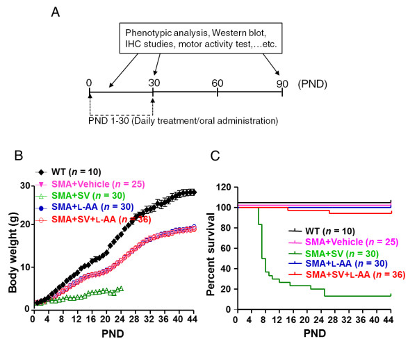
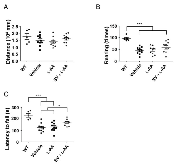
Methods: Sodium vanadate (200 μM), L-ascorbic acid (400 μM), or sodium vanadate combined with L-ascorbic acid (combined treatment) were applied to motor neuron-like NSC34 cells and fibroblasts derived from a healthy donor and a type II SMA patient to evaluate the cellular viability and the efficacy of each treatment in vitro. For the in vivo studies, sodium vanadate (20 mg/kg once daily) and L-ascorbic acid (40 mg/kg once daily) alone or in combination were orally administered daily on postnatal days 1 to 30. Motor performance, pathological studies, and the effects of each treatment (vehicle, L-ascorbic acid, sodium vanadate, and combined treatment) were assessed and compared on postnatal days (PNDs) 30 and 90. The Kaplan-Meier method was used to evaluate the survival rate, with P < 0.05 indicating significance. For other studies, one-way analysis of variance (ANOVA) and Student's t test for paired variables were used to measure significant differences (P < 0.05) between values.

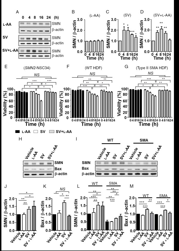
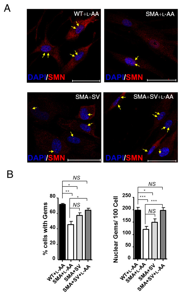
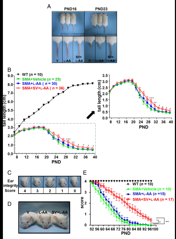
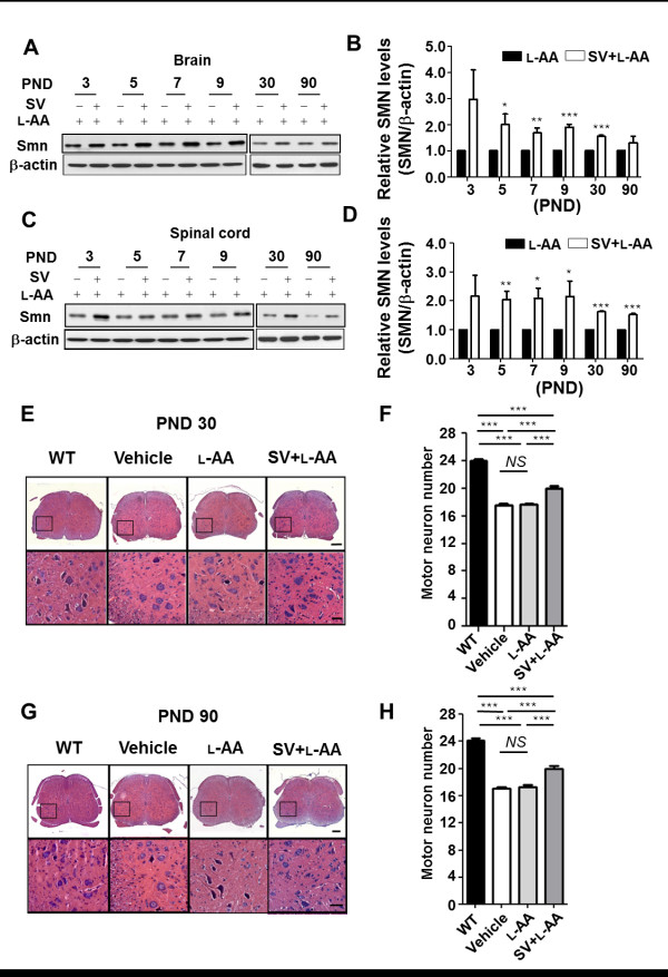
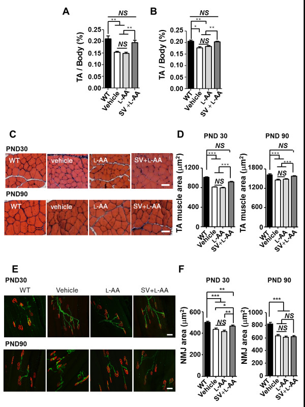
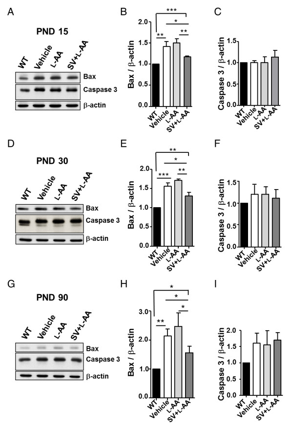
Results: Combined treatment protected cells against vanadate-induced cell death with decreasing B cell lymphoma 2-associated X protein (Bax) levels. A month of combined treatment in mice with late-onset SMA beginning on postnatal day 1 delayed disease progression, improved motor performance in adulthood, enhanced survival motor neuron (SMN) levels and motor neuron numbers, reduced muscle atrophy, and decreased Bax levels in the spinal cord. Most importantly, combined treatment preserved hepatic and renal function and substantially decreased vanadium accumulation in these organs.

Conclusions: Combined treatment beginning at birth and continuing for 1 month conferred protection against neuromuscular damage in mice with milder types of SMA. Further, these mice exhibited enhanced motor performance in adulthood. Therefore, combined treatment could present a feasible treatment option for patients with late-onset SMA.


Figures

Figure 1
Figure 1
L-Ascorbic acid (L-AA) eliminates sodium vanadate (SV)-induced cytotoxicity. (A) Western blots showing survival motor neuron (SMN) expression in SMN2-NSC34 cells treated with 400 μM L-AA, 200 μM SV, or SV combined with L-AA at different time points. β-Actin was used as an internal control. (B-D) Quantification of SMN protein expression in (A). (E-G) Quantification of the viability of SMN2-NSC34 cells (E) and human dermal fibroblasts (HDFs) (F, G) treated with L-AA, SV or SV combined with L-AA. (H, I) Western blots showing SMN and B cell lymphoma 2-associated X protein (Bax) expression in SMN2-NSC34 cells (H) and HDFs (I) treated with vehicle, L-AA, SV or SV combined with L-AA. β-Actin was used as an internal control. (J, K) Quantification of SMN and Bax expression in (H). (L, M) Quantification of SMN and Bax expression in (I). The experiment was repeated at least three times, and the mean ± SEM was calculated. Statistical comparisons were performed by one-way analysis of variance (ANOVA) (E-G) and Student's t test (B-D, and J-M).*P < 0.05, **P < 0.01, and ***P < 0.001.
Figure 2
Figure 2
Combined treatment restores survival motor neuron (SMN) nuclear body gems in fibroblasts of a spinal muscular atrophy (SMA) patient. (A) Immunocytochemical analysis of the nuclear SMN body gems in wild-type (WT) and SMA human dermal fibroblasts (HDFs) treated with L-ascorbic acid (L-AA), sodium vanadate (SV) or L-AA combined with SV. SMN was stained with an SMN antibody (red, indicated by arrows). 4',6-Diamidino-2-phenylindole (DAPI) was used for nuclei staining. Bar: 50 μm. (B) The percentage of nuclei with gems (left) and the number of gems per 100 nuclei (right) in WT and SMA cells after different treatments were evaluated by immunocytochemical analysis. The mean ± SEM was calculated. *P < 0.01, **P < 0.01, and ***P < 0.001, Student's t test. NS = not significant.
Figure 3
Figure 3
L-Ascorbic acid (L-AA) protects against sodium vanadate (SV)-induced toxicity in a mouse model of late-onset spinal muscular atrophy (SMA). (A) Schematic indicating the treatment regimens used in this study. (B) The mean body weight progression of the wild-type (WT) mice (n = 10, black squares) and the SMA mice that received vehicle (n = 25, pink solid triangles), SV (n = 30, green open triangles), L-AA (n = 30, blue solid circles) and SV combined with L-AA (n = 36, red open circles) treatment. Note that SV treatment reduced the rate of body weight gain. (C) Survival analysis of mice that received different treatments. Note that by postnatal day (PND) 25, most of the mice that had received daily SV administration had died.
Figure 4
Figure 4
Combined treatment delays the onset of spinal muscular atrophy (SMA) phenotype. (A) Images of the mice that received different treatments on postnatal days (PNDs) 16 and 23. Note that mice that received combined treatment had longer tails. (B) Plot showing the tail lengths of wild-type mice (n = 10, black triangles) and mice with late-onset SMA that received vehicle (n = 25, green triangles), L-ascorbic acid (L-AA) (n = 30, blue circles) or combined (n = 36, red circles) treatment. The SMA groups are shown in a magnified figure to the right. Lengths were measured every other day from PND 7 to PND 39. A trend of delayed onset of tail necrosis was observed in the mice that received combined treatment. The combined treatment significantly delayed tail loss according to one-way analysis of variance (ANOVA). (C) Ear integrity was scored on a scale of 0 to 4. (D) PND 90 mice that had received different treatments. Note that the mice that received combined treatment retained ear integrity longer. (E) The plot represents the mean score of ear integrity for wild-type mice (n = 10, black triangles) and mice with late-onset SMA mice that received vehicle (n = 10, green solid triangles), L-AA (n = 15, blue solid circles) or combined (n = 17, red open circles) treatment. *P < 0.05 compared with the L-AA-treated group; **P < 0.01 compared with the vehicle-treated group, one-way ANOVA.
Figure 5
Figure 5
Combined treatment improves motor function in adult mice with late-onset spinal muscular atrophy (SMA). (A) Distance traveled in the open-field test during a 60-minute period. No significant differences were observed among the treatment groups. (B) The frequency of rearing events in the first 10 minutes of the 60-minute experiment to examine this behavior independent of habituation. Mice that received combined treatment generally exhibited more rearing events, but this difference did not reach significance. (C) Average latency to fall from the rotarod for mice in response to different treatments. *P < 0.05, one-way analysis of variance (ANOVA). In all motor function tests, each circle, square or triangle represents an individual adult mouse (postnatal day (PND) 90).
Figure 6
Figure 6
Combined treatment increases survival motor neuron (SMN) expression and motor neuron numbers in mice with late-onset spinal muscular atrophy (SMA). (A, C) Western blots showing SMN expression in the brain (A) and spinal cord (C) in mice with late-onset SMA that received L-ascorbic acid (L-AA) or combined (n = 4 in each group) treatment. β-Actin was used as an internal control. (B, D) Quantification of SMN protein expression in (A, C). The mean ± SEM was calculated. (E, G) Histological staining of lumbar spinal cord samples on postnatal days (PNDs) 30 (E) and 90 (G) for the wild-type (WT) mice and mice with late-onset SMA that had received different treatments. Scale bar: 100 μm (E and G, upper panel); 50 μm (E and G, lower panel). (F, H) Quantification of motor neuron numbers in the spinal cords obtained from (E) and (G) (n = 3, 40 sections for each group were quantified). The mean ± SEM was calculated. ***P < 0.001, Student's t test.
Figure 7
Figure 7
Combined treatment improves muscle pathology in mice with late-onset spinal muscular atrophy (SMA). (A, B) The tibialis anterior (TA) muscle weight-to-body weight ratio in mice with late-onset SMA in response to different treatments on postnatal days (PNDs) 30 and 90 (n = 3 in each group). The mean ± SEM was calculated. **P < 0.01 and ***P < 0.001, Student's t test. NS = not significant. (C) Histological assessment of hematoxylin and eosin (H&E)-stained TA muscle from PND 30 (upper panel) and 90 (lower panel) wild-type (WT) mice or mice with late-onset SMA that received different treatments. Scale bar: 50 μm. (D) Quantification of the muscle area (μm2) in the PND 30 (left) and 90 (right) mice (n = 3, >500 myofibers for each group were quantified) obtained in (C). The mean ± SEM was calculated. **P < 0.01 and ***P < 0.001, Student's t test. NS = not significant. (E) Staining with the axonal marker neurofilament H (green) and neuromuscular junction (NMJ) marker α-bungarotoxin (α-BTX) (red) revealed NMJs in the treated PND 30 and 90 WT or SMA mice (n = 3). Bar: 50 μm. (F) Quantification of the NMJ area (μm2) in the PND 30 (left) and 90 (right) mice in each group obtained in (E). The mean ± SEM was calculated. **P < 0.01 and ***P < 0.001, Student's t test. NS = not significant.
Figure 8
Figure 8
Combined treatment decreases B-cell lymphoma 2-associated X protein (Bax) expression in the spinal cord of mice with late-onset spinal muscular atrophy (SMA). (A, D, G) Western blots showing Bax and caspase 3 expression in spinal cord samples from wild-type (WT) mice or mice with late-onset SMA on postnatal days (PNDs) 15 (A), 30 (D) and 90 (G) that received vehicle, L-ascorbic acid (L-AA) or L-AA combined with sodium vanadate (SV) (n = 3 in each group). (B, C, E, F, H, I) Quantification of the western blot results in (A), (D) and (G). The mean ± SEM was calculated. *P < 0.05, **P < 0.01 and ***P < 0.001, Student's t test.

Similar articles

Cited by

References

    1. Sumner CJ. Therapeutics development for spinal muscular atrophy. NeuroRx. 2006;3:235–245. doi: 10.1016/j.nurx.2006.01.010. - DOI - PMC - PubMed
    1. Lunn MR, Wang CH. Spinal muscular atrophy. Lancet. 2008;371:2120–2133. doi: 10.1016/S0140-6736(08)60921-6. - DOI - PubMed
    1. Briese M, Esmaeili B, Sattelle DB. Is spinal muscular atrophy the result of defects in motor neuron processes? BioEssays. 2005;27:946–957. doi: 10.1002/bies.20283. - DOI - PubMed
    1. Hirtz D, Iannaccone S, Heemskerk J, Gwinn-Hardy K, Moxley R, Rowland LP. Challenges and opportunities in clinical trials for spinal muscular atrophy. Neurology. 2005;65:1352–1357. doi: 10.1212/01.wnl.0000183282.10946.c7. - DOI - PubMed
    1. Lefebvre S, Burglen L, Reboullet S, Clermont O, Burlet P, Viollet L, Benichou B, Cruaud C, Millasseau P, Zeviani M, Pasiler D, Frézal J, Cohen D, Weissenbach J, Munnich A, Melki J. Identification and characterization of a spinal muscular atrophy-determining gene. Cell. 1995;80:155–165. doi: 10.1016/0092-8674(95)90460-3. - DOI - PubMed

Publication types

MeSH terms