Review
. 2013 Mar-Apr;58(2):103-26.
doi: 10.1016/j.survophthal.2012.07.004.

Central serous chorioretinopathy: update on pathophysiology and treatment

Affiliations
Review

Central serous chorioretinopathy: update on pathophysiology and treatment

Benjamin Nicholson et al. Surv Ophthalmol. 2013 Mar-Apr.

Abstract

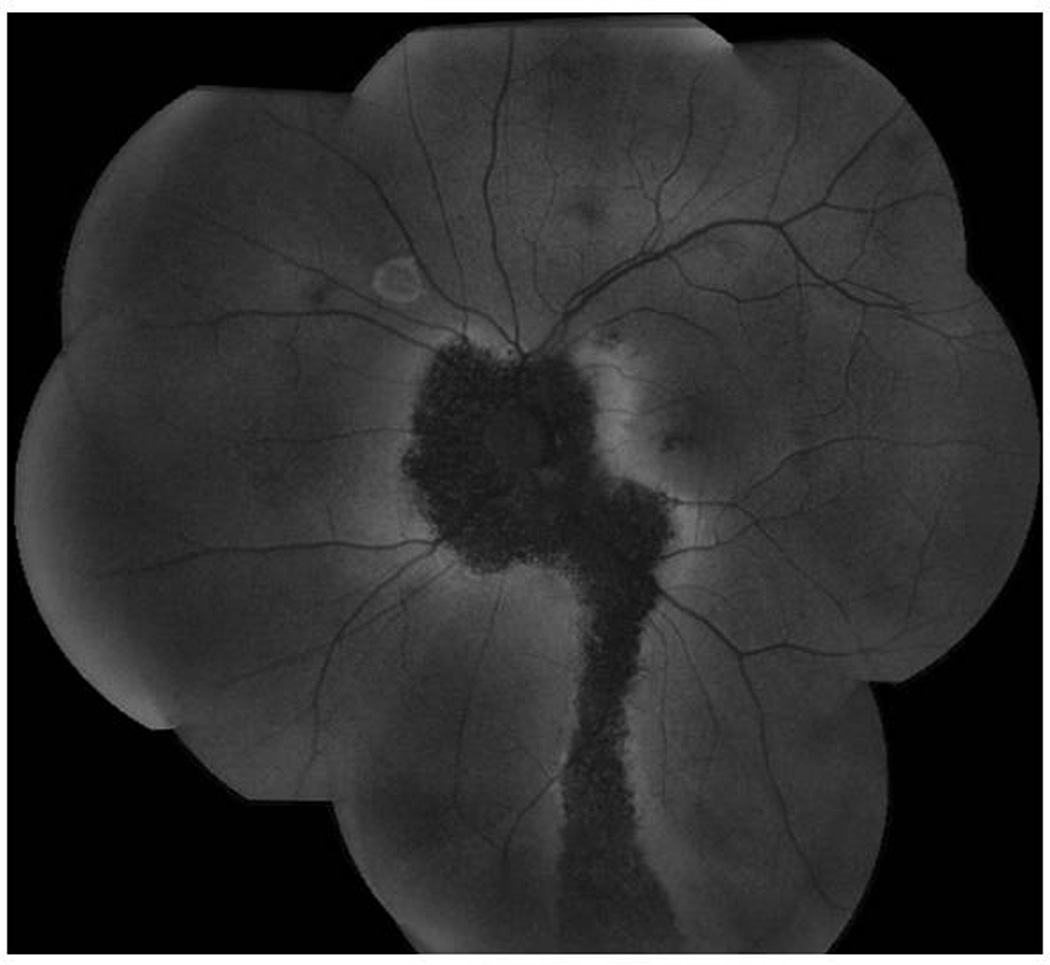
Recent technological advances--new pathophysiological insights, new imaging techniques for diagnosis and management, and new treatments--have led to an improved understanding of central serous chorioretinopathy (CSC). The primary role of the choroid has become more widely accepted with widespread use of indocyanine green angiography. Optical coherence tomography (OCT), and particularly enhanced depth imaging OCT, demonstrate a thickened and engorged choroid. Adaptive optics, fundus autofluorescence, multifocal electroretinography, microperimetry, and contrast sensitivity testing reveal that patients with even a mild course suffer previously undetected anatomic and functional loss. Although focal laser and photodynamic therapy are the current standard of care for persistent subretinal fluid in CSC, they are not appropriate in all cases, and the optimal timing of intervention remains unclear.


Figures

Figure 1
Figure 1
Cystoid macular edema in end-stage CSC. (A) Time-domain OCT of an eye with CSC and subretinal fluid (between arrows). Best-corrected visual acuity is 20/40. (B) SD-OCT of the same eye four years later, now with cystoid macular edema (arrows), subretinal fluid, and best-corrected visual acuity of 20/400. The eye had not responded to two PDT treatments, multiple anti-VEGF injections, and a trial of oral acetazolamide. CNV had been ruled out with fluorescein angiography and indocyanine green angiography. (C) SD-OCT of the same eye one month later. Macular atrophy is apparent after spontaneous resolution of cystoid edema. Best-corrected visual acuity is 20/500.
Figure 2
Figure 2
Fluorescein angiogram demonstrating a single pinpoint leak at twenty seconds (A) and ten minutes (B).
Figure 3
Figure 3
Fluorescein angiogram showing a smokestack leakage pattern at thirty seconds, two minutes, and five minutes.
Figure 4
Figure 4
Fluorescein angiogram of an asymptomatic eye with extramacular evidence of CSC.
Figure 5
Figure 5
Fundus autofluorescence (FAF) of a descending tract in CSC. The marked hypoautofluorescence suggests chronic disease and irreversible RPE damage. There is also a hyperfluorescent ring superiorly due to a retinal pigment epithelial detachment.
Figure 6
Figure 6
Numerous hyperfluorescent plaques on mid-phase indocyanine green (ICG) angiography demonstrating inner choroidal staining.
Figure 7
Figure 7
(A) Enhanced depth imaging (EDI) OCT of an area with CSC-related subretinal fluid. The choroid is abnormally thick (502 microns). (B) Corresponding FAF in which a line shows the location of the EDI OCT. Note central hypoautofluorescence and surrounding hyperautofluorescence suggestive of RPE damage.
Figure 8
Figure 8
Punctate hyperautofluorescence resulting in a granular appearance on FAF (A, arrow) often corresponds to hyperreflective lesions in the outer retina (B, arrow) and subretinal space on OCT. They have been hypothesized to be macrophages engorged with phagocytosed outer segments.
Figure 9
Figure 9
SD-OCT of a fovea-involving a serous retinal detachment in a patient with acute CSC. Note the lack of significant reflective debris on the outer retina or in the subretinal space.
Figure 10
Figure 10
(A) SD-OCT from the initial visit of a patient who had symptoms of CSC for ten months. Note the accumulation of material on the outer retina (arrow). This accumulation is thought to represent shed photoreceptor outer segments, engorged macrophages, and other inflammatory debris such as fibrin. (B) SD-OCT of the same eye two months later showing that the accumulation of this material has increased (arrow), perhaps due to elongation of outer segments.
Figure 11
Figure 11
(A) Fundus autofluorescence (FAF) showing several paramacular areas of autofluorescence abnormalities. The hypoautofluorescent descending tract inferotemporal to the macula represents the area of greatest RPE damage. (B) A microperimetry pattern used to study the inferior macula and mid-periphery shows decreased sensitivity in the areas of autofluorescence abnormality, with a particularly dense scotoma in the area of the descending tract seen in (A). Units are decibels.

References

    1. U.S. Preventive Services Task Force Ratings: Grade Definitions. Guide to Clinical Preventive Services, Third Edition: Periodic Updates. 2000–2003. [Accessed 10/10/2011]. at http://www.uspreventiveservicestaskforce.org/3rduspstf/ratings.htm.
    1. Abu el-Asrar AM. Central serous chorioretinopathy complicating systemic corticosteroid therapy. Eur J Ophthalmol. 1997;7:297–300. - PubMed
    1. Ahnoux-Zabsonre A, Quaranta M, Mauget-Faysse M. Prevalence of Helicobacter pylori in central serous chorioretinopathy and diffuse retinal epitheliopathy: a complementary study. J Fr Ophtalmol. 2004;27:1129–1133. - PubMed
    1. Aliferis K, Petropoulos IK, Farpour B, Matter MA, Safran AB. Should central serous chorioretinopathy be added to the list of ocular side effects of phosphodiesterase 5 inhibitors? Ophthalmologica. 2012;227:85–89. - PubMed
    1. Amalric P, Gourinat P, Rebiere P. Is central serous choroiditis sometimes hereditary? Bull Soc Ophtalmol Fr. 1971;71:163–168. - PubMed