. 2012 Dec 21;1(12):590-601.
doi: 10.1021/sb3000436.

Organization of synthetic alphoid DNA array in human artificial chromosome (HAC) with a conditional centromere

Affiliations

Organization of synthetic alphoid DNA array in human artificial chromosome (HAC) with a conditional centromere

Natalay Kouprina et al. ACS Synth Biol. .

Abstract

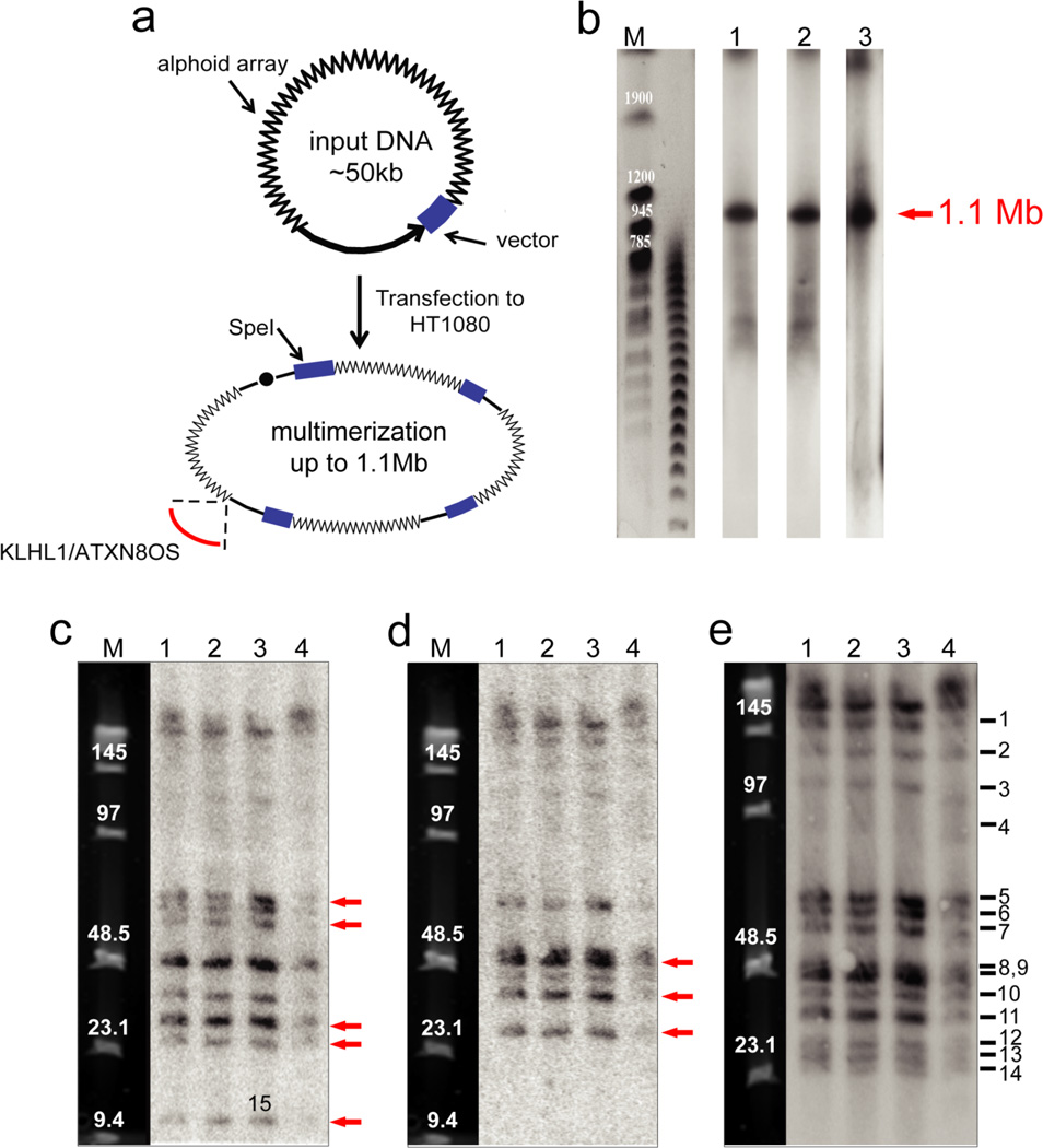
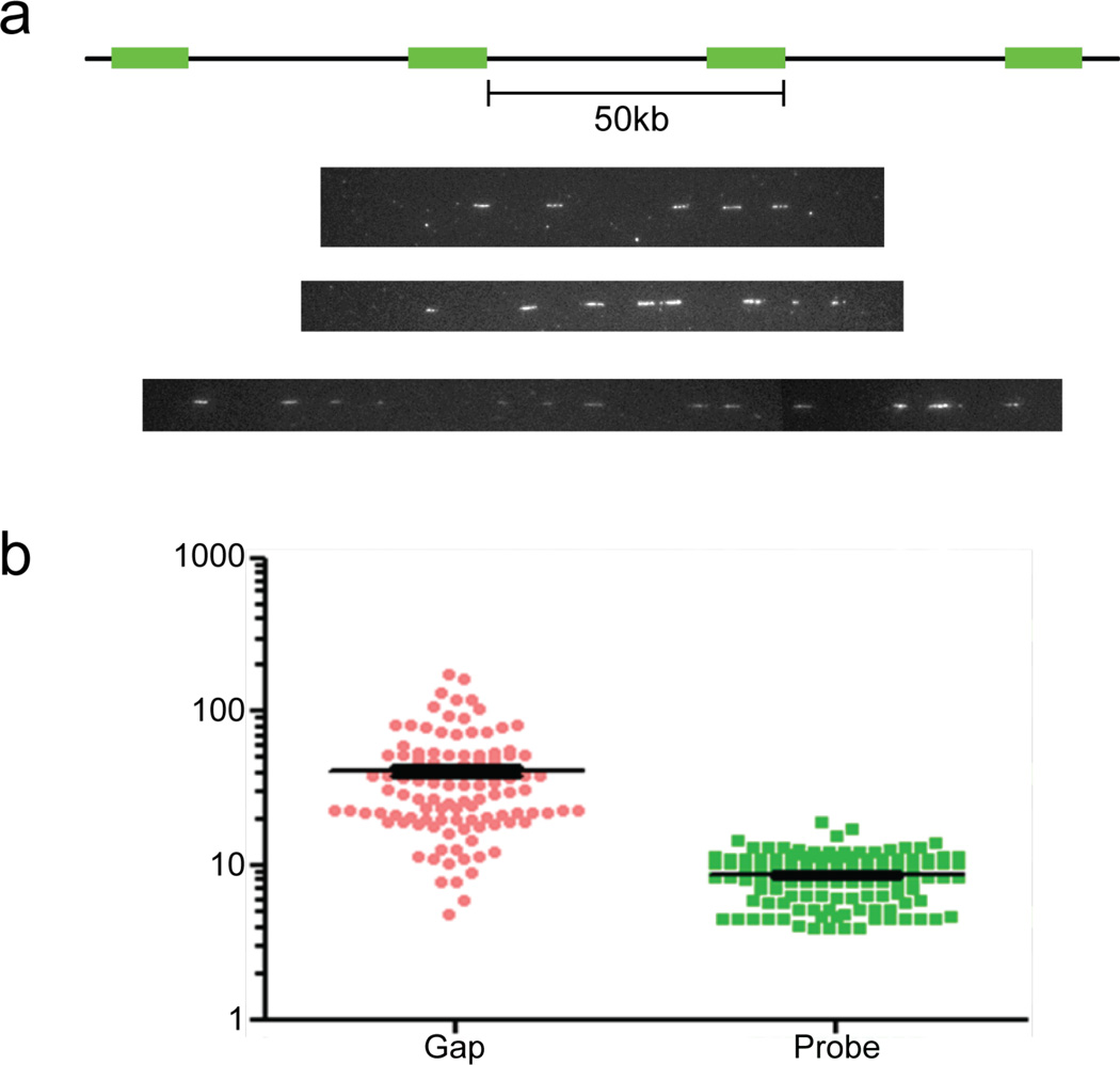
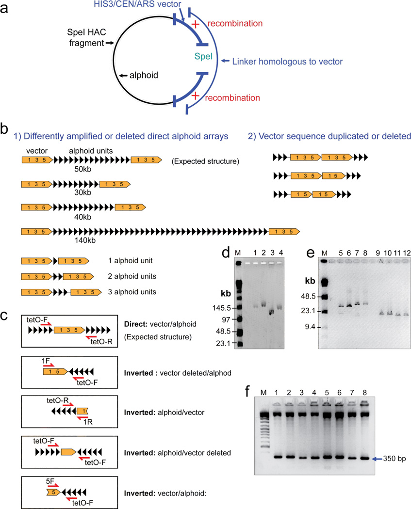
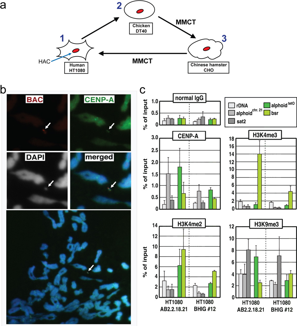
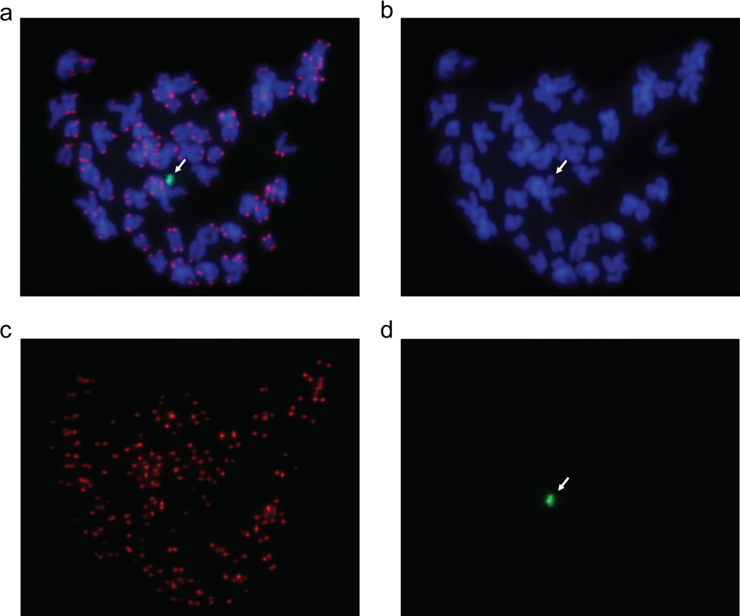
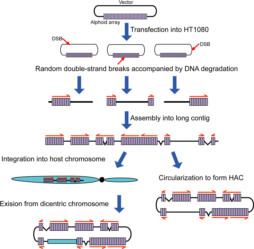
Human artificial chromosomes (HACs) represent a novel promising episomal system for functional genomics, gene therapy, and synthetic biology. HACs are engineered from natural and synthetic alphoid DNA arrays upon transfection into human cells. The use of HACs for gene expression studies requires the knowledge of their structural organization. However, none of the de novo HACs constructed so far has been physically mapped in detail. Recently we constructed a synthetic alphoid(tetO)-HAC that was successfully used for expression of full-length genes to correct genetic deficiencies in human cells. The HAC can be easily eliminated from cell populations by inactivation of its conditional kinetochore. This unique feature provides a control for phenotypic changes attributed to expression of HAC-encoded genes. This work describes organization of a megabase-size synthetic alphoid DNA array in the alphoid(tetO)-HAC that has been formed from a ~50 kb synthetic alphoid(tetO)-construct. Our analysis showed that this array represents a 1.1 Mb continuous sequence assembled from multiple copies of input DNA, a significant part of which was rearranged before assembling. The tandem and inverted alphoid DNA repeats in the HAC range in size from 25 to 150 kb. In addition, we demonstrated that the structure and functional domains of the HAC remains unchanged after several rounds of its transfer into different host cells. The knowledge of the alphoid(tetO)-HAC structure provides a tool to control HAC integrity during different manipulations. Our results also shed light on a mechanism for de novo HAC formation in human cells.


Figures

Figure 1
Figure 1
Structural analysis by restriction enzyme digestion and CHEF of the alphoidtetO-HAC in human, hamster and chicken cells. (a) Diagram illustrating multimerization of input DNA during de novo HAC formation in human cells. Input DNA consists of 40 kb alphoid DNA and 10 kb vector sequence. Chromosome 13 fragment carrying the KLHL1/ATXN8OS genes was captured during formation of the alphoidtetO-HAC. (b) Determination of the size of the multimerized input DNA in the alphoidtetO-HAC in chicken DT40, hamster CHO and human HT1080 cells. Genomic DNA possessing the HAC was digested with PmeI and separated by CHEF gel electrophoresis (range 200–1500 kb). The transferred membranes were hybridized with a radioactively labeled tetO-specific alphoid probe. A single1.1 Mb fragment was detected. Its size was determined by comparison with the DNA size standard, S. cerevisiae chromosomes. Lane 1 – the HAC in chicken cells; lane 2 – the HAC in hamster cells; lane 3 – the HAC in human cells. (c) Analysis of the alphoidtetO-HAC digested by SpeI. Genomic DNA possessing the HAC was digested with SpeI endonuclease and separated by CHEF gel electrophoresis (range 10–70 kb). The transferred membranes were hybridized with either vector probe 3 (c), vector probe 1 (d) or tetO-alphoid (e) probe. Red arrows indicate to the fragments that are specific to either probe 1 or probe 3. Lane 1 – the HAC in DT40 cells; lane 2 - the HAC in CHO cells; lane 3 - the original alphoidtetO-HAC in human HT1080 cells; lane 4 - the HAC transferred back to human cells from CHO cells. M - Pulse Markers.
Figure 2
Figure 2
Analysis of the alphoidtetO-HAC by fiber-FISH. Single DNA fibers from human HT1080 cells containing the HAC were stretched on silanized coverslips. HAC DNA was detected by FISH using the 10 kb RCA/SAT43 vector DNA as a probe. Representative images are showed in (a). All the FISH signals (white short lines) are vector DNA and the gaps between the FISH signals are alphoid-satellite repeat sequences. (b) The length of the FISH signals and gaps are shown as a graph. Ordinate shows the length distribution for vector sequence (probe) and for sequence between neighboring vector signals (gap) in kb.
Figure 3
Figure 3
Structural analysis of the HAC fragments rescued by TAR cloning in yeast. (a) A scheme of rescue of the SpeI HAC fragments as circular molecules by transformation-associated homologous recombination in yeast. The vector part of input DNA contains a YAC cassette with an yeast selectable marker HIS3, ARS and CEN sequences. In vivo recombination between a HAC fragment and a linker results in a circular molecule. The clones with the circularized fragments were selected on SD-His medium. Alphoid DNA is marked in black. Vector DNA is marked in blue. (b) Schematic diagram of the rearranged input DNA molecules rescued in yeast and E. coli. Several examples of SpeI-fragments rescued in yeast are shown. (c) PCR analysis of direct and inverted alphoid sequences in the rescued YAC clones and in the alphoidtetO-HAC propagated in chicken cells. A single tetO-F primer amplifies a part of the vector sequence (positions 2,913–3,300 in the RCA/SAT/43 vector) flanked by inverted alphoid repeats. (d, e) Characterization of the rescued clones in a BAC form. BAC DNAs were isolated from 12 randomly chosen clones, digested by SpeI for linearization, separated by CHEF gel electrophoresis (lanes 1, 2, 3, 4 with range 20–300 kb; lanes 5–12 with range 10–70 kb), and visualized by staining with ethidium bromide. M - Pulse Marker™ 0.1–200 kb (Sigma-Aldrich, USA). (f) The tandem repeat structure of alphoid arrays is confirmed by StuI restriction enzyme digestion (350 bp alphoid dimer unit); the upper bands represent vector fragments.
Figure 4
Figure 4
Integrity of the functional domains in the alphoidtetO-HAC that underwent three rounds of MMCT transfer. (a) The alphoidtetO-HAC was first transferred to chicken DT40 from human HT1080. A loxP cassette was inserted into the HAC by homologous recombination in DT40 cells. Then the HAC was transferred to CHO cells. From CHO cells the HAC was transferred back to HT1080. (b) Immuno-FISH analysis of metaphase chromosome spreads containing the alphoidtetO-HAC in human HT1080 cells. Cells with the alphoidtetO-HAC (clone BHIG#12) were used for analysis. Immunolocalization of the centromeric protein CENP-A on metaphases was performed by indirect immunofluorescence with anti–CENP-A antibody and Alexa 488-conjugated secondary antibody (green). HAC-specific DNA sequence (RCA/SAT43 vector) was used as a FISH probe to detect the HAC (red). CENP-A and BAC signals on the HAC overlap one another. (c) ChIP analysis of CENP-A assembly and modified histone H3 at the alphoidtetO-HAC. The results of ChIP analysis using normal mouse IgG (top left panel), antibodies against CENP-A (middle left panel), dimethylated histone H3 Lys4 (H3K4me2, bottom left panel), trimethylated histone H3 Lys4 (H3K4me3, middle right panel) and trimethylated histone H3 Lys9 (H3K9me3, bottom right panel). The assemblies of these proteins on the original alphoidtetO-HAC in the AB2.2.18.21 cell line (left), the alphoidtetO-HAC in the BHIG#12 cell line (right) are shown. The bars show the percentage recovery of the various target DNA loci by immunoprecipitation with each antibody to input DNA. Error bars indicate s.d. (n= 2 or 3). Analyzed loci were rDNA (5S ribosomal DNA), alphoidchr.21 (centromeric alphoid DNA of chromosome 21), sat2 (pericentromeric satellite 2), alphoidtetO (alphoid DNA with tetO motif on tetO alphoid HAC), Bsr (the marker gene in BAC vector region of tetO alphoid HAC). Comparison of the enrichment of tetO-alphoid DNA and the endogenous chromosome 21 alphoid DNA by CENP-A, H3K4me2, H3K4me3 in BHIG#12 and AB2.2.18.21 cells was carried out by calculations of the ratio between IPed tetO-alphoid DNA in the HAC and IPed endogenous chromosome 21 alphoid DNA for each cell line. As seen in Figure S1 (Supporting Information), a relative enrichment of CENP-A, H3K4me2, H3K4me3 and H3K9me3 on tetO-alphoid DNAs in BHIG#12 cells is not different from that observed in AB2.2.18.21 cells. This indicates that kinetochore regions in the HAC did not change after multiple steps of HAC transfer via MMCT.
Figure 5
Figure 5
FISH analysis of the alphoidtetO-HAC in HT1080 cells using the telomere probe after several steps of MMCT transfer. FISH analysis was performed using PNA labeled probes for telomeric (red) and tetO-alphoid sequences (green). Panels c and d represent metaphase spreads hybridized with telomeric and tetO-alphoid probes, correspondingly. Panel a represents merged images of panels b, c, and d.
Figure 6
Figure 6
Mechanism of de novo HAC formation in human cells from the input DNA. Input DNA contains ~40 kb of alphoid DNA and ~10 kb vector DNA. Full lines represent input alphoid DNA. Multiple molecules of input DNA penetrate into a single human cell. Red arrows indicate to randomly introduced DSBs in input DNA during or after penetration into a human cell. The ends of input DNA molecules are underwent degradation. Degradation of either alphoid DNA or vector sequence takes place. The resulting fragments are assembled by either NHEJ or homologous recombination into long contig. The final linear concatamers either are circularized by NHEJ to form a HAC or are inserted into a host chromosome. In the latter case, the dicentric chromosome underwent breakage in a subsequent mitosis, with further breakage and circularization events leading to the HAC.

References

    1. Ren X, Tahimic CG, Katoh M, Kurimasa A, Inoue T, Oshimura M. Human artificial chromosome vectors meet stem cells: new prospects for gene delivery. Stem Cell Review. 2006;2:43–50. - PubMed
    1. Grimes BR, Monaco ZL. Artificial and engineered chromosomes: Developments and prospects for gene therapy. Chromosoma. 2005;114:230–241. - PubMed
    1. Basu J, Willard HF. Artificial and engineered chromosomes: Non-integrating vectors for gene therapy. Trends Mol. Med. 2005;11:251–258. - PubMed
    1. Monaco ZL, Moralli D. Progress in artificial chromosome technology. Biochem. Soc. Trans. 2006;34:324–327. - PubMed
    1. Macnab S, Whitehouse A. Progress and prospects: human artificial chromosomes. Gene Ther. 2009;16:1180–1188. - PubMed

Publication types