

Enlarged perivascular spaces as a marker of underlying arteriopathy in intracerebral haemorrhage: a multicentre MRI cohort study
- PMID: 23412074
- PMCID: PMC3905629
- DOI: 10.1136/jnnp-2012-304434
Enlarged perivascular spaces as a marker of underlying arteriopathy in intracerebral haemorrhage: a multicentre MRI cohort study
Abstract
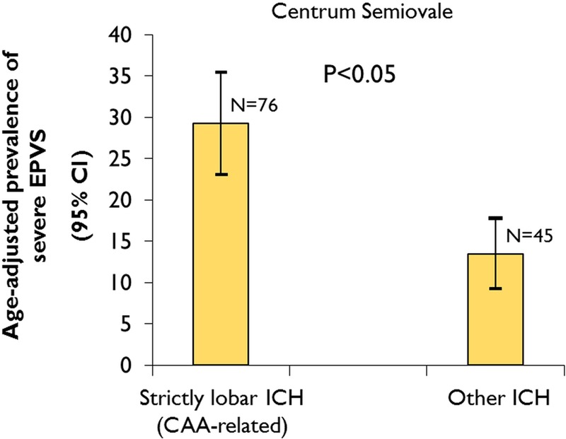
Background and purpose: Small vessel disease (mainly hypertensive arteriopathy and cerebral amyloid angiopathy (CAA)) is an important cause of spontaneous intracerebral haemorrhage (ICH), a devastating and still poorly understood stroke type. Enlarged perivascular spaces (EPVS) are a promising neuroimaging marker of small vessel disease. Based on the underlying arteriopathy distributions, we hypothesised that severe centrum semiovale EPVS are more common in lobar ICH attributed to CAA than other ICH. We evaluated EPVS prevalence, severity and distribution, and their clinical-radiological associations.
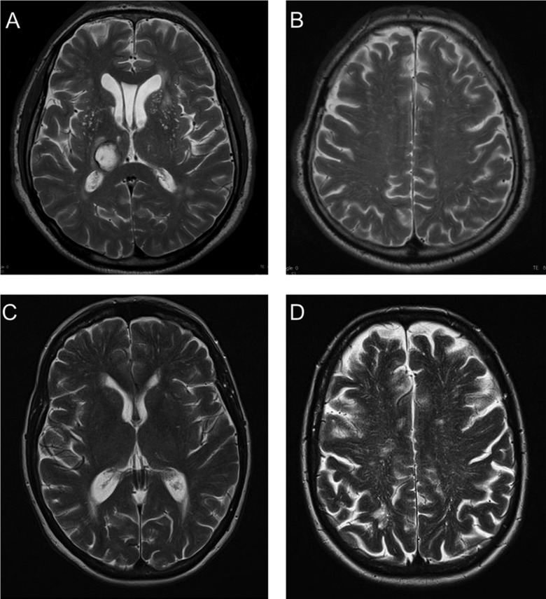
Methods: Retrospective multicentre cohort study of 121 ICH patients. Clinical information was obtained using standardised forms. Basal ganglia and centrum semiovale EPVS on T2-weighted MRI (graded 0-4 (>40 EPVS)), white-matter changes, cerebral microbleeds (CMBs) and lacunes were rated using validated scales.
Results: Patients with probable or possible CAA (n=76) had a higher prevalence of severe (>40) centrum semiovale EPVS compared with other ICH patients (35.5% vs 17.8%; p=0.041). In logistic regression age (OR: 1.43; 95% CI 1.01 to 2.02; p=0.045), deep CMBs (OR: 3.27; 95% CI 1.27 to 8.45; p=0.014) and mean white-matter changes score (OR: 1.29; 95% CI 1.17 to 1.43; p<0.0001) were independently associated with increased basal ganglia EPVS severity; only age was associated with increased centrum semiovale EPVS severity (OR: 1.50; 95% CI 1.08 to 2.10; p=0.017).
Conclusions: EPVS are common in ICH. Different mechanisms may account for EPVS according to their anatomical distribution. Severe centrum semiovale EPVS may be secondary to, and indicative of, CAA with value as a new neuroimaging marker. By contrast, basal ganglia EPVS severity is associated with markers of hypertensive arteriopathy.
Keywords: Amyloid; Cerebrovascular Disease; Clinical Neurology; MRI.
Figures
References
-
- Charidimou A, Gang Q, Werring DJ. Sporadic cerebral amyloid angiopathy revisited: recent insights into pathophysiology and clinical spectrum. J Neurol Neurosurg Psychiatry 2012;83:124–37 - PubMed
-
- Ozturk MH, Aydingoz U. Comparison of MR signal intensities of cerebral perivascular (Virchow-Robin) and subarachnoid spaces. J Comput Assist Tomogr 2002;26:902–4 - PubMed
-
- Wardlaw JM. Blood-brain barrier and cerebral small vessel disease. J Neurol Sci 2010;299:66–71 - PubMed
Publication types
MeSH terms
Substances
Grants and funding
LinkOut - more resources
Full Text Sources
Other Literature Sources