

Relationship between changes in the cochlear blood flow and disorder of hearing function induced by blast injury in guinea pigs
- PMID: 23412965
- PMCID: PMC3563195
Relationship between changes in the cochlear blood flow and disorder of hearing function induced by blast injury in guinea pigs
Abstract
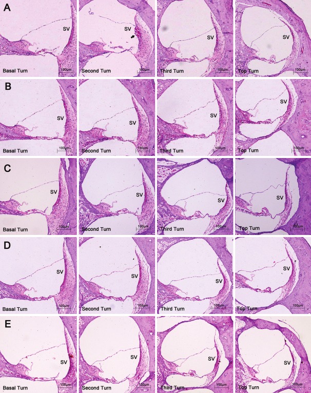
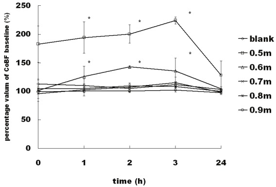
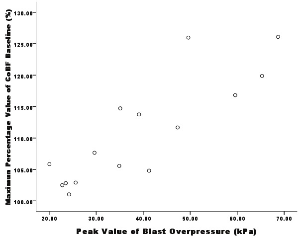
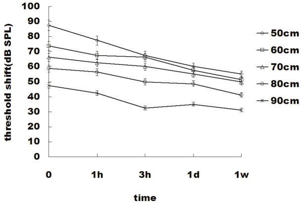
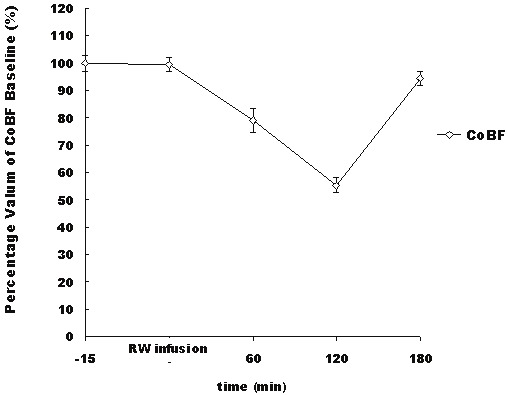
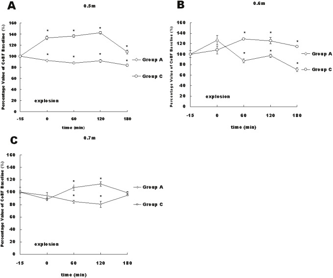
The auditory system is the most susceptible to damages from blast waves. Blast injuries always lead to varying degrees of hearing impairment. Although a disorder of the cochlear blood flow (CoBF) has been considered to be related to many pathological processes of the auditory system and to contribute to various types of hearing loss, changes in the CoBF induced by blast waves and the relationship between such changes and hearing impairment are undefined. To observe the changes in the cochlear microcirculation after exposure to an explosion blast, investigate the relationship between changes in the CoBF and hearing impairment and subsequently explore the mechanism responsible for the changes in the CoBF, we detected the perfusion of the cochlear microcirculation and hearing threshold shift after exposure to an explosion blast. Then, an N-nitro-L-arginine-methyl ester (L-NAME, NO synthase inhibitor) solution and artificial perilymph were applied to the round window (RW) of the cochlea before the blast exposure, followed by an evaluation of the CoBF and hearing function. The results indicated that the changes in the CoBF were correlated to the strength of the blast wave. The cochlear blood flow significantly increased when the peak value of the blast overpressure was greater than approximately 45 kPa, and there was no significant change in the cochlear blood flow when the peak value of the blast overpressure was less than approximately 35 kPa. Following local administration of the NO synthase inhibitor L-NAME, the increase in the CoBF induced by the blast was inhibited, and this reduction was significantly associated with the hearing threshold.
Keywords: Cochlea; blast injury; blood flow; guinea pigs; hearing function.
Figures
References
-
- Matsumoto Y, Hatano B, Matsushita Y, Nawashiro H, Shima K. [The characteristics of blast traumatic brain injury] . No Shinkei Geka. 2010;38:695–702. - PubMed
-
- Phillips YY, Zajtchuk JT. Blast injuries of the ear in military operations. Ann Otol Rhinol Laryngol Suppl. 1989;140:3–4. - PubMed
-
- Lockhart P, Cronin D, Williams K, Ouellet S. Investigation of head response to blast loading. J Trauma. 2011;70:E29–36. - PubMed
-
- Chandler DW, Edmond CV. Effects of blast overpressure on the ear: case reports. J Am Acad Audiol. 1997;8:81–88. - PubMed
-
- Chen YS, Tseng FY, Liu TC, Lin-Shiau SY, Hsu CJ. Involvement of nitric oxide generation in noise-induced temporary threshold shift in guinea pigs. Hear Res. 2005;203:94–100. - PubMed
Publication types
MeSH terms
Substances
LinkOut - more resources
Full Text Sources
Medical
Research Materials