

A mouse model for human osteogenesis imperfecta type VI
- PMID: 23413146
- PMCID: PMC3688658
- DOI: 10.1002/jbmr.1892
A mouse model for human osteogenesis imperfecta type VI
Abstract
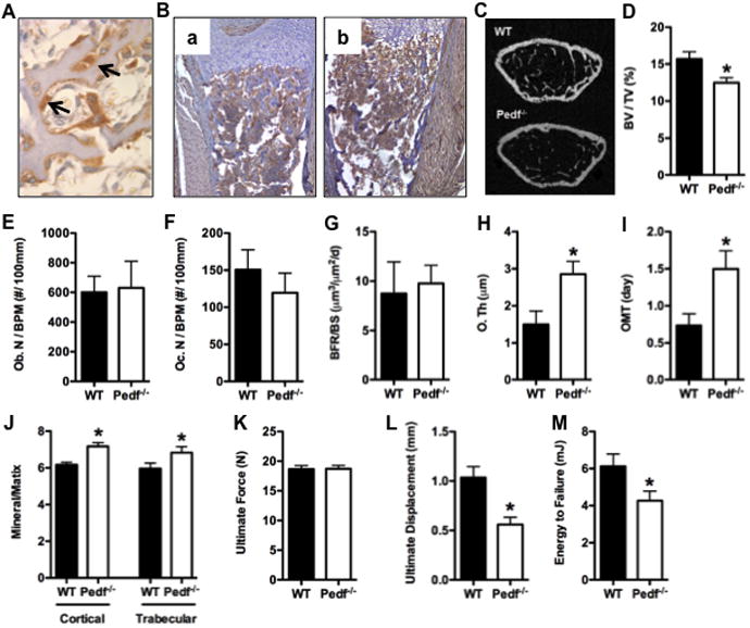
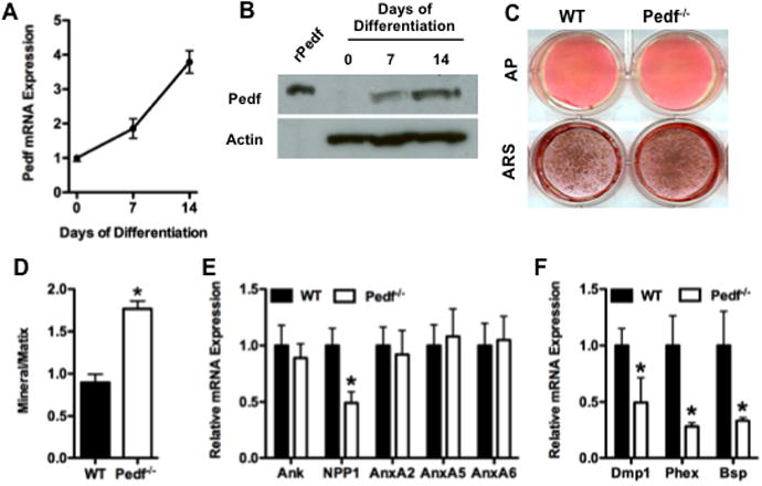
Osteogenesis imperfecta type VI (OI type VI) has recently be linked to a mutation in the SERPINF1 gene, which encodes pigment epithelium-derived factor (PEDF), a ubiquitously expressed protein originally described for its neurotrophic and antiangiogenic properties. In this study, we characterized the skeletal phenotype of a mouse with targeted disruption of Pedf. In normal mouse bone, Pedf was localized to osteoblasts and osteocytes. Micro-computed tomography (µCT) and quantitative bone histomorphometry in femurs of mature Pedf null mutants revealed reduced trabecular bone volume and the accumulation of unmineralized bone matrix. Fourier transform infrared microscopy (FTIR) indicated an increased mineral:matrix ratio in mutant bones, which were more brittle than controls. In vitro, osteoblasts from Pedf null mice exhibited enhanced mineral deposition as assessed by Alizarin Red staining and an increased mineral:matrix determined by FTIR analysis of calcified nodules. The findings in this mouse model mimic the principal structural and biochemical features of bone observed in humans with OI type VI and consequently provide a useful model with which to further investigate the role of PEDF in this bone disorder.
Copyright © 2013 American Society for Bone and Mineral Research.
Figures
Comment in
-
Osteogenesis imperfecta, an ever-expanding conundrum.J Bone Miner Res. 2013 Jul;28(7):1519-22. doi: 10.1002/jbmr.1982. J Bone Miner Res. 2013. PMID: 23696068 No abstract available.
References
-
- Marini JC, Forlino A, Cabral WA, Barnes AM, San Antonio JD, Milgrom S, Hyland JC, Korkko J, Prockop DJ, De Paepe A, Coucke P, Symoens S, Glorieux FH, Roughley PJ, Lund AM, Kuurila-Svahn K, Hartikka H, Cohn DH, Krakow D, Mottes M, Schwarze U, Chen D, Yang K, Kuslich C, Troendle J, Dalgleish R, Byers PH. Consortium for osteogenesis imperfecta mutations in the helical domain of type I collagen: regions rich in lethal mutations align with collagen binding sites for integrins and proteoglycans. Hum Mutat. 2007;28:209–221. - PMC - PubMed
-
- Prockop DJ, Kivirikko KI. Collagens: molecular biology, diseases, and potentials for therapy. Annu Rev Biochem. 1995;64:403–434. - PubMed
-
- Morello R, Bertin TK, Chen Y, Hicks J, Tonachini L, Monticone M, Castagnola P, Rauch F, Glorieux FH, Vranka J, Bachinger HP, Pace JM, Schwarze U, Byers PH, Weis M, Fernandes RJ, Eyre DR, Yao Z, Boyce BF, Lee B. CRTAP is required for prolyl 3- hydroxylation and mutations cause recessive osteogenesis imperfecta. Cell. 2006;127:291–304. - PubMed
-
- Barnes AM, Chang W, Morello R, Cabral WA, Weis M, Eyre DR, Leikin S, Makareeva E, Kuznetsova N, Uveges TE, Ashok A, Flor AW, Mulvihill JJ, Wilson PL, Sundaram UT, Lee B, Marini JC. Deficiency of cartilage-associated protein in recessive lethal osteogenesis imperfecta. N Engl J Med. 2006;355:2757–2764. - PMC - PubMed
Publication types
MeSH terms
Substances
Supplementary concepts
Grants and funding
LinkOut - more resources
Full Text Sources
Other Literature Sources
Medical
Molecular Biology Databases
Miscellaneous
