

Super-resolution track density imaging of glioblastoma: histopathologic correlation
- PMID: 23413250
- PMCID: PMC4017075
- DOI: 10.3174/ajnr.A3400
Super-resolution track density imaging of glioblastoma: histopathologic correlation
Abstract
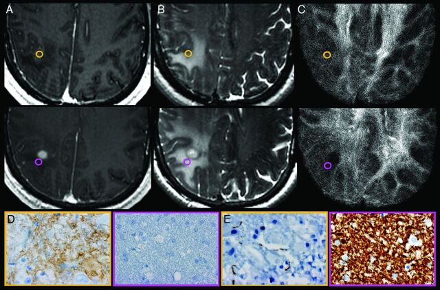
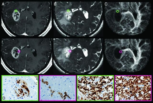
Background and purpose: Super-resolution track density imaging generates anatomic images with submillimeter voxel resolution by using high-angular-resolution diffusion imaging and fiber-tractography. TDI within the diseased human brain has not been previously described. The purpose of this study was to correlate TDI with histopathologic features of GBM.
Materials and methods: A total of 43 tumor specimens (24 contrast-enhancing, 12 NE, and 7 centrally necrotic regions) were collected from 18 patients with treatment-naïve GBM by use of MR imaging-guided neurosurgical techniques. Immunohistochemical stains were used to evaluate the following histopathologic features: hypoxia, architectural disruption, microvascular hyperplasia, and cellular proliferation. We reconstructed track density maps at a 0.25-mm isotropic spatial resolution by using probabilistic streamline tractography combined with constrained spheric deconvolution (model order, 8; 0.1-mm step size; 1 million seed points). Track density values were obtained from each tissue site. A P value of .05 was considered significant and was adjusted for multiple comparisons by use of the false discovery rate method.
Results: Track density was not significantly different between contrast-enhancing and NE regions but was more likely to be elevated within regions demonstrating aggressive histopathologic features (P < .05). Significant correlation between relative track density and hypoxia (odds ratio, 3.52; P = .01), architectural disruption (odds ratio, 3.49; P = .03), and cellular proliferation (odds ratio, 1.70; P = .05) was observed irrespective of the presence or absence of contrast enhancement.
Conclusions: Numeric values of track density correlate with GBM biologic features and may be clinically useful for identification of regions of tumor infiltration within both enhancing and NE components of GBM.
Figures
References
-
- Calamante F, Tournier JD, Jackson GD, et al. Track-density imaging (TDI): super-resolution white matter imaging using whole-brain track-density mapping. Neuroimage 2010;53:1233–43 - PubMed
-
- Calamante F, Tournier JD, Heidemann RM, et al. Track density imaging (TDI): validation of super resolution property. Neuroimage 2011;56:1259–66 - PubMed
-
- Calamante F, Tournier JD, Kurniawan ND, et al. Super-resolution track-density imaging studies of mouse brain: comparison to histology. Neuroimage 2012;59:286–96 - PubMed
-
- Hess CP, Mukherjee P. Visualizing white matter pathways in the living human brain: diffusion tensor imaging and beyond. Neuroimaging Clin N Am 2007;17:407–26, vii - PubMed
-
- von Morze C, Xu D, Hess CP. Development and evaluation of a robust and efficient computational pipeline for track density imaging for use in a clinical research environment. In: Proceedings of the International Society for Magnetic Resonance Medicine, Montreal, Canada; May 12, 2011. Abstract 4025
Publication types
MeSH terms
Substances
Grants and funding
LinkOut - more resources
Full Text Sources
Other Literature Sources
Medical