Review
. 2013:82:693-721.
doi: 10.1146/annurev-biochem-072711-164732. Epub 2013 Feb 13.

Signal recognition particle: an essential protein-targeting machine

Affiliations
Review

Signal recognition particle: an essential protein-targeting machine

David Akopian et al. Annu Rev Biochem. 2013.

Abstract

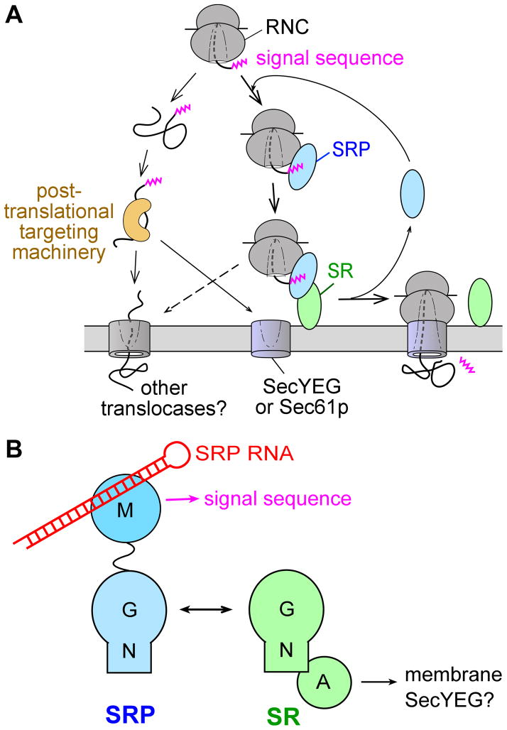
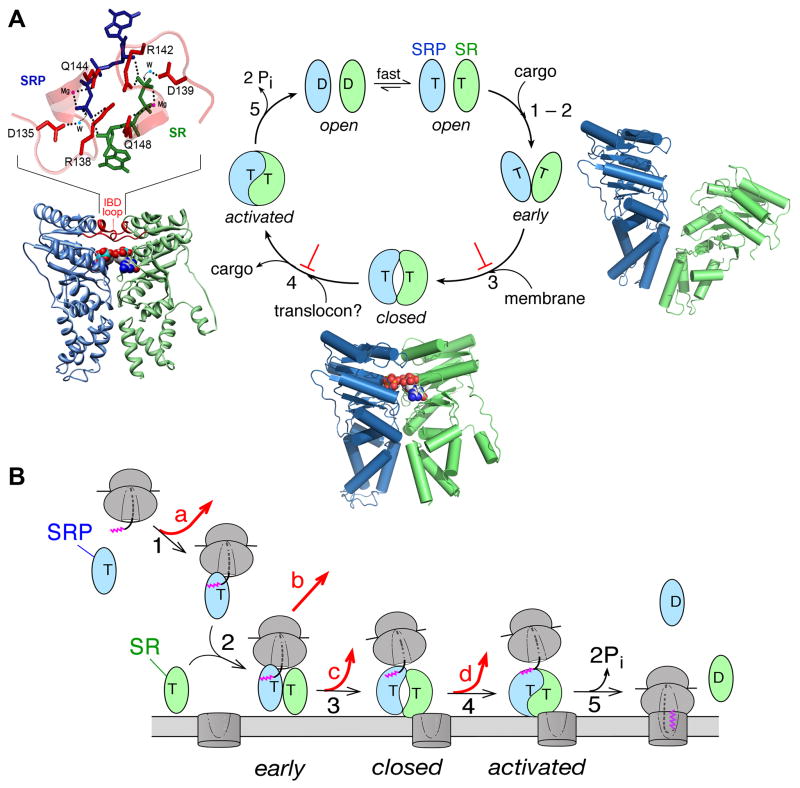
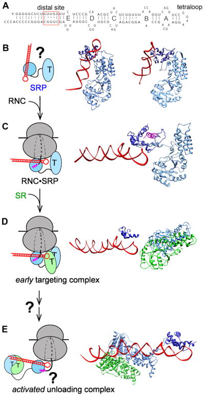
The signal recognition particle (SRP) and its receptor compose a universally conserved and essential cellular machinery that couples the synthesis of nascent proteins to their proper membrane localization. The past decade has witnessed an explosion in in-depth mechanistic investigations of this targeting machine at increasingly higher resolutions. In this review, we summarize recent work that elucidates how the SRP and SRP receptor interact with the cargo protein and the target membrane, respectively, and how these interactions are coupled to a novel GTPase cycle in the SRP·SRP receptor complex to provide the driving force and enhance the fidelity of this fundamental cellular pathway. We also discuss emerging frontiers in which important questions remain to be addressed.


Figures

Figure 1
Figure 1
Overview of the pathways and components of SRP. (A) Multiple pathways deliver newly synthesized proteins to the ER or plasma membrane, with the SRP pathway mediating the co-translational targeting of translating ribosomes (right) and post-translational targeting machineries mediating the targeting of proteins released from the ribosome. (B) Domain structures of the ribonucleoprotein core of SRP, comprised of the SRP54 (or Ffh) protein and the SRP RNA (left), and the bacterial SRP receptor (right).
Figure 2
Figure 2
(A) Molecular model for interaction of the bacterial SRP with the translating ribosome (gray; PDB 2J28), derived from cryoEM reconstruction and docking of the crystal structures of individual protein fragments as described in (44). The M- and NG-domains of the SRP are in dark and light blue, respectively, the SRP RNA is in red, and the signal sequence is in magenta. (B) Crystal structure of the bacterial FtsY (NG+1) construct (PDB 2QY9) highlighting its lipid-binding helix at the N-terminus (orange).
Figure 3
Figure 3
Conformational changes in the SRP and SR GTPases ensure the efficiency and fidelity of protein targeting. The steps are numbered to be consistent between parts (A) and (B). The Ffh and FtsY NG domains are in blue and green, respectively. T and D denote GTP and GDP, respectively. (A) A series of discrete rearrangements drive the SRP•SR GTPase cycle and are regulated by the cargo and target membrane. ⊥ denotes the pausing effect of cargo in disfavoring the conformational rearrangements. Right panel: molecular model of the early intermediate (PDB 2XKV). Bottom panel: Co-crystal structure of the Ffh-FtsY NG-domain complex in the closed/activated conformation (PDB 1RJ9). The two GTP analogues are in spacefill. Left panel: Zoom-in of the composite active site formed at the dimer interface required for GTPase activation, with the GMPPCP molecules from Ffh and FtsY in blue and green, respectively, active site Mg2+ in magenta, nucleophilic waters (W) in blue, and catalytic residues in the IBD loops in red. (B) GTPase rearrangements provide the driving force and ensure the fidelity of protein targeting. Step 1, RNC with a signal sequence (magenta) binds the SRP. Step 2, cargo-loaded SRP forms a stabilized early targeting complex with FtsY. Step 3, membrane association of FtsY drives rearrangement to the closed state, which weakens SRP’s affinity for the cargo. Step 4, interaction of SR with the SecYEG translocon is proposed to drive GTPase rearrangements to the activated state required for cargo handover. Step 5, the cargo is unloaded from the SRP onto SecYEG, and GTP hydrolysis drives the disassembly and recycling of SRP and SR. At each step, the cargo can be either retained in (black arrows) or rejected from (red arrows) the SRP pathway.
Figure 4
Figure 4
RNA-mediated global reorganization of the SRP couples the GTPase cycle to the cargo loading and unloading events during protein targeting. (A) Secondary structure of the E. coli 4.5S SRP RNA. The internal loops A–E, the GGAA tetraloop and the distal site near the 5′,3′-end of this RNA are denoted. (B) Free SRP exist in a variety of ‘latent’ conformations in which the SRP RNA tetraloop is not positioned to contact SR. Two representative structures of SRP from Methanococcus jannaschii (left; PDB 2V3C) and Sulfolobus solfataricus (right; PDB 1QZW) are shown. (C) Binding of the RNC induces SRP into a more active conformation, in which the SRP RNA tetraloop is properly positioned to interact with the G-domain of the incoming SR to form a stabilized early targeting complex in (D). Both panels show the molecular model derived from cryo-EM reconstructions of the RNC•SRP or RNC•SRP•FtsY early complex; the ribosome is not shown for clarity. (E) GTPase activation is potentially coupled to relocalization of the SRP•SR NG-domain complex to the distal end of the SRP RNA, a conformation that is more conducive to cargo unloading (PDB 2XXA). The structures in (B) and (C) are aligned with respect to the SRP54-NG domain, and the structures in (C) – (E) are aligned with respect to the SRP RNA. Color codings are the same as in Figure 2.
Figure 5
Figure 5
Organization of the mammalian SRP. (A) Comparison of the RNA secondary structure and composition of the mammalian and bacterial SRP. The SRP54 M- and NG-domains are in dark and light blue, respectively, SRP19 is in cyan, SRP9 is in brown, SRP14 is in orange, and the SRP68/72 complex, which lacks a crystal structure, is represented as a gray sphere. (B) Cryo-EM reconstruction of the mammalian SRP bound to the RNC (left; EMD-1063), and molecular model of the mammalian SRP derived from the cryo-EM and docking of the crystal structures of the individual proteins (right; PDB 1RY1). The S- and Alu-domains of the SRP RNA are in red and yellow, respectively, the protein subunits are colored as in (A).
Figure 6
Figure 6
(A) Similarity and differences between the bacterial (left) and chloroplast (right) SRP systems. The SRP54 M- and NG-domains, FtsY, and the SRP RNA are colored as in Figure 2. The LHC protein (LHCP) is in green, and cpSRP43 is in magenta. The red arrows denote stimulatory effects of the SRP RNA (left) and the cpSRP54 M-domain (right) on assembly of the GTPase complex. (B) A molecular model of cpSRP43, obtained from small angle x-ray reconstructions of its 3-D shape (envelope; (183)) and rigid body docking of the structures of the CD1-Ank(1-4)-CD2 (PDB 3UI2) and CD3 (PDB 1X3P) fragments.

References

    1. Cross BCS, Sinning I, Luirink J, High S. Delivering proteins for export from the cytosol. Nature Rev Mol Cell Biol. 2009;10:255–64. - PubMed
    1. Driessen AJ, Nouwen N. Protein translocation across the bacterial cytoplasmic membrane. Annu Rev Biochem. 2008;77:643–67. - PubMed
    1. Pool MR. Signal recognition particles in chloroplasts, bacteria, yeast and mammals. Mole Membr Biol. 2005;22:3–15. - PubMed
    1. Shao S, Hegde RS. Membrane protein insertion at the endoplasmic reticulum. Ann Rev Cell Dev Biol. 2011;27:25–56. - PMC - PubMed
    1. Gilmore R, Blobel G, Walter P. Protein translocation across the endoplasmic reticulum: 1. Detection in the microsomal membrane of a receptor for the signal recognition particle. J Cell Biol. 1982a;95:463–9. - PMC - PubMed

Publication types

LinkOut - more resources