. 2013 Feb 15:10:26.
doi: 10.1186/1742-2094-10-26.

Treatment with an anti-CD11d integrin antibody reduces neuroinflammation and improves outcome in a rat model of repeated concussion

Affiliations

Treatment with an anti-CD11d integrin antibody reduces neuroinflammation and improves outcome in a rat model of repeated concussion

Sandy R Shultz et al. J Neuroinflammation. .

Abstract

Background: Concussions account for the majority of traumatic brain injuries (TBI) and can result in cumulative damage, neurodegeneration, and chronic neurological abnormalities. The underlying mechanisms of these detrimental effects remain poorly understood and there are presently no specific treatments for concussions. Neuroinflammation is a major contributor to secondary damage following more severe TBI, and recent findings from our laboratory suggest it may be involved in the cumulative properties of repeated concussion. We previously found that an anti-CD11d monoclonal antibody that blocks the CD11d/CD18 integrin and adhesion molecule interaction following severe experimental TBI reduces neuroinflammation, oxidative activity, and tissue damage, and improves functional recovery. As similar processes may be involved in repeated concussion, here we studied the effects of the anti-CD11d treatment in a rat model of repeated concussion.

Methods: Rats were treated 2 h and 24 h after each of three repeated mild lateral fluid percussion injuries with either the CD11d antibody or an isotype-matched control antibody, 1B7. Injuries were separated by a five-day inter-injury interval. After the final treatment and either an acute (24 to 72 h post-injury) or chronic (8 weeks post-injury) recovery period had elapsed, behavioral and pathological outcomes were examined.

Results: The anti-CD11d treatment reduced neutrophil and macrophage levels in the injured brain with concomitant reductions in lipid peroxidation, astrocyte activation, amyloid precursor protein accumulation, and neuronal loss. The anti-CD11d treatment also improved outcome on tasks of cognition, sensorimotor ability, and anxiety.

Conclusions: These findings demonstrate that reducing inflammation after repeated mild brain injury in rats leads to improved behavioral outcomes and that the anti-CD11d treatment may be a viable therapy to improve post-concussion outcomes.


Figures

Figure 1
Figure 1
Experimental timeline. Rats received either three sham injuries or three mild lateral fluid percussion injuries. After each injury rats were injected with their assigned treatment at 2 h and 24 h post-injury. After the final treatment, rats had either a short- (24 h) or long- (8 weeks) recovery period before behavioral testing. After behavioral testing, rats were sacrificed for histochemical and biochemical analyses.
Figure 2
Figure 2
Anti-CD11d treatment improves recovery in the acute period after repeated mild fluid percussion injury. Treatment group of rats in panels A-C is indicated by the legend in panel A (n = 27/group). (A) Plot of duration of apnea following the final (third) injury. (B) Plot of duration of unconsciousness following the final injury. (C) Plot of time required for self-righting following the final injury. Histogram bars represent means (± standard error of the mean). *Different from sham-injured group; #different from 1B7 group. For post hoc comparisons of means, all P ≤0.05. For additional statistical details see Results.
Figure 3
Figure 3
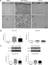
Neutrophil infiltration after repeated mild fluid percussion injury is reduced by the anti-CD11d treatment. (A) Photomicrographs of brain sections at the cortical injury site, or a comparable site in the sham-injured rats, immunostained by an anti-neutrophil antibody. The sections from sham-injured, 1B7 and anti-CD11d-treated rats at 72 h after injury are shown at lower power in panels 1 to 3 and higher power in panels 4 to 6. Scale bars = 100 μm. Scale bar in panel 3 refers to panels 1 to 3. Scale bar in panel 6 refers to panels 4 to 6. (B) MPO activity in brain homogenates in sham-injured groups (n = 6, both times), 1B7 groups (n = 7 and 6, respectively), and CD11d groups (n = 6 and 5 respectively) at 72 h and at 8 weeks post-injury. A.U., arbitrary units; *different from sham-injured group; #different from 1B7 group; all P ≤0.05 except that the gray *indicates P = 0.067. (C). Neutrophil protein, identified by western blotting in brain homogenates expressed as mean values ± standard error of the mean, with a representative autoradiogram shown above each set of histograms (n = 6 for each group).
Figure 4
Figure 4
Macrophages in the brain after repeated mild fluid percussion injury are reduced by the anti-CD11d treatment. (A) Photomicrographs of brain sections at the injury or equivalent site at 72 h after injury in the cortex in sham-injured, 1B7 control and anti-CD11d-treated rats immunostained by an anti-ED1 antibody. Format is similar to that described for Figure 3. Scale bars = 100 μm. (B) Macrophage protein, identified by western blotting in brain homogenates expressed as mean values (± standard error of the mean) with a representative autoradiogram shown above each set of histograms. Macrophage protein is shown at 72 h and 8 weeks in sham-injured, 1B7 and anti-CD11d groups (n = 6 all groups). A.U., arbitrary units; *different from sham group; #different from 1B7 group; all P ≤ 0.05 with the exception that gray # indicates P = 0.073.
Figure 5
Figure 5
Astrogliosis assessed by the expression of glial fibrillary acidic protein (GFAP) in the brain after repeated mild fluid percussion injury is reduced by the anti-CD11d treatment. (A) Photomicrographs of brain sections at the location of cortical injury or equivalent site, at 72 h after injury, immunostained by an antibody to GFAP. Format is as in Figure 3. Scale bars = 100 μm. (B) GFAP, identified by western blotting in brain homogenates, shown at 72 h and 8 weeks in sham-injured, 1B7, and anti-CD11d groups (n = 6, all groups). A.U., arbitrary units; *different from sham-injured group; #different from 1B7 group; all P ≤0.05.
Figure 6
Figure 6
Neuronal loss assessed by expression of the neuronal protein NeuN in the brain after repeated mild fluid percussion injury is decreased by the anti-CD11d treatment. (A) Photomicrographs of brain sections at the cortical injury site or equivalent region, at 72 h after injury, immunostained by an antibody to NeuN. Format is as in Figure 3. Scale bars = 100 μm. (B) NeuN, identified by western blotting in brain homogenates, is shown at 72 h and 8 weeks in sham-injured, 1B7, and anti-CD11d groups (n = 6, all groups). A.U., arbitrary units; *different from sham-injured group; #different from 1B7 group; all P ≤0.05 with the exception that gray # indicates P = 0.066.
Figure 7
Figure 7
White matter injury measured by the increase in amyloid precursor protein (APP) expression in the brain after repeated mild fluid percussion injury is reduced by the anti-CD11d treatment. (A) Photomicrographs of brain sections at the center of the corpus callosum, 72 h after injury, immunostained by an antibody to APP. Format is as in Figure 3. Scale bars = 100 μm. (B) APP, identified by western blotting in brain homogenates, is shown at 72 h and 8 weeks in sham-injured, 1B7, and anti-CD11d groups (n = 6, all groups). A.U., arbitrary units; *different from sham-injured group; #different from 1B7 group; all P ≤0.05.
Figure 8
Figure 8
Anti-CD11d treatment improves cognitive performance in the water maze in the short recovery period (24 h) after repeated mild fluid percussion injury. Treatment groups for panels A-D are defined in the legend in panel A (n = 15/group). (A) Plot of search time vs. sequential trial during acquisition training in the water maze test. Each block is the average of two trials for each rat and data points are mean ± standard error of the mean (SEM) for the group of rats. (B) Plot of percent of direct and circle swims during acquisition training. Histogram bars represent means of data for the groups of rats collected during the 10 trials (± SEM). (C) Plot of search time with respect to number of trials (as in A) during reversal training. (D) Plot of percent of direct and circle swims during reversal training. *Different from sham-injured group; #different from 1B7 group; for post hoc comparisons of means, all P ≤0.05. For additional statistical details see Results.
Figure 9
Figure 9
Anti-CD11d treatment improves cognitive performance in water maze in the long recovery period (8 weeks) after repeated mild fluid percussion injury. Format of figure is identical to that of Figure 8 (n = 12/group). (A) Plot of search time vs. sequential trial during acquisition training in the water maze test. (B) Plot of percent of direct and circle swims during acquisition training. (C) Plot of search time with respect to number of trials (as in A) during reversal training. (D) Plot of percent of direct and circle swims during reversal training. *Different from sham-injured group; #different from 1B7 group; for post hoc comparisons of means, all P ≤0.05. For additional statistical details see Results.

References

    1. Cassidy JD, Carroll LJ, Peloso PM, Borg J, von Holst H, Holm L, Kraus J, Coronado VG. Incidence, risk factors and prevention of mild traumatic brain injury: results of the WHO Collaborating Centre Task Force on Mild Traumatic Brain Injury. J Rehabil Med. 2004;43(Suppl):28–60. - PubMed
    1. Hoge CW, McGurk D, Thomas JL, Cox AL, Engel CC, Castro CA. Mild traumatic brain injury in U.S. Soldiers returning from Iraq. N Engl J Med. 2008;358:453–463. doi: 10.1056/NEJMoa072972. - DOI - PubMed
    1. McCrory P, Meeuwisse W, Johnston K, Dvorak J, Aubry M, Molloy M, Cantu R. Consensus statement on concussion in sport–the 3rd International Conference on concussion in sport, held in Zurich, November 2008. J Clin Neurosci. 2009;16:755–763. doi: 10.1016/j.jocn.2009.02.002. - DOI - PubMed
    1. Prins ML, Alexander D, Giza CC, Hovda DA. Repeated mild traumatic brain injury: mechanisms of cerebral vulnerability. J Neurotrauma. 2013;30:30–38. doi: 10.1089/neu.2012.2399. - DOI - PMC - PubMed
    1. Shultz SR, Bao F, Omana V, Chiu C, Brown A, Cain DP. Repeated mild lateral fluid percussion brain injury in the rat causes cumulative long-term behavioral impairments, neuroinflammation, and cortical loss in an animal model of repeated concussion. J Neurotrauma. 2012;29:281–294. doi: 10.1089/neu.2011.2123. - DOI - PubMed

Publication types