. 2013 Feb 13;13(2):181-92.
doi: 10.1016/j.chom.2013.01.007.

A cis-acting element in retroviral genomic RNA links Gag-Pol ribosomal frameshifting to selective viral RNA encapsidation

Affiliations

A cis-acting element in retroviral genomic RNA links Gag-Pol ribosomal frameshifting to selective viral RNA encapsidation

Mastooreh Chamanian et al. Cell Host Microbe. .

Abstract

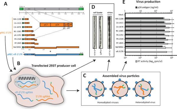
During retroviral RNA encapsidation, two full-length genomic (g) RNAs are selectively incorporated into assembling virions. Packaging involves a cis-acting packaging element (Ψ) within the 5' untranslated region of unspliced HIV-1 RNA genome. However, the mechanism(s) that selects and limits gRNAs for packaging remains uncertain. Using a dual complementation system involving bipartite HIV-1 gRNA, we observed that gRNA packaging is additionally dependent on a cis-acting RNA element, the genomic RNA packaging enhancer (GRPE), found within the gag p1-p6 domain and overlapping the Gag-Pol ribosomal frameshift signal. Deleting or disrupting the two conserved GRPE stem loops diminished gRNA packaging and infectivity >50-fold, while deleting gag sequences between Ψ and GRPE had no effect. Downregulating the translation termination factor eRF1 produces defective virus particles containing 20 times more gRNA. Thus, only the HIV-1 RNAs employed for Gag-Pol translation may be specifically selected for encapsidation, possibly explaining the limitation of two gRNAs per virion.


Figures

Figure 1
Figure 1. Complementation system used for packaging studies and infectious virus production
(A) Series of pREC-5'LTR HIV-1 vectors were constructed to express different lengths of HIV-1 sub-genomic (sg) RNA. The plasmid depicted in orange harbors the 5'LTR followed by various HIV-1 coding sequence. The pREC-nfl-3'LTR (near full length or nfl) HIV-1 vector (in blue) lacks the 5'LTR, and is used to co-transfect 293T cells and complement the pREC-5'LTR. (B) Both vectors express 5' capped, 3' polyA HIV-1 mRNA species for the full complement HIV-1 proteins in the cells and produce virus particles indistinguishable from those derived for transfection with full length proviral DNA constructs (e.g. pNL4-3) (Dudley et al., 2009). 293T transfectants produce three virus types (C) containing either two 5'LTR sgRNAs (x), two nfl-3'LTR sgRNAs (y), or one of each (heterodiploid or xy). As described in Figure S2, the 5'LTR and nfl-3'LTR sgRNAs in the heterodiploid virus can complement each other during reverse transcription to generate a wt, full-length proviral DNA where the coding sequence is only derived from the nfl-3'LTR. At 48-hours post-transfection, virus produced from 293T cells transfected with pREC-nfl-3'LTR and pREC-5'LTR was monitored by Western blots using anti-p24 and anti-β actin (as control for cellular protein expression) in cell lysates or in virus-containing supernatants (D). (E) Virus production was also measured in supernatants by measuring RT activity using a radiolabelled assay or by quantifying CA p24 using an antigen capture assay. Data are presented as mean ± SEM. See also Figure S2.
Figure 2
Figure 2. Comparing the expected and actual infectious titers of virus derived from cotransfected 293T cells
(A) Wild type NL4-3 HIV-1 was serially diluted (1:4) and used to measure RT activity and to infect U87.CD4.CXCR4. (B) RT activity was plotted against the level of virus production for each dilution to determine a linear regression formula and to obtain as a surrogate of infectious titer (Marozsan et al., 2004). Virus produced from the co-transfected 293T cells were diluted to measure RT activity (C) or to infect U87.CD4.CXCR4 cells (D). Panel E provides the estimated infectious titers based on analyses from panels B and C. The actual TCID50 values (F) were derived from the serial dilution/infections of the U87.CD4.CXCR4 cells (D) using the standard limiting protocol (Marozsan et al., 2004). The values are plotted at log10 infectious units/ml. See also Figure S1.
Figure 3
Figure 3. Relative packaging of the HIV-1 sub-genomic RNA in virus derived from co-transfected cells
pREC-5'LTR plasmids were used to transfect 293T cells along with pREC-nfl-3'LTR. (A) Schematic representation of primers and probes used to specifically PCR amplify and quantify the 5'LTR sgRNA (orange) and nfl-3'LTR sgRNA (blue) following cDNA synthesis (See Table S2 for primer details). Viral RNA was extracted from cell-free supernatants of transfected cells. Copy number of 5'LTR sgRNA (B) and nfl-3'LTR sgRNA (C) were determined by qRT-PCR, compared to qRT-PCR amplification of in vitro transcribed HIV-1 RNA of known copy number (104 to 1010 copies), and presented as relative to the viral RT activity. (Figure S3 presents the cytoplasmic and cellular levels of sgRNAs prior to packaging. (D) RNA packaging efficiency was not influenced by deletion of a Gag AUG start codon in the 5'LTR sgRNAs. Gag AUG initiation codon was deleted in the pREC-5'LTR-CA1878, NC2085, RT2845, and Nef8902. Viral RNA was extracted from cell-free supernatants of transfected cells. Copy number of wt and ΔAUG 5'LTR sgRNAs were determined by qRT-PCR and normalized to the viral RT activity. (E) Virus infectivity was measured by first normalizing for RT activity, serially diluted as described in Figure 2D, and then added to U87.CD4.CXCR4 cells, i.e. a standard TCID50 assay. The level of infectious virus is presented as log10 infectious units/ml. Data in (B–D) are presented as mean ± SEM. See also Figure S3.
Figure 4
Figure 4. Effect of deleting (i) the putative GRPE element and (ii) the region separating GRPE and Ψ on packaging of the 5'LTR sgRNA
(A) The putative GRPE was deleted as a 1819–2251 and 1956–2188 nt region in the p1–p6 coding regions of gag within the pREC-5'LTR-RT2845 which was then co-transfected with pREC-nfl-3'-LTR in 293T cells. A similar GRPE region was also deleted from the wt pNL4-3 construct and co-transfected with the pCMVDR8.91 vector (D). Finally, four regions were deleted separating the putative GRPE from Ψ (E). The gRNAs were quantified in virus particles by qRT-PCR as described in Figure 3 and presented as copies/ml in panel B, D and F for the 5'LTR sgRNAs (or NL4-3 gRNA) and for the nfl-3'LTR sgRNA in Figures S4. Virus infectivity was measured by first normalizing for RT activity, serially diluting as described in Figure 2C, and then adding to U87.CD4.CXCR4 cells (Figure 2D). The level of infectious virus is presented as log10 infectious units/ml in panel C and G. The structure of the sgRNA was analyzed using SHAPE (Watts et al., 2009) and presented in panel H using the modeling algorithm, RNAstructure (Mathews et al., 2004). Full-length in vitro transcribed 5'LTR sgRNA (E) were used for structural probing but only the region proximal to GRPE/RFS was presented in panel H. Data in (B), (D), and (F) are presented as mean ± SEM. See also Figure S4.
Figure 5
Figure 5. The role of RNA secondary structure in the GRPE/RFS region on HIV-1 RNA packaging
(A) Multiple point mutations were introduced to the predicted P2 and P3 stem-loops within the GRPE/RFS sequence. (B) The RNA secondary structures in the GRPE/RFS region of these five mutants were predicted by Mfold (http://mfold.rna.albany.edu/). The five mutant 5'LTR sgRNA templates were expressed in co-transfected 293T cells. Supernatants from these transfected cells were harvested to measure 5'LTR sgRNA (C) and nfl-3'LTR sgRNA levels (D) by q-RT-PCR. The same virus-containing supernatant was then equalized for RT activity, serially diluted and then used to infect U87.CD4.CXCR4 cells. The relative infectivity, as measured by TCID50 assay, is presented as Log10 infectious units/ml (E). Data in (C) and (D) are presented as mean ± SEM.
Figure 6
Figure 6. The effect of eRF1 knockdown on virus production and gRNA encapsidation
A pool of three siRNAs against eRF1 effectively reduces cellular eRF1 expresion level by ~70% prior to 293T transfections as indicated by the Western blot analysis (A and C). These siRNA-treated 293T cells were co-transfected with the pREC-nfl-3'LTR along with either pREC-5'LTR-MA1208, pREC-5'LTR-p6Δ868 and pREC-5'LTR-RT2845 or transfected with HIV Gag-iGFP. It is important to note that eRF1 knockdown reduced all protein levels in the cell 2-fold within 72 h. Virus produced from these siRNA-treated, co-transfected cells were non-infectious for U87.CD4.CXCR4 cells. The 5'LTR sgRNA or NL4-3 gRNA in virus particles was measured by qRT-PCR and presented as relative to p24 antigen content (B and D). In the presence of wild type levels of eRF1 (SD siRNA treatment), the virus produced from the co-transfected cells harbored approximately 1.6 5'LTR sgRNA copies per 2000 molecule of p24 (or the estimated size of one HIV-1 particle). With the eRF1 knockdown and the wt pREC-5'LTR-RT2845, there is approximately 19 5'LTR sgRNA copies per 2000 p24 molecules. Virus derived from transfections with the HIV Gag-iGFP molecular clone (eRF1 knockdown – E; wt eRF1 - F) was sucrose-cushion purified, spread on poly-L-lysine coverslips, and images captured on a Deltavision RT epifluorescent microscope system.
Figure 7
Figure 7. Model for translation and packaging of the unspliced HIV-1 RNA
Unspliced mRNA are destined for (1) Gag translation and subsequent substrate for NMD pathway or (2) may serve as a template Gag-Pol translation following ribosomal frameshift and thus avoiding NMD (<5%). Ribosomal translocation across the unspliced mRNA used for Gag-Pol translation may clear the RNA of cellular factors (e.g. eRF1) and thus promote secondary/tertiary HIV-1 RNA resulting in RNA-protein interactions necessary for gRNA dimerization following by packaging. MA: Matrix, CA: Capsid, NC: Nucleocapsid. See also Figure S5.

Comment in

  • HIV-1: packaging a shifty genome?
    Durney MA, D'Souza VM. Durney MA, et al. Cell Host Microbe. 2013 Feb 13;13(2):123-5. doi: 10.1016/j.chom.2013.01.015. Cell Host Microbe. 2013. PMID: 23414752

References

    1. Abbink TE, Berkhout B. A novel long distance base-pairing interaction in human immunodeficiency virus type 1 RNA occludes the Gag start codon. J. Biol. Chem. 2003;278:11601–11611. - PubMed
    1. Aldovini A, Young RA. Mutations of RNA and protein sequences involved in human immunodeficiency virus type 1 packaging result in production of noninfectious virus. J. Virol. 1990;64:1920–1926. - PMC - PubMed
    1. Amarasinghe GK, De Guzman RN, Turner RB, Chancellor KJ, Wu ZR, Summers MF. NMR structure of the HIV-1 nucleocapsid protein bound to stem-loop SL2 of the psi-RNA packaging signal. Implications for genome recognition. J. Mol. Biol. 2000;301:491–511. - PubMed
    1. Berkowitz R, Fisher J, Goff SP. RNA packaging. Curr. Top. Microbiol. Immunol. 1996;214:177–218. - PubMed
    1. Burniston MT, Cimarelli A, Colgan J, Curtis SP, Luban J. Human immunodeficiency virus type 1 Gag polyprotein multimerization requires the nucleocapsid domain and RNA and is promoted by the capsid-dimer interface and the basic region of matrix protein. J. Virol. 1999;73:8527–8540. - PMC - PubMed

Publication types

MeSH terms