

Accuracy of PET/CT Scan in the diagnosis of the focal form of congenital hyperinsulinism
- PMID: 23414871
- PMCID: PMC3597386
- DOI: 10.1016/j.jpedsurg.2012.11.025
Accuracy of PET/CT Scan in the diagnosis of the focal form of congenital hyperinsulinism
Abstract
Purpose: The purpose of the study was to determine the sensitivity of the (18)fluoro-dihydroxyphenylalanine positron emission tomography/computed tomography scan (18F-PET/CT) in the diagnosis of focal congenital hyperinsulinism (HI).
Methods: A retrospective review of children with HI who underwent a preoperative 18F-PET/CT scan was performed.
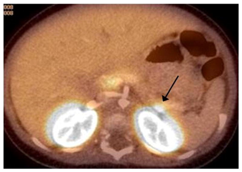
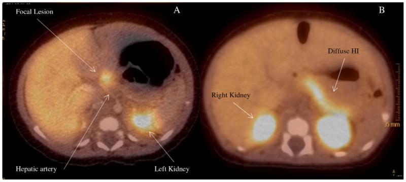
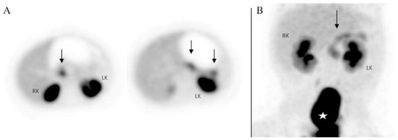
Results: Between 1/2008 and 2/2012 we performed 105 consecutive 18F-PET/CT scans on infants with HI. Fifty-three patients had focal HI. Of those fifty-three patients, eight had a preoperative 18F-PET/CT scan read as "diffuse disease". The sensitivity of the study in the diagnosis of focal HI was 85%. The location of the eight missed focal lesions was: head (3), body (2), and tail (3). The 18F-PET/CT of the missed head lesions showed homogeneous tracer uptake (n =2) or heterogeneous uptake throughout the pancreas (n=1). The 18F-PET/CT of the 2 missed body lesions and 1 missed tail lesion showed heterogeneous uptake throughout the pancreas. The 18F-PET/CT of the other 2 missed tail lesions showed lesions adjacent to and obscured by the signal of the upper renal pole, identified retrospectively by closer observation. Fifty-two of the 105 patients had diffuse HI. Two of them had 18F-PET/CT studies read as "focal disease". Therefore, the specificity of the study was 96%. Of the forty-seven 18F-PET/CT studies read as "focal disease", forty-five had true focal HI. Therefore, the positive predictive value of the study in the diagnosis of focal HI was 96%.
Conclusion: The sensitivity and specificity of 18 F-PET/CT can be affected by certain anatomic features of the pancreas, by the location of the lesion, and by the reader's experience.
Copyright © 2013 Elsevier Inc. All rights reserved.
Figures

References
-
- Kirchner PT, Ryan J, Zalutsky M, et al. Positron emission tomography for the evaluation of pancreatic disease. Semin Nucl Med. 1980;4:374–91. - PubMed
-
- Gazdar AF, Helman LJ, Israel MA, et al. Expression of neuroendocrine cell markers L-dopa decarboxylase, chromogranin A, and dense core granules in human tumors of endocrine and nonendocrine origin. Cancer Res. 1988;48:4078–82. - PubMed
-
- Eriksson B, Bergström M, Sundin A, et al. The role of PET in localization of neuroendocrine and adrenocortical tumors. Ann N Y Acad Sci. 2002;970:159–69. - PubMed
-
- Ribeiro MJ, De Lonlay P, Delzescaux T, et al. Characterization of hyperinsulinism in infancy assessed with PET and 18F-fluoro-L-DOPA. J Nucl Med. 2005;46:560–6. - PubMed
-
- Otonkoski T, Näntö-Salonen K, Seppänen M, et al. Noninvasive diagnosis of focal hyperinsulinism of infancy with [18F]-DOPA positron emission tomography. Diabetes. 2006;55:13–8. - PubMed
MeSH terms
Grants and funding
LinkOut - more resources
Full Text Sources
Other Literature Sources
Medical
