. 2013 Feb 14;152(4):884-94.
doi: 10.1016/j.cell.2013.01.042.

Tit-for-tat: type VI secretion system counterattack during bacterial cell-cell interactions

Affiliations

Tit-for-tat: type VI secretion system counterattack during bacterial cell-cell interactions

Marek Basler et al. Cell. .

Abstract

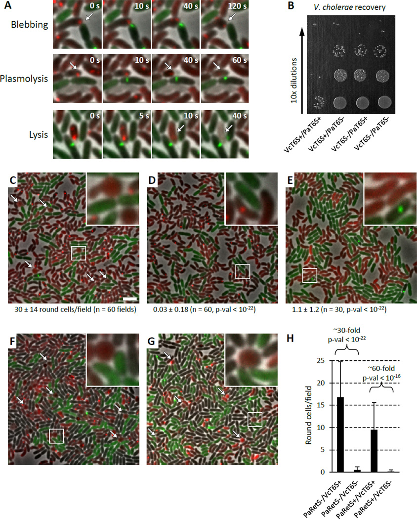
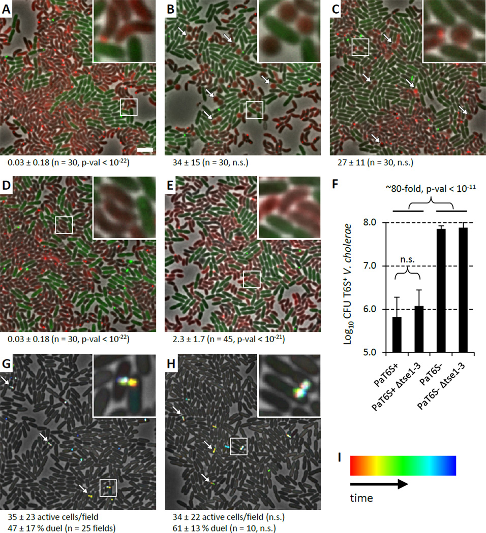
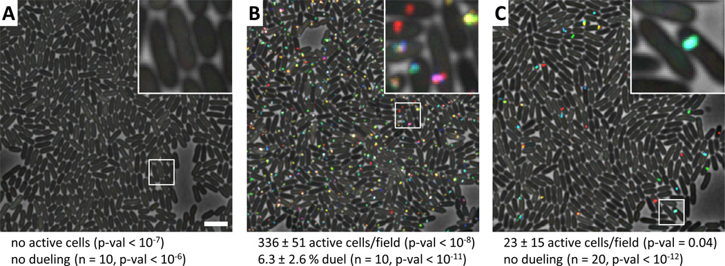
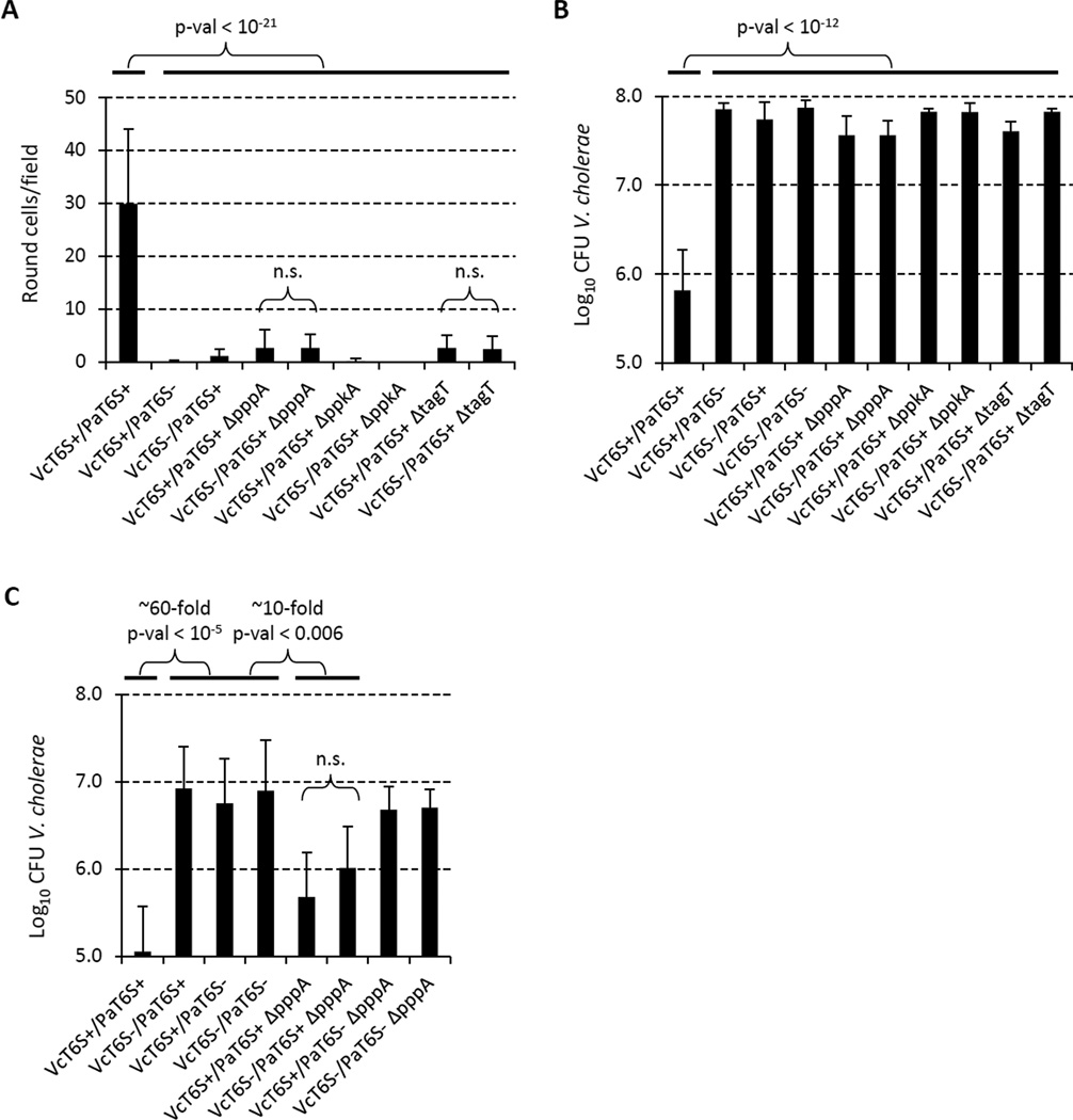
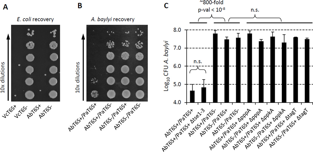
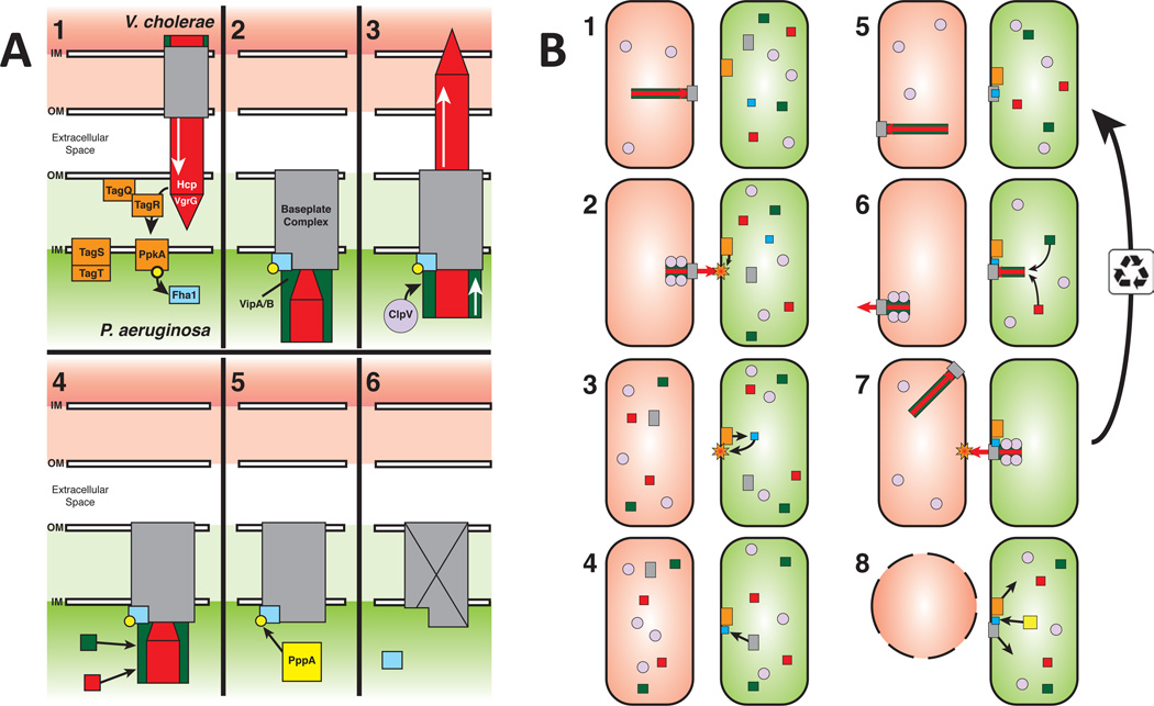
The bacterial type VI secretion system (T6SS) is a dynamic organelle that bacteria use to target prey cells for inhibition via translocation of effector proteins. Time-lapse fluorescence microscopy has documented striking dynamics of opposed T6SS organelles in adjacent sister cells of Pseudomonas aeruginosa. Such cell-cell interactions have been termed "T6SS dueling" and likely reflect a biological process that is driven by T6SS antibacterial attack. Here, we show that T6SS dueling behavior strongly influences the ability of P. aeruginosa to prey upon heterologous bacterial species. We show that, in the case of P. aeruginosa, T6SS-dependent killing of either Vibrio cholerae or Acinetobacter baylyi is greatly stimulated by T6SS activity occurring in those prey species. Our data suggest that, in P. aeruginosa, T6SS organelle assembly and lethal counterattack are regulated by a signal that corresponds to the point of attack of the T6SS apparatus elaborated by a second aggressive T6SS(+) bacterial cell. PAPERFLICK:


Figures

Figure 1
Figure 1. P. aeruginosa T6SS preferentially targets T6SS positive V. cholerae
See also Movie S1, S2, Table S1, S2. VcT6S+ indicates V. cholerae clpV-mCherry2, VcT6S indicates V. cholerae ΔvipA clpV-mCherry2, PaT6S+ indicates P. aeruginosa ΔretS clpV1-gfp, PaT6S indicates P. aeruginosa ΔretS ΔvipA1 clpV1-gfp. PaRetS indicates P. aeruginosa ΔretS, PaRetS+ indicates P. aeruginosa. (A) Examples of morphological changes of V. cholerae seen in mixtures of P. aeruginosa ΔretS clpV1-gfp (T6SS+, green) and V. cholerae clpV-mCherry2 (T6SS+, red). 4.5x4.5 µm fields are shown. (B) Example of a dilution series used to enumerate V. cholerae recovery from a competition with P. aeruginosa. (C−G) 30x30 µm representative field of cells with a 4x magnified 3x3 µm inset (marked by box). Bar in C is 3 µm and applies to C–G. Arrows point to examples of round V. cholerae cells. (C−E) Average number of round V. cholerae cells per 30x30 µm field (± standard deviation) is shown for each mixture (n fields were analyzed), p-val compared to mixture in C. (C) P. aeruginosa ΔretS clpV1-gfp (T6SS+, green) mixed with V. cholerae clpV-mCherry2 (T6SS+, red), (D) P. aeruginosa ΔretS ΔvipA1 clpV1-gfp (T6SS, green) mixed with V. cholerae clpV-mCherry2 (T6SS+, red), (E) P. aeruginosa ΔretS clpV1-gfp (T6SS+, green) mixed with V. cholerae ΔvipA clpV-mCherry2 (T6SS, red). (F, G) V. cholerae clpV-mCherry2 (T6SS+, red), and V. cholerae ΔvipA clpV-gfp (T6SS, green) strains were mixed at equal ratios with (F) P. aeruginosa ΔretS (T6SS+, unlabeled), (G) P. aeruginosa (T6SS+, unlabeled). (H) Quantification of number of round V. cholerae cells detected per 30x30 µm field (n = 60) for mixtures shown in F and G.
Figure 2
Figure 2. P. aeruginosa T6SS effector Tse1 responsible for V. cholerae cell rounding but Tse effectors dispensable for dueling and V. cholerae growth inhibition
See also Movie S3, Table S1, S3. 30x30 µm representative field of cells with a 4x magnified 3x3 µm inset (marked by box) is shown for A–E, G, H. Bar in A is 3 µm and applies to A–E, G, H. Strain abbreviations were as used in Figure 1. n.s. – not statistically significant (p-val > 0.01). (A−E) Arrows point to examples of round V. cholerae cells. Average number of round V. cholerae cells per 30x30 µm field (± standard deviation) is shown for each mixture (n fields were analyzed), p-val compared to mixture in Figure 1C. For A–D V. cholerae clpV-mCherry2 strain was mixed with (A) P. aeruginosa ΔretS Δtse1 clpV1-gfp, (B) P. aeruginosa ΔretS Δtse2 clpV1-gfp, (C) P. aeruginosa ΔretS Δtse3 clpV1-gfp, (D) P. aeruginosa ΔretS Δtse1-3 clpV1-gfp. (E) P. aeruginosa ΔretS clpV1-gfp was mixed with V. cholerae / pBAD24-Tsi1-mCherry2 strain. (F) Summary of competition assays for P. aeruginosa and V. cholerae mixtures. Data are presented as mean of Log10CFU of recovered V. cholerae with error bar representing standard deviation (n = 8 – 19). (G, H) ClpV1-GFP localization was followed for 3 minutes and temporally color coded. Arrows point to examples of dueling P. aeruginosa cells. Average number of active cells and dueling cells per 30x30 µm field (± standard deviation) is shown (n fields were analyzed), p-val compared to strain in G. (G) P. aeruginosa ΔretS clpV1-gfp, (H) P. aeruginosa ΔretS Δtse1-3 clpV1-gfp. (I) Color scale used to temporal-color code ClpV1-GFP signal.
Figure 3
Figure 3. T6SS dueling depends on PpkA, PppA and TagT
See also Movie S3, Table S3. ClpV1-GFP localization was followed for 3 minutes and temporally color coded (color scale in Figure 2I). 30x30 µm representative field of cells with a 4x magnified 3x3 µm inset (marked by box) is shown for A–C. Bar in A is 3 µm and applies to A–C. Average number of active cells and dueling cells per 30x30 µm field (± standard deviation) is shown (n fields were analyzed), p-val compared to parental strain in Figure 2G. (A) P. aeruginosa ΔretS ΔppkA clpV1-gfp, (B) P. aeruginosa ΔretS ΔpppA ΔclpV1-gfp, (C) P. aeruginosa ΔretS ΔtagT clpV1-gfp.
Figure 4
Figure 4. PpkA, PppA and TagT important for P. aeruginosa targeting of the prey
See also Table S1. Strain abbreviations were as used in Figure 1. n.s. – not statistically significant (p-val > 0.01). (A) Quantification of number of round V. cholerae cells per 30x30 µm field for indicated mixtures (n = 60 for VcT6S+/PaT6S+ and VcT6S+/PaT6S mixtures, n = 30 for all other indicated mixtures). (B) Summary of competition assays for P. aeruginosa and V. cholerae mixtures. Data are presented as mean of Log10CFU of recovered V. cholerae with error bar representing standard deviation (n = 6 – 19). (C) Competition assays for P. aeruginosa and V. cholerae mixtures at 10:1 ratio. Data are presented as mean of Log10CFU of recovered V. cholerae with error bars representing standard deviation (n = 5 – 10).
Figure 5
Figure 5. A. baylyi has functional T6SS and is targeted by P. aeruginosa
Strain abbreviations were as used in Figure 1. AbT6S+ indicates A. baylyi parental strain, AbT6Sindicates A. baylyi ΔT6SS. (A) Example of a dilution series used to enumerate E. coli survival in mixtures with A. baylyi or V. cholerae. (B) Example of a dilution series used to enumerate A. baylyi survival in mixtures with P. aeruginosa. (C) Summary of competition assays for P. aeruginosa and A. baylyi mixtures. Data are presented as mean of Log10CFU of recovered A. baylyi with error bar representing standard deviation (n = 3 – 8).
Figure 6
Figure 6. Model for TagQRST-mediated T6SS aiming
(A) Regulation of the T6SS response in P. aeruginosa. (A1) T6SS assault from V. cholerae is sensed by the TagQRST/PpkA signal cascade (orange) to phosphorylate Fha1 (blue). (A2) Phosphorylated Fha1 interacts with the baseplate complex (grey) locking it in place and allowing assembly of the Hcp/VgrG tube/spike (red) and VipA/B sheath (green). (A3) Sheath contraction fires the retaliatory P. aeruginosa tube/spike complex at V. cholerae. ClpV (light purple) then disassembles the contracted sheath. (A4) The baseplate can then be reused to assemble an additional tube/spike/sheath complex, or (A5) PppA (yellow) can dephosphorylate Fha1, (A6) inactivating or disassembling the baseplate complex. The P. aeruginosa T6SS is now ready to respond to new T6SS assaults. (B) T6SS interactions between P. aeruginosa and V. cholerae. (B1) V. cholerae T6SS will spontaneously fire occasionally hitting a nearby P. aeruginosa cell. (B2-6) P. aeruginosa sense the assault and builds its T6SS organelle at the location of the assault. (B7) P. aeruginosa fires its T6SS organelle back at the V. cholerae cell. The baseplate is then recycled to allow for multiple firing events or (B8) the Fha1 complex is dephosphorylated by PppA and the baseplate complex is disassembled and free to reform at a new location. (B5) Meanwhile, V. cholerae continues to fire its T6SS organelle arbitrarily in a different location and direction. (B8) After the retaliatory attack from P. aeruginosa, the V. cholerae cell dies.

Comment in

References

    1. Aoki SK, Malinverni JC, Jacoby K, Thomas B, Pamma R, Trinh BN, Remers S, Webb J, Braaten BA, Silhavy TJ, et al. Contact-dependent growth inhibition requires the essential outer membrane protein BamA (YaeT) as the receptor and the inner membrane transport protein AcrB. Mol Microbiol. 2008;70:323–340. - PMC - PubMed
    1. Aoki SK, Pamma R, Hernday AD, Bickham JE, Braaten BA, Low DA. Contact-dependent inhibition of growth in Escherichia coli. Science. 2005;309:1245–1248. - PubMed
    1. Aoki SK, Webb JS, Braaten BA, Low DA. Contact-dependent growth inhibition causes reversible metabolic downregulation in Escherichia coli. J Bacteriol. 2009;191:1777–1786. - PMC - PubMed
    1. Axelrod R, Hamilton WD. The evolution of cooperation. Science. 1981;211:1390–1396. - PubMed
    1. Basler M, Mekalanos JJ. Type 6 secretion dynamics within and between bacterial cells. Science. 2012;337:815. - PMC - PubMed

Publication types

MeSH terms

Substances