. 2013 May 27;425(10):1683-96.
doi: 10.1016/j.jmb.2013.02.011. Epub 2013 Feb 14.

An unusual dimeric small heat shock protein provides insight into the mechanism of this class of chaperones

Affiliations

An unusual dimeric small heat shock protein provides insight into the mechanism of this class of chaperones

Eman Basha et al. J Mol Biol. .

Abstract

Small heat shock proteins (sHSPs) are virtually ubiquitous stress proteins that are also found in many normal tissues and accumulate in diseases of protein folding. They generally act as ATP-independent chaperones to bind and stabilize denaturing proteins that can be later reactivated by ATP-dependent Hsp70/DnaK, but the mechanism of substrate capture by sHSPs remains poorly understood. A majority of sHSPs form large oligomers, a property that has been linked to their effective chaperone action. We describe AtHsp18.5 from Arabidopsis thaliana, demonstrating that it is dimeric and exhibits robust chaperone activity, which adds support to the model that suboligomeric sHSP forms are a substrate binding species. Notably, like oligomeric sHSPs, when bound to substrate, AtHsp18.5 assembles into large complexes, indicating that reformation of sHSP oligomeric contacts is not required for assembly of sHSP-substrate complexes. Monomers of AtHsp18.5 freely exchange between dimers but fail to coassemble in vitro with dodecameric plant cytosolic sHSPs, suggesting that AtHsp18.5 does not interact by coassembly with these other sHSPs in vivo. Data from controlled proteolysis and hydrogen-deuterium exchange coupled with mass spectrometry show that the N- and C-termini of AtHsp18.5 are highly accessible and lack stable secondary structure, most likely a requirement for substrate interaction. Chaperone activity of a series of AtHsp18.5 truncation mutants confirms that the N-terminal arm is required for substrate protection and that different substrates interact differently with the N-terminal arm. In total, these data imply that the core α-crystallin domain of the sHSPs is a platform for flexible arms that capture substrates to maintain their solubility.


Figures

Figure 1
Figure 1. AtHsp18.5 and homologues are unusual cytosolic plant sHSPs lacking β-strand 6
Alignment of AtHsp18.5 (A.t.18.5) with homologs from Corylus avellana (C.a.), Populus trichocarpa (P.t.), Gossypium hirsutum (G.h.), Citrus aurantium (C.au.), Brassica rapa (B.r.), and Brassica napus (B.n.) compared to other cytosolic sHSPs from class I, II and III represented by Arabidopsis thaliana Hsp17.4-CI, Hsp17.7-CII and Hsp17.4-CIII. Secondary structural elements are based on alignment with TaHsp16.9. The ACD comprises β2 through β9 (delimited by arrowheads). Consensus sequences for AtHsp18.5 and homologues, or for all proteins are shown. Residues corresponding to the beginning and end of the truncated proteins as discussed in the text are underlined.
Figure 2
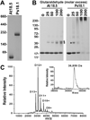
Figure 2. Native AtHsp18.5 is dimeric
A) Non-denaturing PAGE of 15 µl of 24 µM sHSP. B) 24 µM sHSPs were crosslinked with glutaraldehyde at the indicated molar ratios and 15 µl analyzed by SDS-PAGE. Asterisks indicate position of the dimeric crosslinked species. C) Nano- ESIMS of native AtHsp18.5. Inset shows dimer mass as determined from the deconvoluted spectrum.
Figure 3
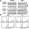
Figure 3. Dimeric AtHsp18.5 protects substrates from aggregation in large sHSP-substrate complexes
3 µM MDH or 1 µM Luc were heated with either PsHsp18.1 or AtHsp18.5 at the indicated molar ratios. Heat treatments were 45°C for 1 h for MDH and 42°C for 8.5 min for Luc. A) SDS-PAGE of MDH and Luc from the soluble or pellet fractions after heating at the indicated molar ratio of sHSP to substrate. Gels were stained with Coomassie Blue. B) SEC analysis of the soluble fractions from Panel A. Asterisk indicates position of sHSP-substrate complexes.
Figure 4
Figure 4. AtHsp18.5 monomers exchange between dimers at a temperature-dependent rate
A) Nondenaturing -PAGE separation of wild type (WT) and N- or C-terminally Strep tagged AtHsp18.5 either alone, or after incubation together as indicated. B) Representative spectra of subunit exchange between WT (*) and AtHsp18.5 carrying a C-terminal Strep tag (C). Spectra from 90 sec, 45 min, and 18 hrs show an increase in the relative amount of heterodimer species (gray bars). C) Rate of appearance of the heterodimer between AtHsp18.5 and C-terminal Strep tag as a function of temperature. Inset: calculated rate constants.
Figure 5
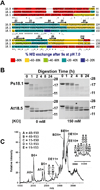
Figure 5. The AtHsp18.5 ACD forms a stable core flanked by flexible N- and C-termini
A) Percent HDX for peptides of AtHsp18.5 compared to TaHsp16.9 and PsHsp18.1 after a 5 sec pulse labeling at pD 7.5 at room temperature. Each peptide is represented by a colored bar, with the color indicating percentage of amide HDX as shown in the legend. B) 24 µM AtHsp18.5 or PsHsp18.1 was incubated with trypsin at a 400:1 molar ratio at room temperature for times indicated with or without KCl. Products were analyzed by SDS-PAGE. C) Nano-ESIMS of the 4 hr trypsin digest in 150 mM KCl (asterisk Panel B). Inset shows expansion of the higher m/z data.
Figure 6
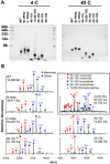
Figure 6. AtHsp18.5 truncated at both the N- and C-terminus retains the ability to dimerize
A) Blue native PAGE of the indicated proteins separated at 4 °C (left) or at 45 °C (right). The 48–150 truncation was obtained by trypsin digestion of the WT protein in the absence of KCl as in Fig. 5B B) Nano-ESIMS of the indicated proteins at 100 µM, showing detection of different amounts of dimer and monomer forms in all samples.
Figure 7
Figure 7. Truncations of the N- and C-termini alter substrate protection efficiency and the ACD alone is ineffective as a chaperone
3 uM MDH or 1 µM Luc were heat denatured with the indicated molar ratios of wild type or truncated AtHsp18.5 proteins. Denaturation of MDH was for 1 hr at 45 °C and 8.5 min at 42°C for Luc. A) SDS-PAGE of MDH from the soluble or pellet fractions. B) SDS-PAGE of Luc from the soluble or pellet fractions. Gels were stained with Coomassie Blue.

References

    1. van Montfort R, Slingsby C, Vierling E. Structure and function of the small heat shock protein/alpha-crystallin family of molecular chaperones. Adv Protein Chem. 2001;59:105–156. - PubMed
    1. McHaourab HS, Godar JA, Stewart PL. Structure and mechanism of protein stability sensors: chaperone activity of small heat shock proteins. Biochemistry. 2009;48:3828–3837. - PMC - PubMed
    1. Basha E, O’Neill H, Vierling E. Small heat shock proteins and α-crystallins: dynamic proteins with flexible functions. Trends Biochem Sci. 2012 - PMC - PubMed
    1. Gusev NB. The Role of Intrinsically Disordered Regions in the Structure and Functioning of Small Heat Shock Proteins. Curr Protein Pept Sci. 2011 - PubMed
    1. Clark JI, Muchowski PJ. Small heat-shock proteins and their potential role in human disease. Curr Opin Struct Biol. 2000;10:52–59. - PubMed

Publication types

MeSH terms

LinkOut - more resources