. 2013 May;15(5):578-88.
doi: 10.1093/neuonc/not004. Epub 2013 Feb 14.

miR-92b controls glioma proliferation and invasion through regulating Wnt/beta-catenin signaling via Nemo-like kinase

Affiliations

miR-92b controls glioma proliferation and invasion through regulating Wnt/beta-catenin signaling via Nemo-like kinase

Kun Wang et al. Neuro Oncol. 2013 May.

Erratum in

  • Neuro Oncol. 2013 Jul;15(7):970

Abstract

Background: Nemo-like kinase (NLK) is an evolutionarily conserved protein kinase involved in Wnt/beta-catenin signaling, which has been reported to be associated with gliomagenesis. In the present study, we aimed to identify a concrete mechanism of Wnt/beta-catenin pathway regulation by microRNAs (miRNAs) in glioma.

Methods: Quantitative reverse-transcription polymerase chain reaction and in situ hybridization were conducted to detect the expression of miR-92b. The cell proliferation rate and cell cycle kinetics were detected using 3-(4,5)-dimethylthiahiazo (-z-y1)-3,5-di-phenytetrazoliumromide (MTT) assay and flow cytometry, cell invasion and migration were evaluated using Transwell assay and wound healing assay, and cell apoptosis was detected using annexin V staining. Furthermore, the relevant molecules regulating proliferation and invasion were examined using Western blot analysis, immunohistochemistry, and immunofluorescence staining. Luciferase reporter assay was used to identify the direct regulation of NLK by miR-92b and beta-catenin/TCF4 activity.

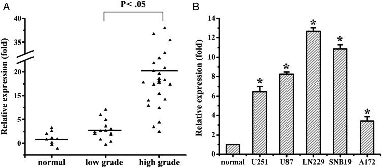
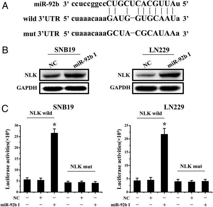
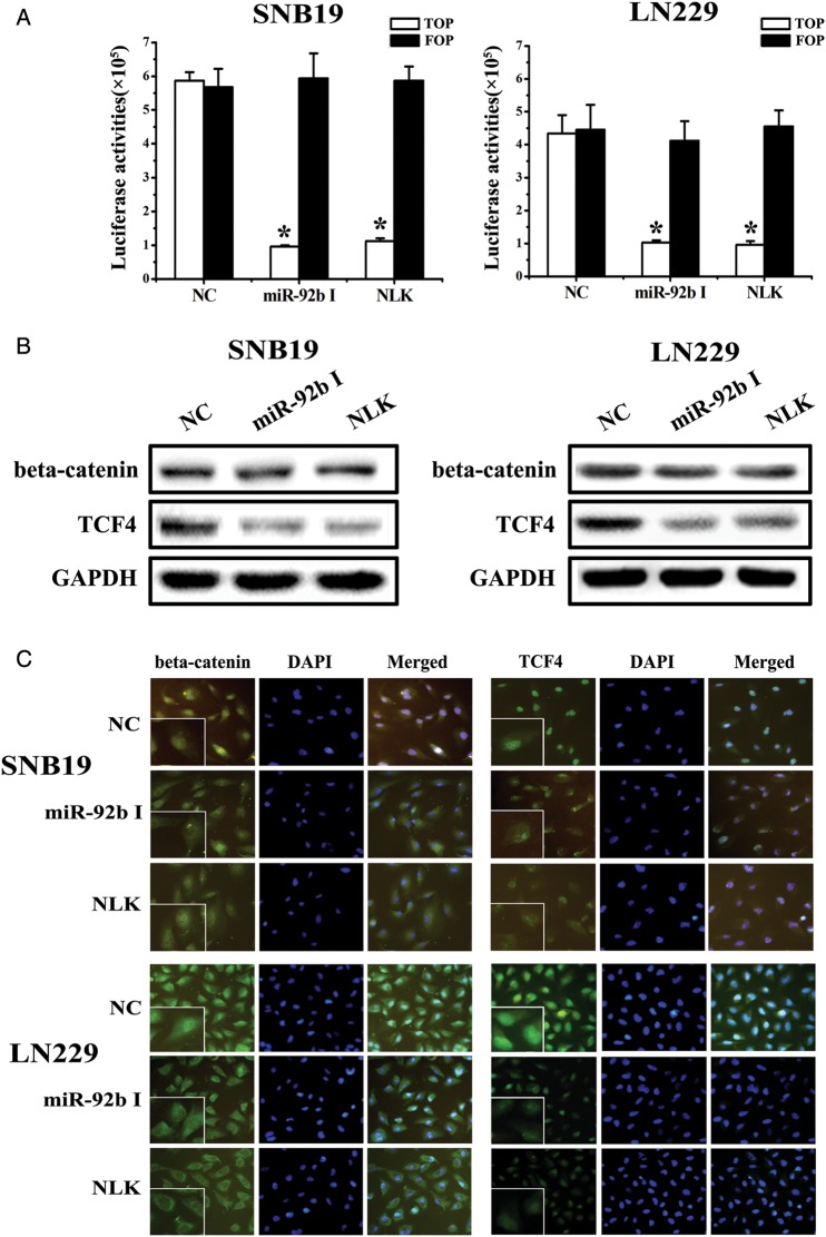
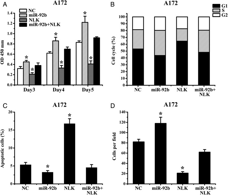
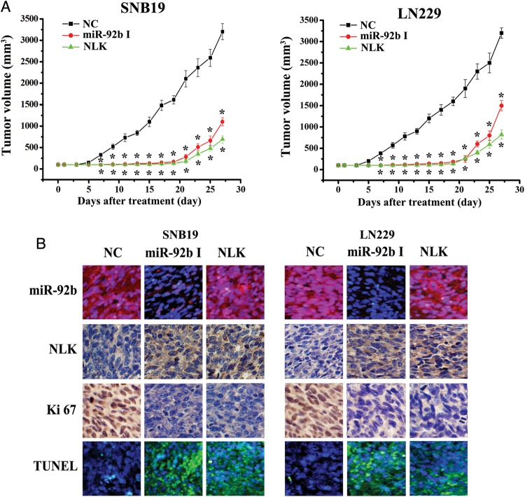
Results: We first showed that the expression of miR-92b was elevated in both glioma samples and glioma cells. Furthermore, down-regulation of miR-92b triggered growth inhibition, induced apoptosis, and suppressed invasion of glioma in vitro and in vivo. Luciferase assay and Western blot analysis revealed that NLK is a direct target of miR-92b. Restoring expression of NLK inhibited glioma proliferation and invasion. Mechanistic investigation revealed that miR-92b deletion suppressed beta-catenin/TCF-4 transcription activity by targeting NLK. Moreover, expression of NLK was inversely correlated with miR-92b in glioma samples and was predictive of patient survival in a retrospective analysis.

Conclusions: Our findings identify a role for miR-92b in glioma proliferation and invasion after activation of Wnt/beta-catenin signaling via NLK.


Figures

Fig. 1.
Fig. 1.
miR-92b is upregulated in glioma samples and glioma cells. (A) miR-92b expression in different grade glioma and normal brain tissues by qRT-PCR. (B) qRT-PCR analysis showed that U251, U87, LN229, SNB19, and A172 glioma cells express high levels of miR-92b, compared with normal brain tissues. *P < .05.
Fig. 2.
Fig. 2.
miR-92b I suppresses glioma cell proliferation and invasion. (A) miR-92b expression was quantified by qRT-PCR analysis. miR-92b inhibitor significantly suppressed miR-92b expression in both SNB19 and LN229 cells (P < .05), relative to the NC. (B) Expression of cell proliferation and invasion-related molecules was analyzed by Western blot 48 h after transfection with miR-92b inhibitor and NC. (C and D) Representative cartogram showing the proliferation, cell cycle, apoptosis, and invasion regulated by miR-92b inhibitor or NC in glioma cells. *P < .05.
Fig. 3.
Fig. 3.
miR-92b directly targets NLK. (A) Bioinformatic analysis identifed NLK as a potential target for miR-92b. NLK wild and NLK mut luciferase report plasmids were constructed. (B) Western blotting for NLK at 48 h after transfection of miR-92b inhibitor in SNB19 and LN229 cells. (C) Luciferase reporter assays confirmed that NLK was the direct target of miR-92b in SNB19 and LN229 cells. *P < .05.
Fig. 4.
Fig. 4.
Effect of miR-92b on the activity the Wnt/beta-catenin signaling pathway. (A) The luciferase reporter assay using TopFlash and FopFlash vectors was used to study beta-catenin TCF/LEF promoter activity. The TopFlash reporter vector has 3-TCF binding sites, and FopFlash has mutated TCF binding sites. SNB19 and LN229 cells were cotransfected with different expression vectors as indicated. MiR-92b inhibitor and NLK treatment decreased beta-catenin TCF/LEF promoter activity. (B) Down-regulation of miR-92b or upregulation of NLK did not affect beta-catenin expression, but decreased TCF-4 expression. (C) Immunofluorescence assay for beta-catenin indicated that the location of beta-catenin in cells shifted from nucleus to cytoplasm when the expression of miR-92b was decreased or NLK was overexpressed and that TCF-4 expression decreased in the nucleus, compared with NC. *P < .05.
Fig. 5.
Fig. 5.
Expression of NLK abrogates miR-92b survival function. (A) Proliferation of A172 cells after Bim transfection with or without miR-92b was significantly reduced, compared with NC. (B) A172 cells were transfected with NLK (not including the 3′UTR) and miR-92b, and cell proliferation and invasion were measured by MTT, flow cytometry, annexin V, and Transwell assay. *P < .05.
Fig. 6.
Fig. 6.
Knock-down of miR-92b inhibits glioma growth in vivo. (A) When subcutaneous tumors were established, miR-92b inhibitor was injected in a multisite injection manner every 2 days for 14 days. Tumor volumes were measured every 2 days during treatment. (B) Fluorescence in situ hybridization showed that miR-92b inhibitor effectively inhibited the expression of miR-92b; NLK and Ki67 expressions were detected by immunohistochemistry assay in xenograft tumor sections; TUNEL assay in xenograft tumor sections revealed that miR-92b inhibitor induced cell apoptosis.
Fig. 7.
Fig. 7.
NLK expression inversely correlated with miR-92b and low NLK expression correlated with poor survival in GBM. (A) Expression of NLK in resected glioma specimen was assessed by IHC assay. ISH assay showed the expression of miR-92b in the homologous specimens. (B) χ2-test analysis of miR-92b and NLK expressions. The inverse correlation is significant. (C) Kaplan-Meier survival curves indicating cumulative survival as a function of time for those patients with low expression of NLK versus high expression of NLK. The patients with low NLK expression experienced significantly worse outcome.

References

    1. Hadjipanayis CG, Van Meir EG. Tumor initiating cells in malignant gliomas: biology and implications for therapy. J Mol Med. 2009;87:363–374. - PMC - PubMed
    1. Purow B, Schiff D. Advances in the genetics of glioblastoma: are we reaching critical mass? Nat Rev Neurol. 2009;5:419–426. - PMC - PubMed
    1. Polakis P. Wnt signaling and cancer. Genes Dev. 2000;14:1837–1851. - PubMed
    1. Giles RH, van Es JH, Clevers H. Caught up in a Wnt storm: Wnt signaling in cancer. Biochim Biophys Acta. 2003;1653:1–24. - PubMed
    1. Willert K, Jones KA. Wnt signaling: is the party in the nucleus? Genes Dev. 2006;20:1394–1404. - PubMed

Publication types

MeSH terms