. 2013 Mar;20(3):311-6.
doi: 10.1038/nsmb.2510. Epub 2013 Feb 17.

Naive pluripotency is associated with global DNA hypomethylation

Affiliations

Naive pluripotency is associated with global DNA hypomethylation

Harry G Leitch et al. Nat Struct Mol Biol. 2013 Mar.

Abstract

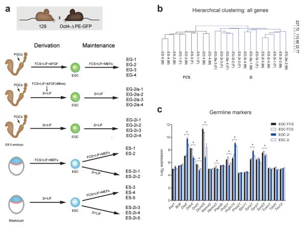
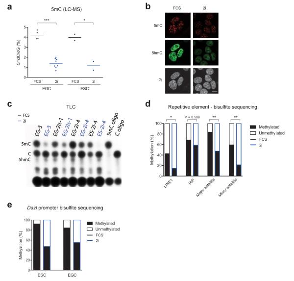
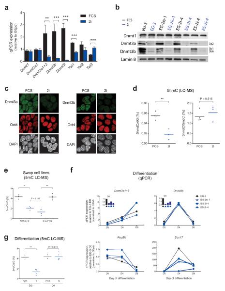
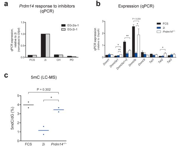
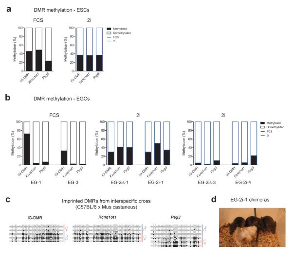
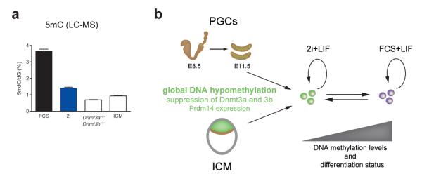
Naive pluripotent embryonic stem cells (ESCs) and embryonic germ cells (EGCs) are derived from the preimplantation epiblast and primordial germ cells (PGCs), respectively. We investigated whether differences exist between ESCs and EGCs, in view of their distinct developmental origins. PGCs are programmed to undergo global DNA demethylation; however, we find that EGCs and ESCs exhibit equivalent global DNA methylation levels. Inhibition of MEK and Gsk3b by 2i conditions leads to pronounced reduction in DNA methylation in both cell types. This is driven by Prdm14 and is associated with downregulation of Dnmt3a and Dnmt3b. However, genomic imprints are maintained in 2i, and we report derivation of EGCs with intact genomic imprints. Collectively, our findings establish that culture in 2i instills a naive pluripotent state with a distinctive epigenetic configuration that parallels molecular features observed in both the preimplantation epiblast and nascent PGCs.


Figures

Figure 1
Figure 1. Culture environment defines the transcriptional profile of pluripotent stem cells
(a) Summary of derivation procedure and culture conditions for ESC and EGC lines analyzed in this study; (b) Unsupervised hierarchical clustering of ESC and EGC lines demonstrates the dominant effect of culture condition on the transcriptional profile of pluripotent cells; (c) Transcript levels of germline marker genes, microarray log2 expression intensity. * FDR < 0.05, fold change > 1.5 for FCS versus 2i (see online methods for details), ≥ four independent cell lines for each condition (see Fig. 1a), error bars represent standard error of mean (SEM).
Figure 2
Figure 2. Global DNA hypomethylation in 2i
a) Analysis of global 5mC content by LC-MS in EGC and ESC lines cultured in FCS or 2i. Each data point represents the average of two technical replicates for an independent cell line. * P < 0.05, *** P < 0.001, as determined by unpaired t-tests; (b) Immunofluorescence staining for 5mC and 5hmC in EGC line EG-2i-1 cultured in FCS or 2i. DNA is counterstained with propidium iodide (PI). Scale bar, 10 μm; (c) Thin layer chromatography (TLC) analysis of methylation levels in DNA of ESC and EGC lines. Each cell line was grown side-by-side in 2i or FCS; Control oligonucleotides (oligo) containing 5mC or C are shown (d) Summary of bisulphite sequencing of repetitive elements in ESC line ES-2i-4 grown in FCS or 2i. * P < 0.05, ** P < 0.01, Mann-Whitney U-tests, ≥ eight clones for each condition; (e) Summary of bisulfite sequencing for Dazl promoter DNA methylation in ESC line ES-2i-4 and EGC line EG-3, both of which were cultured side-by-side in FCS or 2i.
Figure 3
Figure 3. Characteristics of DNA methylation changes between culture conditions
(a) qPCRs of Dnmt and Tet expression in EGCs maintained in 2i or FCS. ** P < 0.01, *** P < 0.001, unpaired t-tests, ≥ four independent cell lines for each condition, error bars represent SEM; (b) Western blot of Dnmt expression in ESCs and EGCs cultured in 2i or FCS for five passages; (c) Immunofluorescence staining for Dnmt3a and Dnmt3b in EGC line EG-2i-1 cultured in FCS or 2i. DNA counterstained with DAPI. Scale bar, 10 μm; (d) Global 5hmC content as a proportion of dG (left) or 5mdC (right) by LC-MS in a subset of ESC and EGC lines grown in FCS or 2i. Each data point represents the average of two technical replicates for an independent cell line. ** P < 0.01, unpaired t-tests; (e) 5mC abundance by LC-MS in ESCs and EGCs swapped from FCS to 2i conditions and vice versa for five passages. Each data point represents the average of two technical replicates for each cell line. * P < 0.05, ** P < 0.01, paired t-tests for swaps, unpaired t-test for 2i comparison; (f) qPCRs of Dnmt expression during embryoid body differentiation of cells grown in FCS or 2i. Oct4 and Sox17 expression shown for progression of differentiation; (g) Global 5mC levels by LC-MS in undifferentiated (D0) and day 4 embryoid bodies (D4) derived from pluripotent cells grown in FCS or 2i. Each data point represents the average of two technical replicates for one sample derived from an independent cell line. ** P < 0.01, unpaired t-tests.
Figure 4
Figure 4. Prdm14 regulates DNA methylation in pluripotent stem cells
(a) Prdm14 expression as assessed by qPCR in cells grown in FCS, 2i or in the presence of a single Erk (PD) or Gsk3b (CH) inhibitor (LIF was added in all conditions); (b) qPCR analysis of Dnmt and Tet expression levels in Prdm14−/− ESCs (two independent cell lines) in comparison to representative ESC lines grown in FCS or 2i (three independent cell lines for each condition). * P < 0.05, ** P < 0.01, results of unpaired t-tests are shown for comparison to Prdm14−/− ESCs only, error bars represent SEM; (c) Analysis of global 5mC levels in Prdm14−/− ESCs in comparison to representative ESC lines grown in FCS or 2i. Each data point represents the average of two technical replicates for an independent cell line. * P < 0.05, unpaired t-tests.
Figure 5
Figure 5. Imprinted DMR methylation status of ESC and EGC lines
(a) Summary of bisulphite sequencing of imprinted DMRs in ESC line ES-2i-4 cultured in 2i or FCS; (b) Summary of bisulphite analyses of imprinted DMRs in EGCs derived in 2i or FCS. Note that EGCs derived in 2i exhibit two distinct methylation patterns; (c) Bisulphite sequencing of imprinted DMRs in an EGC line derived in 2i following an interspecific cross, allowing the parental origin of each allele to be determined. Blue line represents the paternal allele and red the maternal allele. Filled and open circles represent methylated and unmethylated CpGs, respectively; (d) Image of chimeras generated by injection of EG-2i-1 (agouti) into C57BL/6 blastocysts (black).
Figure 6
Figure 6. Naïve pluripotency is characterized by DNA hypomethylation
(a) LC-MS analysis of global 5mC levels in E4.0 inner cell mass (ICM) cells in comparison to ESC lines grown in FCS or 2i, and Dnmt3a and 3b double knockout ESCs. Two technical replicates were analyzed for each sample, error bars represent SEM; (b) Model depicting the capture of the naïve pluripotent state in 2i culture conditions. This state is characterized by DNA hypomethylation, suppression of de novo methyltransferases and expression of Prdm14, and thus has clear mechanistic parallels to the in vivo sources of naïve pluripotent stem cells. Although pluripotent cells cultured in FCS conditions are still categorized as naïve we propose that they are somewhat more advanced in their differentiation status. This is evidenced by decreased Prdm14 levels, increased expression of the de novo methyltransferases and the accumulation of DNA methylation.

Comment in

  • DNA methylation: a matter of culture.
    Bock C, Wutz A. Bock C, et al. Nat Struct Mol Biol. 2013 Mar;20(3):249-51. doi: 10.1038/nsmb.2531. Nat Struct Mol Biol. 2013. PMID: 23463307 No abstract available.

References

    1. Gardner RL, Beddington RS. Multi-lineage ‘stem’ cells in the mammalian embryo. J. Cell Sci. Suppl. 1988;10:11–27. - PubMed
    1. Nichols J, Smith A. Pluripotency in the Embryo and in Culture. Cold Spring Harbor Perspectives in Biology. 2012;4:a008128–a008128. - PMC - PubMed
    1. Evans MJ, Kaufman MH. Establishment in culture of pluripotential cells from mouse embryos. Nature. 1981;292:154–156. - PubMed
    1. Martin GR. Isolation of a pluripotent cell line from early mouse embryos cultured in medium conditioned by teratocarcinoma stem cells. Proc Natl Acad Sci USA. 1981;78:7634–7638. - PMC - PubMed
    1. Brook FA, Gardner RL. The origin and efficient derivation of embryonic stem cells in the mouse. Proc Natl Acad Sci USA. 1997;94:5709–5712. - PMC - PubMed

Publication types

MeSH terms

Associated data