. 2014 Feb 20;33(8):986-95.
doi: 10.1038/onc.2013.33. Epub 2013 Feb 18.

Engineering a single ubiquitin ligase for the selective degradation of all activated ErbB receptor tyrosine kinases

Affiliations

Engineering a single ubiquitin ligase for the selective degradation of all activated ErbB receptor tyrosine kinases

F Kong et al. Oncogene. .

Abstract

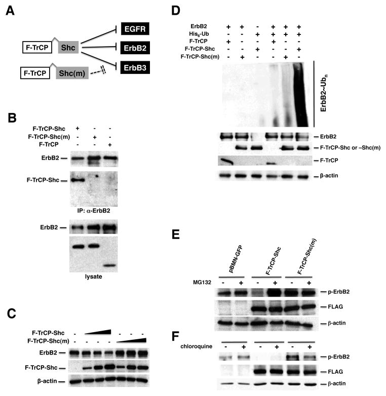
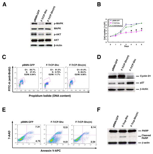
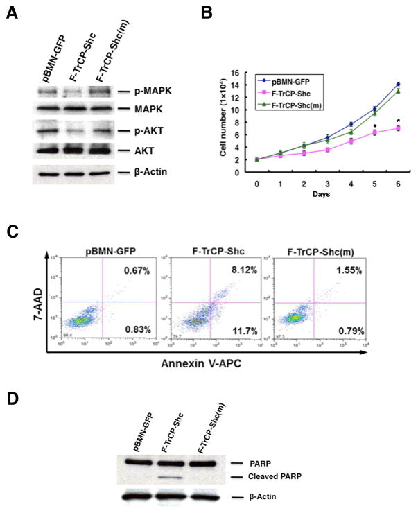
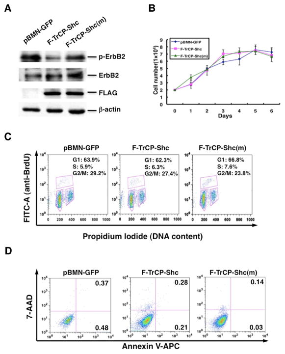
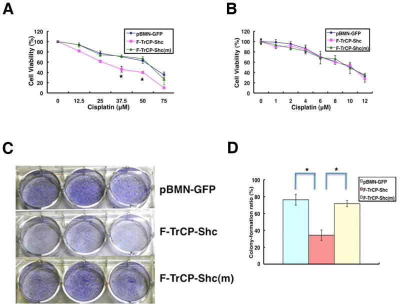
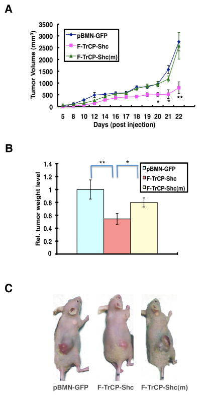
Interrogating specific cellular activities often entails the dissection of posttranslational modifications or functional redundancy conferred by protein families, which demands more sophisticated research tools than simply eliminating a specific gene product by gene targeting or RNA interference. We have developed a novel methodology that involves engineering a single SCF(βTrCP)-based ubiquitin ligase that is capable of not only simultaneously targeting the entire family of ErbB receptor tyrosine kinases for ubiquitination and degradation, but also selectively recruiting only activated ErbBs. The engineered SCF(βTrCP) ubiquitin ligase effectively blocked ErbB signaling and attenuated oncogenicity in breast cancer cells, yet had little effect on the survival and growth of non-cancerous breast epithelial cells. Therefore, engineering ubiquitin ligases offers a simple research tool to dissect the specific traits of tumorigenic protein families, and provides a rapid and feasible means to expand the dimensionality of drug discovery by assessing protein families or posttranslational modifications as potential drug targets.


Figures

Figure 1
Figure 1. The engineered F-TrCP-Shc E3 ligase recruits and induces the dose-dependent degradation of ErbB2
A. Schematic diagram of the engineered F-TrCP-Shc ubiquitin ligase to target the ErbB RTKs. B. F-TrCP-Shc binds to ErbB2. 293T cells were transiently transfected with the indicated plasmids. ErbB2 was immunoprecipitated with the anti-ErbB2 antibody and immunoblotted with the ErbB2, FLAG and β-actin antibodies. C. Degradation of endogenous ErbB2 by the engineered F-TrCP-Shc. 293T cells were transiently transfected with increasing doses of F-TrCP-Shc or F-TrCP-Shc(m), and levels of endogenous ErbB2, F-TrCP fusions and β-actin were determined by Western blotting. The experiment was repeated three times. D. F-TrCP-Shc promotes ubiquitination of ErbB2. 293T cells were transfected with the indicated plasmids for in vivo ubiquitination assays. E–F. F-TrCP-Shc-induced ErbB2 degradation is proteasome-dependent. SKBR3 and BT474 cells expressing F-TrCP-Shc, F-TrCP-Shc(m) or pBMN-GFP were treated with 50 μM MG132 or 100 μM choloroquine. The levels of endogenous ErbB2, F-TrCP fusions and β-actin were determined by Western blotting.
Figure 2
Figure 2. The engineered F-TrCP-Shc E3 ligase selectively targets the activated ErbB family members for degradation and depletes ErbB2 RTK on the cell surface
SKBR3 (A) and BT474 (B) cells infected with the indicated recombinant retroviruses were lysed and 50 μg of each cell lysate was analyzed by SDS-PAGE and Western blotting with the indicated antibodies. C. SKBR3 and BT474 cells were plated in 6-well plates for 24 hours, trypsinized and washed in FACS buffer. The live cells were stained with anti-ErbB2 antibody (Santa Cruz) against the extracellular domain of ErbB2. Results are the mean for three replicate experiments. D. Dose-dependent degradation of endogenous ErbB2 upon infection of adenoviral F-TrCP-Shc was determined by Western blotting with the indicated antibodies. E. Depletion of ErbB2 on the cell membrane by adenoviral F-TrCP-Shc. ErbB2 (red) was detected by immunofluorescent staining with the anti-ErbB2 antibody (R&D Systems). Infected cells were marked by GFP expression from the adenoviral vector. The nuclei were visualized by DAPI staining (blue). White arrow, a low F-TrCP-Shc expressing SKBR3 cell.
Figure 3
Figure 3. Targeted degradation of ErbB RTKs by F-TrCP-Shc led to the attenuation of MAPK and PI3K signaling, cell cycle arrest and inhibition of proliferation in SKBR3 breast cancer cells
A. SKBR3 cells were infected with pBMN-F-TrCP-Shc, pBMN-F-TrCP-Shc(m) or pBMN-GFP. Infected cells were sorted, lysed and probed with antibodies against p-MAPK, MAPK, p-AKT, AKT and β-actin. B. SKBR3 cells were infected with the same retroviruses as in (A) and were plated in 12-well plates for up to 6 days. The number of cells in each group was counted at the indicated time points. Results are the mean for three replicate experiments. * indicates P<0.05 for pBMN-F-TrCP-Shc-infected cells compared to those with pBMN-GFP or pBMN-F-TrCP-Shc(m). C. SKBR3 cells were infected with pBMN-F-TrCP-Shc, pBMN-F-TrCP-Shc(m) or pBMN-GFP, labeled with BrdU and propidium iodide, and subjected to FACS analysis. Results are represented as the mean ± standard deviation (n=3). D. Western blotting was performed to measure the expression of G1 cell cycle markers cyclin D1 and p27 using the appropriate antibodies. E–F. Apoptotic induction of SKBR3 cells by F-TrCP-Shc, as determined by Annexin V staining-FACS analysis, and PARP1 cleavage by Western blotting.
Figure 4
Figure 4. Targeted degradation of ErbB RTKs triggered apoptosis in BT474 cells
A. BT474 cells were infected with pBMN-F-TrCP-Shc, pBMN-F-TrCP-Shc(m) or pBMN-GFP, sorted and analyzed for inhibition of MAPK and PI3K signaling by Western blotting as in Fig. 4A. B. Retroviral expression of F-TrCP-Shc in BT474 cells inhibited cell growth. Results are the mean for three replicate experiments. * indicates P<0.05 for pBMN-F-TrCP-Shc-infected cells compared to those with pBMN-GFP or pBMN-F-TrCP-Shc(m). C–D. Apoptotic induction of BT474 cells by F-TrCP-Shc, as determined by Annexin V-FACS analysis and PARP1 cleavage by Western blotting. For FACS analysis, cells were stained with Annexin-V-APC and 7-AAD, and the percentage of early (right bottom) and late apoptotic (right top) populations are indicated.
Figure 5
Figure 5. The engineered F-TrCP-Shc E3 ligase had no effect on the non-cancerous MCF-10A mammary epithelial cells
MCF-10A cells were infected with pBMN-F-TrCP-Shc, pBMN-F-TrCP-Shc(m) or pBMN-GFP, and assayed for (A) ErbB2 degradation by Western blotting, (B) growth rate, (C) cell cycle by BrdU labeling and FACS analysis, and (D) apoptosis by Annexin V/7-AAD staining and FACS analysis as in Figures 3 and 4.
Figure 6
Figure 6
The engineered F-TrCP-Shc E3 ligase sensitizes BT474 breast cancer cells to cytotoxic killing by cisplatin and decreases their transforming ability. A–B. BT474 or MCF-10A cells infected with pBMN-F-TrCP-Shc, pBMN-F-TrCP-Shc(m) or pBMN-GFP retroviruses were treated with the indicated amounts of cisplatin for 48 hours, and the cell viability was measured by the XTT assay. Results are represented as the mean ± standard deviation (n=3), for BT474 cells. C–D. Focus formation assay. Retroviral vector infected BT474 cells were seeded in 6-well plates and cultured for 10 days to allow for colony formation. The colonies were fixed, stained and photographed. The colony numbers were counted and the colony formation ratio was calculated according to the formula: colonyformationratio(%)=(numberofcolonies/numberofcellsseeded)×100. Results are represented as the mean ± standard deviation (n=3). * indicates P<0.05 for pBMN-F-TrCP-Shc-infected cells compared to those with pBMN-GFP or pBMN-F-TrCP-Shc(m).
Figure 7
Figure 7. The engineered F-TrCP-Shc E3 ligase suppressed BT474 xenograft tumor growth in nude mice
A. Nude mice were injected subcutaneously in the right flank with 5×10⁶ BT474 cells infected with pBMN-F-TrCP-Shc, pBMN-F-TrCP-Shc(m) or pBMN-GFP retroviruses. Tumor volumes were measured over a 4-week period and calculated by the formula: (length×width2)/2. B. Tumor weight (g) was recorded at the end of the experiment. Columns, mean value of tumor weight; bars, SD. C. Representative images of tumor-bearing mice. * P<0.05 or **P<0.01 indicate tumors expressing pBMN-F-TrCP-Shc compared to those infected with pBMN-GFP or pBMN-F-TrCP-Shc(m).

References

    1. Guy CT, Webster MA, Schaller M, Parsons TJ, Cardiff RD, Muller WJ. Expression of the neu protooncogene in the mammary epithelium of transgenic mice induces metastatic disease. Proc Natl Acad Sci U S A. 1992 Nov 15;89(22):10578–82. - PMC - PubMed
    1. Hynes NE, Stern DF. The biology of erbB-2/neu/HER-2 and its role in cancer. Biochim Biophys Acta. 1994 Dec 30;1198(2–3):165–84. - PubMed
    1. Muller WJ, Sinn E, Pattengale PK, Wallace R, Leder P. Single-step induction of mammary adenocarcinoma in transgenic mice bearing the activated c-neu oncogene. Cell. 1988 Jul 1;54(1):105–15. - PubMed
    1. Olayioye MA, Neve RM, Lane HA, Hynes NE. The ErbB signaling network: receptor heterodimerization in development and cancer. EMBO J. 2000 Jul 3;19(13):3159–67. - PMC - PubMed
    1. Yarden Y, Sliwkowski MX. Untangling the ErbB signalling network. Nat Rev Mol Cell Biol. 2001 Feb;2(2):127–37. - PubMed

Publication types

Substances