. 2014 Feb 13;33(7):842-50.
doi: 10.1038/onc.2013.18. Epub 2013 Feb 18.

CLIC4 regulates TGF-β-dependent myofibroblast differentiation to produce a cancer stroma

Affiliations

CLIC4 regulates TGF-β-dependent myofibroblast differentiation to produce a cancer stroma

A Shukla et al. Oncogene. .

Abstract

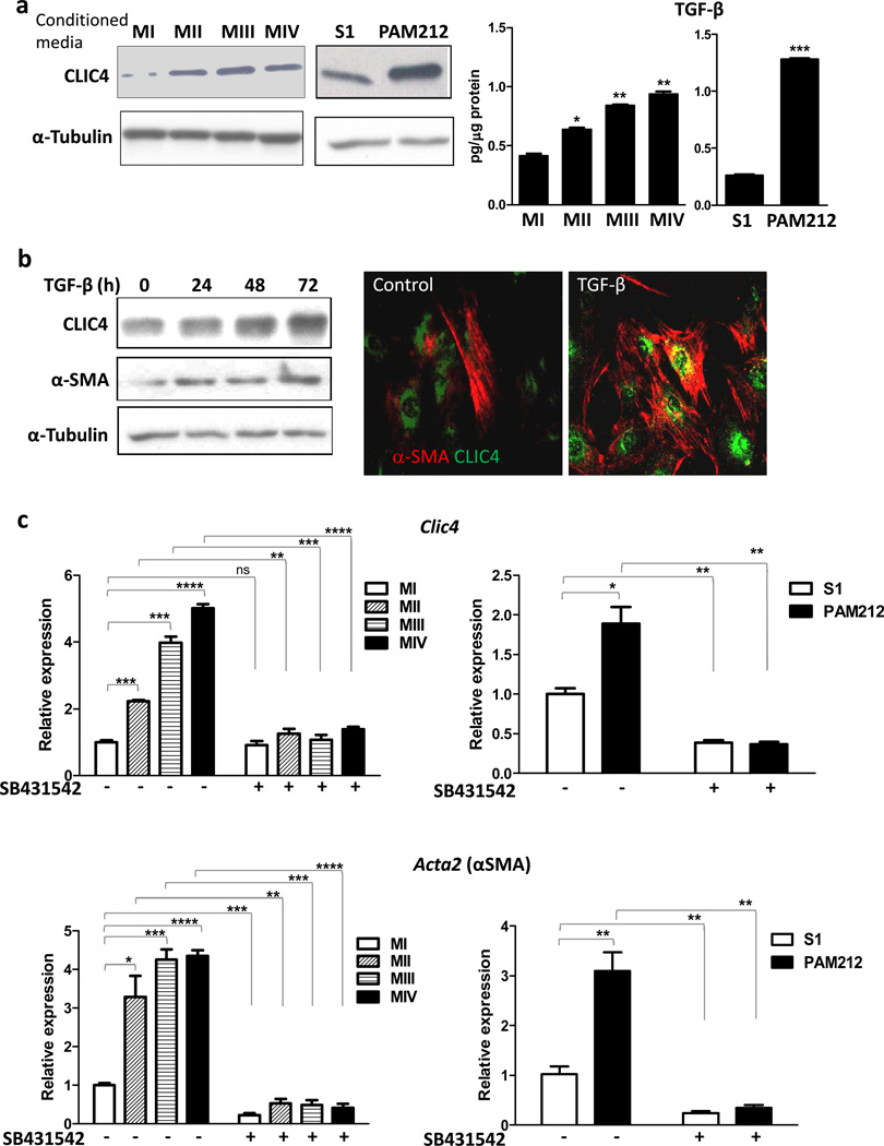
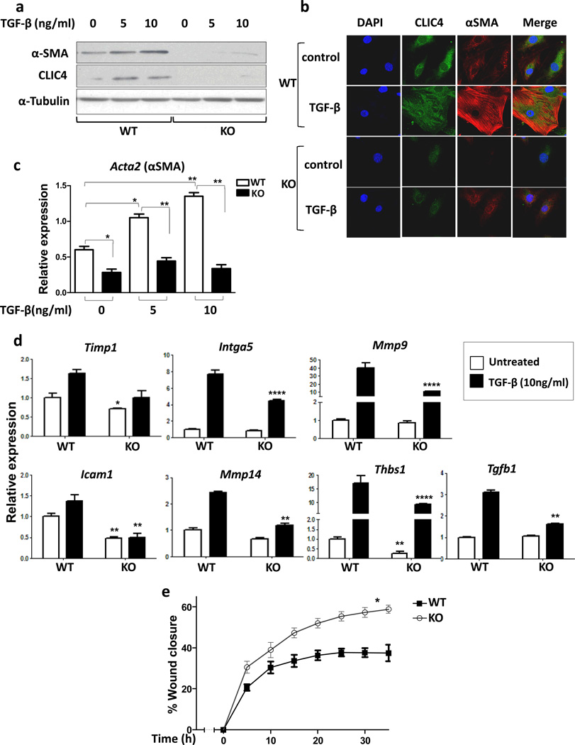
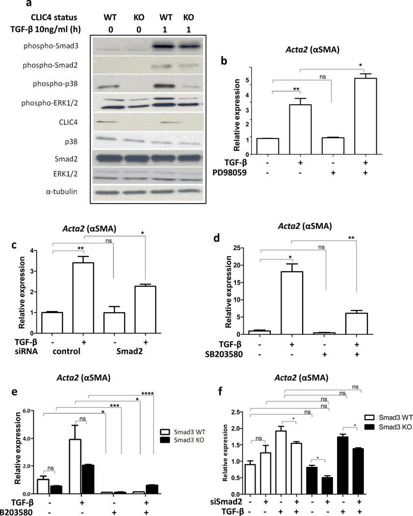
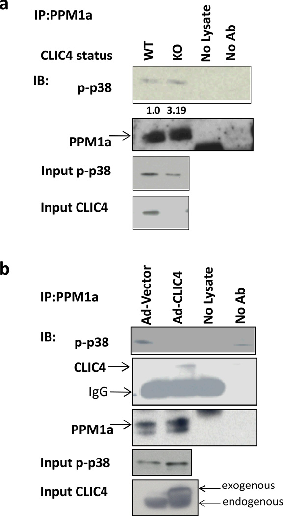
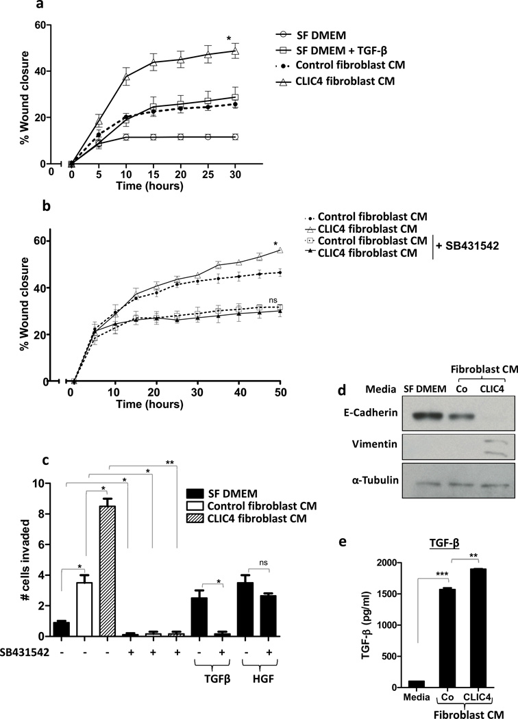
Cancer stroma has a profound influence on tumor development and progression. The conversion of fibroblasts to activated myofibroblasts is a hallmark of reactive tumor stroma. Among a number of factors involved in this conversion, transforming growth factor (TGF)-β has emerged as a major regulator. CLIC4, an integral protein in TGF-β signaling, is highly upregulated in stroma of multiple human cancers, and overexpression of CLIC4 in stromal cells enhances the growth of cancer xenografts. In this study, we show that conditioned media from tumor cell lines induces expression of both CLIC4 and the myofibroblast marker alpha smooth muscle actin (α-SMA) in stromal fibroblasts via TGF-β signaling. Genetic ablation of CLIC4 in primary fibroblasts prevents or reduces constitutive or TGF-β-induced expression of α-SMA and extracellular matrix components that are markers of myofibroblasts. CLIC4 is required for the activation of p38 map kinase by TGF-β, a pathway that signals myofibroblast conversion in stromal cells. This requirement involves the interaction of CLIC4 with PPM1a, the selective phosphatase of activated p38. Conditioned media from fibroblasts overexpressing CLIC4 increases tumor cell migration and invasion in a TGF-β-dependent manner and promotes epithelial to mesenchymal transition indicating that high stromal CLIC4 serves to enhance tumor invasiveness and progression. Thus, CLIC4 is significantly involved in the development of a nurturing tumor microenvironment by enhancing TGF-β signaling in a positive feedback loop. Targeting CLIC4 in tumor stroma should be considered as a strategy to mitigate some of the tumor enhancing effects of the cancer stroma.


Conflict of interest statement

Conflicts of interest

The authors have no conflicts of interest.

Figures

Figure 1
Figure 1
Tumor cells induce CLIC4 and α-SMA expression in fibroblasts via TGF-β signaling. (a) Left Primary mouse dermal fibroblasts were treated with serum free conditioned media from either of the human breast cell lines MI, MII, MIII or MIV or the murine squamous cell lines S1 or PAM 212 cells. Expression of CLIC4 was analyzed by immunoblotting. Right TGF-β concentrations in conditioned media from human and mouse cell lines were determined by ELISA and normalized to total protein content. Data sets were compared for statistical significance with MI or S1(non-tumorigenic lines). (b) Primary dermal fibroblasts were treated for different time periods with TGF-β (10ng/ml) and immunoblotted (left) for CLIC4 and α-SMA. (Right) Co-immunofluorescence for CLIC4 (green) and α-SMA (red) in primary untreated fibroblasts or fibroblasts treated with TGF-β for 48h. (c) Primary dermal fibroblasts were treated with conditioned media as in A with or without pretreatment with the ALK5 inhibitor SB431542 (5µM). Expression of CLIC4 and α-SMA (gene name Acta2) was analyzed by real time PCR normalized to respective GAPDH levels and plotted as relative to MI or S1. Data sets were compared as indicated by lines for statistical significance.
Figure 2
Figure 2
CLIC4 is required for TGF-β dependent conversion of fibroblasts to myofibroblasts. (a) Adenoviral Cre recombinase transduced CLIC4 wild-type (WT) and floxed fibroblasts (KO) were treated with TGF-β for 48h at varying concentrations and immunoblotted for CLIC4 and α-SMA. α-Tubulin was used as loading control. (b) Co-immunofluorescence for CLIC4 and α-SMA in CLIC4 WT and KO fibroblasts treated with or without TGF-β (10ng/ml). (c) CLIC4 WT and KO fibroblasts treated with TGF-β for 24h at varying concentrations and analyzed for α-SMA by real time PCR. Data sets were compared as indicated by lines for statistical significance. (d) Real time PCR analysis of various ECM genes in CLIC4 WT and KO fibroblasts with or without treatment with 10ng/ml TGF-β for 24h. For statistical analysis, untreated KO were compared to untreated WT. TGF-β treated KO were compared to TGF-β treated WT. (e) Scratch assay on WT and KO fibroblasts in media containing 0.2% serum with 10ng/ml TGF-β. Migration was recorded and quantified using Incucyte technology. The two curves were compared for statistical significance by Mann Whitney t-test.
Figure 3
Figure 3
CLIC4 dependent p38 and Smad activation contribute to increased α-SMA expression in primary dermal fibroblasts. (a) Lysates of CLIC4 WT and KO fibroblasts untreated or treated with TGF-β (10ng/ml) for 1h were immunoblotted for proteins involved in TGF-β signaling. α-Tubulin was used as loading control. (b–d) Primary dermal fibroblasts were pretreated with (b) 60µM MEK1 inhibitor PD98059 for 30 min, (c) Smad2 siRNA for 48h or (d) 10µM p38 inhibitor SB 203580 for 30 min before TGF-β treatment (10ng/ml) for 24h. α-SMA expression was analyzed by real time PCR, normalized to respective GAPDH levels and expressed as relative to untreated control.(e,f) Primary dermal fibroblasts from Smad3 wild-type or knockout mice were (e) pretreated with 10µM p38 inhibitor SB203580 for 30min, or (f) transfected with nonsilencing control (NS) or Smad2 siRNA for 48h before TGF-β treatment for 24h. α-SMA expression was analyzed by real time PCR, normalized to respective GAPDH levels and expressed as relative to untreated control. b-f, Data sets were compared as indicated by lines for statistical significance. ns=not significant.
Figure 4
Figure 4
CLIC4 prolongs p38 phosphorylation by inhibiting interaction of p-p38 and its phosphatase PPM1a. (a) Lysates from CLIC4 WT and KO fibroblasts were immunoprecipitated with anti-PPM1a antibody and immunoblotted for phospho-p38 and PPM1a. Non-immunoprecipitated (input) lysates were immunoblotted for phospho-p38 and CLIC4. Immunoprecipitated p-p38 bands were quantified using Image J software, normalized to their respective input p-p38 bands and expressed relative to WT. (b) Primary dermal fibroblasts were transduced with vector or CLIC4 expressing adenovirus. Lysates were immunoprecipitated with anti-PPM1a antibody and immunoblotted for phospho-p38, CLIC4 and PPM1a. Non-immunoprecipitated (input) lysates were immunoblotted for phospho-p38 and CLIC4. A,B-Samples undergoing the immunoprecipitation process without any protein lysate (no lysate) or without antibody (no Ab) were used as controls.
Figure 5
Figure 5
Stromal CLIC4 enhances tumor cell migration and invasion via TGF-β signaling. (a) Scratch assay on PAM212 cells treated with either serum free media (SF DMEM) +/− TGF-β (10ng/ml) or serum free conditioned media from fibroblasts transduced with adeno-vector (Control fibroblast CM)) or adeno-CLIC4 (CLIC4 fibroblast CM). Control CM curve was compared to CLIC4 CM curve for statistical analysis using Mann Whitney t-test. (b) Scratch assay on PAM212 cells treated with serum free conditioned media from fibroblasts transduced with adeno-vector control or adeno-CLIC4 +/− ALK5 blocker SB431542 (5µM). For statistical analysis, untreated control CM curve was compared to untreated CLIC4 CM curve while SB431542 treated control CM curve was compared to treated CLIC4 CM curve using Mann Whitney t-test. A,B-Time lapse monitoring of migration of PAM212 cells was conducted using Incucyte technology. (c) Matrigel invasion assay on PAM212 cells plated in matrigel coated upper chambers of a 24-transwell plate. Serum free media (SF DMEM)or serum free conditioned media from fibroblasts transduced with adeno-vector (Control fibroblast CM) or adeno-CLIC4 (CLIC4 fibroblast CM) was placed in the lower chamber. ALK5 blocker SB431542 (5µM) was added to the upper and lower chambers and TGF-β or HGF added to the lower chambers as indicated. Invasion through matrigel was analyzed after 24h. Data sets were compared as indicated by lines for statistical significance. (d) PAM212 cells were treated with SF DMEM or serum free conditioned media from fibroblasts transduced with adeno-vector (Co) or adeno-CLIC4 (CLIC4) for 6 days. Whole cell lysates were immunoblotted for E-Cadherin, Vimentin and α-Tubulin. (e) ELISA analysis of TGF-β levels in SF DMEM (Media) or serum free conditioned media from vector transduced (Co) or CLIC4 overexpressing (CLIC4) fibroblasts. For statistical analysis, Co data set was compared to SF DMEM and CLIC4 to Co.

References

    1. Suh KS, Yuspa SH. Intracellular chloride channels: critical mediators of cell viability and potential targets for cancer therapy. Curr Pharm Des. 2005;11(21):2753–2764. - PubMed
    1. Littler DR, Harrop SJ, Goodchild SC, Phang JM, Mynott AV, Jiang L, et al. The enigma of the CLIC proteins: Ion channels, redox proteins, enzymes, scaffolding proteins? FEBS Lett. 2010 May 17;584(10):2093–2101. - PubMed
    1. Littler DR, Harrop SJ, Fairlie WD, Brown LJ, Pankhurst GJ, Pankhurst S, et al. The intracellular chloride ion channel protein CLIC1 undergoes a redox-controlled structural transition. J Biol Chem. 2004 Mar 5;279(10):9298–9305. - PubMed
    1. Littler DR, Assaad NN, Harrop SJ, Brown LJ, Pankhurst GJ, Luciani P, et al. Crystal structure of the soluble form of the redox-regulated chloride ion channel protein CLIC4. FEBS J. 2005 Oct;272(19):4996–5007. - PubMed
    1. Shorning BY, Wilson DB, Meehan RR, Ashley RH. Molecular cloning and developmental expression of two Chloride Intracellular Channel (CLIC) genes in Xenopus laevis. Dev Genes Evol. 2003 Oct;213(10):514–518. - PubMed

Publication types

Substances