. 2013 Apr;19(4):539-51.
doi: 10.1261/rna.035691.112. Epub 2013 Feb 15.

Tertiary structure-based analysis of microRNA-target interactions

Affiliations

Tertiary structure-based analysis of microRNA-target interactions

Hin Hark Gan et al. RNA. 2013 Apr.

Abstract

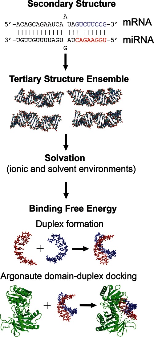
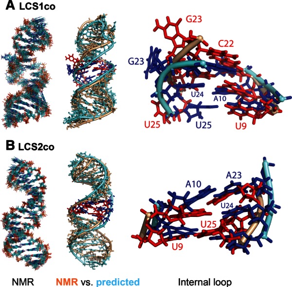
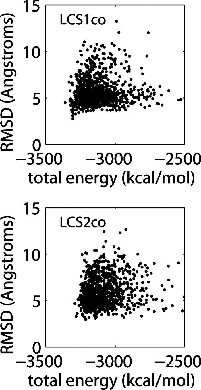
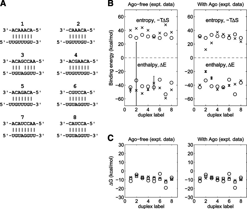
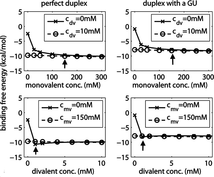
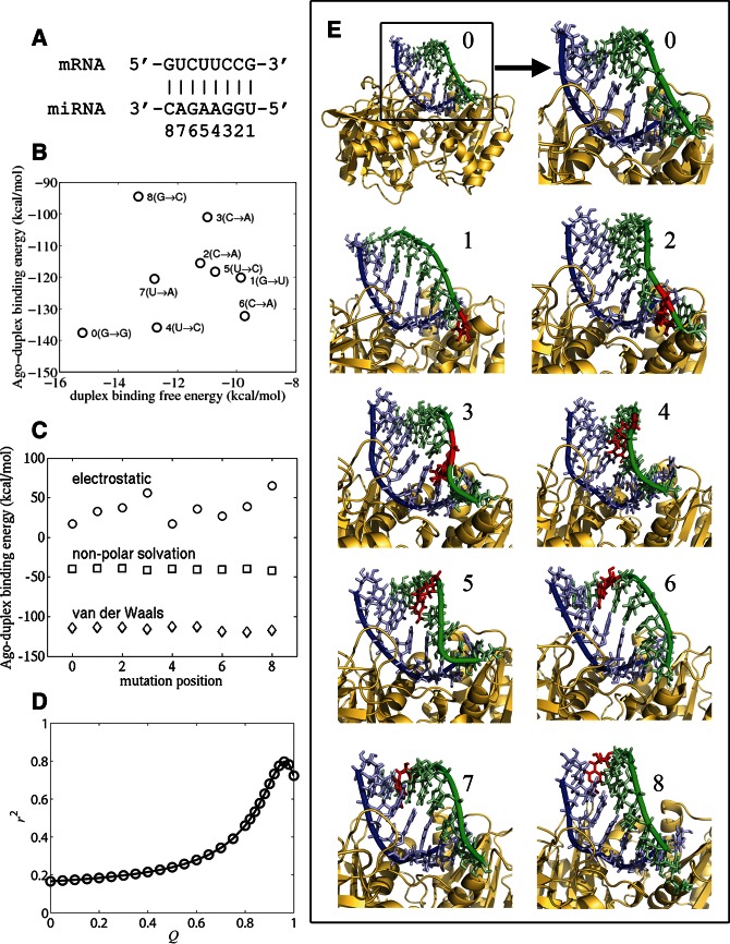
Current computational analysis of microRNA interactions is based largely on primary and secondary structure analysis. Computationally efficient tertiary structure-based methods are needed to enable more realistic modeling of the molecular interactions underlying miRNA-mediated translational repression. We incorporate algorithms for predicting duplex RNA structures, ionic strength effects, duplex entropy and free energy, and docking of duplex-Argonaute protein complexes into a pipeline to model and predict miRNA-target duplex binding energies. To ensure modeling accuracy and computational efficiency, we use an all-atom description of RNA and a continuum description of ionic interactions using the Poisson-Boltzmann equation. Our method predicts the conformations of two constructs of Caenorhabditis elegans let-7 miRNA-target duplexes to an accuracy of ∼3.8 Å root mean square distance of their NMR structures. We also show that the computed duplex formation enthalpies, entropies, and free energies for eight miRNA-target duplexes agree with titration calorimetry data. Analysis of duplex-Argonaute docking shows that structural distortions arising from single-base-pair mismatches in the seed region influence the activity of the complex by destabilizing both duplex hybridization and its association with Argonaute. Collectively, these results demonstrate that tertiary structure-based modeling of miRNA interactions can reveal structural mechanisms not accessible with current secondary structure-based methods.


Figures

FIGURE 1.
FIGURE 1.
Computational pipeline for generating, solvating, and computing binding energies of 3D RNA structures, starting from a secondary duplex structure. The guide (red) and target (blue) strands in the seed region are highlighted. First, a conformational ensemble is generated using the MC-Sym algorithm. Second, the RNA interaction energies are computed at specific ionic conditions using a continuum electrostatic model. Third, the binding free energy is obtained by evaluating the enthalpy and entropy changes associated with either duplex formation (vs. free strands) or Argonaute–duplex formation (vs. free duplex), as illustrated here for docking of the PIWI/MID domain of Thermus thermophilus Argonaute to the given seed duplex.
FIGURE 2.
FIGURE 2.
NMR models and predicted structures for single-stranded constructs of C. elegans let-7 miRNA–target site duplexes (A) LCS1co and (B) LCS2co. (Left) Superposition of 10 lowest-energy NMR structures for each construct. (Middle) Structure alignments of individual representative NMR (wheat color) and predicted (light blue) structures, highlighting bases in the internal loop (NMR, red; predicted, blue). (Right) Magnified view of internal loops from aligned structures, with bases labeled. For substructure alignments, structures were partitioned into internal loop, hairpin (upper), and stem (lower) regions. The predicted structures shown have average RMSD values of 4.2 Å for LCS1co and 3.3 Å for LCS2co (relative to the 10 lowest-energy NMR structures for the corresponding construct).
FIGURE 3.
FIGURE 3.
Total energy versus RMSD for LCS1co (top) and LCS2co (bottom). Plotted are individual data points for 1000 computed structures, each representing an individual structure’s total energy and its average RMSD value obtained from superposition with the 10 low-energy NMR structures.
FIGURE 4.
FIGURE 4.
Comparison of experimental and computed binding free energy terms for different RNA duplexes. (A) The eight structures analyzed, labeled 1–8. (B,C) Enthalpy (ΔE), entropy (−T ΔS), and free energy (ΔG) of free (left) or Argonaute-bound (right) duplexes, as determined from titration calorimetry experiments (crosses) versus tertiary (circles) and secondary (squares) structure computational methods (Supplemental Table S1 also summarizes all numerical values). Experiments and 3D calculations were performed under a constant solvent condition (150 mM KCl and 10 mM MgCl2 at 20°C); 2D calculations assumed 1 M NaCl. All entropies were computed at experimental guide miRNA concentrations. Error bars represent the standard deviation (SD) of experimental uncertainty in enthalpy.
FIGURE 5.
FIGURE 5.
Concentration dependence and saturation behavior of the binding free energy as a function of monovalent (top) and divalent (bottom) ions for a perfect duplex (left; duplex 1 in Fig. 4) and the same duplex but with a GU wobble (right; duplex 4 in Fig. 4). In each plot, the concentration of the other ion species is held constant either at 0 mM (crosses) or above its own saturation (circles). (Arrow) Concentration at which the binding free energy plateaus (solid line).
FIGURE 6.
FIGURE 6.
Analysis of interactions between the T. thermophilus Argonaute MID/PIWI domain and seed duplexes of D. melanogaster miR-7 with single point mutations in mRNA at base positions 1–8; 0 indicates the wild-type duplex with no mutation in panels B, C, and E. (A) Composition of wild-type duplex (duplex 0), with labeled base-pair positions 1–8. (B) Duplex binding free energy versus Argonaute–duplex binding energy for each duplex, indicating the specific substitutions in the mRNA at each position. (C) Interaction energy components (van der Waals, nonpolar solvation, and electrostatic) of Argonaute–duplex binding energy for all mutation positions. (D) r2 statistics versus the Q value (weight of the duplex energy term) derived from linear least squares fit between miRNA activity and an effective Argonaute–duplex binding free energy (Eq. 1). (E) 3D structure models of Argonaute–seed duplexes, illustrating wild-type duplex (0) and duplexes with point mutations (highlighted by red nucleotides) at each of the eight positions in the mRNA strand (1–8). Significant structural distortions occur in the mRNA strand (green) but only minor distortions in the miRNA strand (blue). These distortions depend on the mutation position, and they weaken the duplex’s binding affinity for Argonaute.

References

    1. Abu Almakarem AS, Petrov AI, Stombaugh J, Zirbel CL, Leontis NB 2012. Comprehensive survey and geometric classification of base triples in RNA structures. Nucleic Acids Res 40: 1407–1423 - PMC - PubMed
    1. Ambros V 2004. The functions of animal microRNAs. Nature 431: 350–355 - PubMed
    1. Baker NA, Sept D, Joseph S, Holst MJ, McCammon JA 2001. Electrostatics of nanosystems: Application to microtubules and the ribosome. Proc Natl Acad Sci 98: 10037–10041 - PMC - PubMed
    1. Balasubramanian C, Ojha RP, Maiti S, Desideri A 2010. Sampling the structure of the noncanonical lin-4:lin-14 microRNA:mRNA complex by molecular dynamics simulations. J Phys Chem B 114: 16443–16449 - PubMed
    1. Bartel DP 2009. MicroRNAs: Target recognition and regulatory functions. Cell 136: 215–233 - PMC - PubMed

Publication types

MeSH terms