. 2013 Feb 28;494(7438):443-8.
doi: 10.1038/nature11871. Epub 2013 Feb 17.

Crystal structure of the entire respiratory complex I

Affiliations

Crystal structure of the entire respiratory complex I

Rozbeh Baradaran et al. Nature. .

Abstract

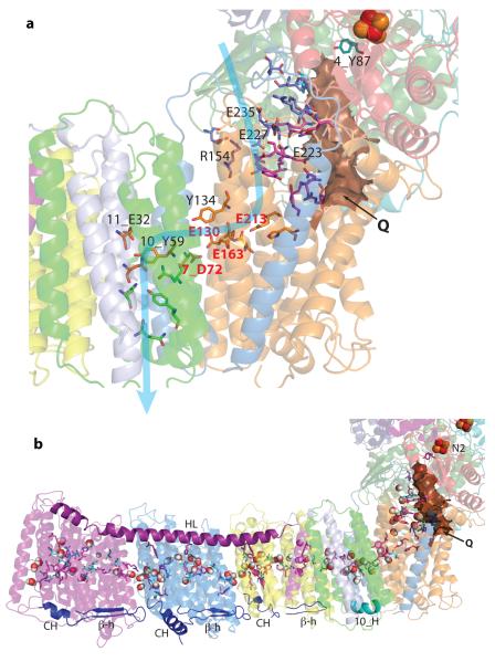
Complex I is the first and largest enzyme of the respiratory chain and has a central role in cellular energy production through the coupling of NADH:ubiquinone electron transfer to proton translocation. It is also implicated in many common human neurodegenerative diseases. Here, we report the first crystal structure of the entire, intact complex I (from Thermus thermophilus) at 3.3 Å resolution. The structure of the 536-kDa complex comprises 16 different subunits, with a total of 64 transmembrane helices and 9 iron-sulphur clusters. The core fold of subunit Nqo8 (ND1 in humans) is, unexpectedly, similar to a half-channel of the antiporter-like subunits. Small subunits nearby form a linked second half-channel, which completes the fourth proton-translocation pathway (present in addition to the channels in three antiporter-like subunits). The quinone-binding site is unusually long, narrow and enclosed. The quinone headgroup binds at the deep end of this chamber, near iron-sulphur cluster N2. Notably, the chamber is linked to the fourth channel by a 'funnel' of charged residues. The link continues over the entire membrane domain as a flexible central axis of charged and polar residues, and probably has a leading role in the propagation of conformational changes, aided by coupling elements. The structure suggests that a unique, out-of-the-membrane quinone-reaction chamber enables the redox energy to drive concerted long-range conformational changes in the four antiporter-like domains, resulting in translocation of four protons per cycle.


Figures

Fig. 1
Fig. 1
a) Structure of the entire complex I from T. thermophilus. FMN and Fe-S clusters are shown as magenta and red-orange spheres, respectively, with cluster N2 labelled. Key helices around the entry point (Q) into the quinone reaction chamber, and approximate membrane position are indicated. b) Putative proton translocation channels in the antiporter-like subunits. Polar residues lining the channels are shown as sticks with carbon in dark blue for the first (N-terminal) half-channel, in green for the second (C-terminal) half-channel and in orange for connecting residues. Key residues, GluTM5 and LysTM7 from the first half-channel, Lys/HisTM8 from the connection and Lys/GluTM12 from the second half-channel, are labelled. Approximate proton translocation paths are indicated by blue arrows.
Fig. 2
Fig. 2. Fold of subunit Nqo8
Coloured blue to red from N to C terminus. Neighbouring subunits Nqo7/10 are shown in light/dark grey. a) View from the cytoplasm. TM helices are numbered, with helices corresponding to the antiporter half-channel in bold. The conserved salt bridge Arg36-Asp62, supporting amphipathic helix AH1, is shown. b) Side view. Charged residues from the conserved 3rd cytoplasmic loop, mainly lining the Q cavity, are shown as sticks. c) Alignment of TM helices 2-6 of Nqo8 (orange) with TM helices 4-8 of Nqo13 (blue). LysTM7 from Nqo13 and Glu213 from Nqo8 TM5 are shown as sticks.
Fig. 3
Fig. 3
a) E-channel (fourth proton translocation channel). Charged and polar residues constituting the channel are shown as sticks. Central residues are shown with carbon in yellow, those forming a link to the Q site in magenta, link to the cytoplasm in blue, link to the periplasm in green and those interacting with quinone headgroup in cyan. Key residues are labelled, with Glu/Asp quartet in red. Approximate proton translocation path is indicated by blue arrow. Quinone cavity is shown with surface in brown. b) Central axis of charged and polar residues. Residues shown are either central to half-channels or are forming the connection between them (charged residues have carbon in magenta, polar in cyan). Most of them are located near the breaks in key helices TM7/8/12 (antiporters), 10_TM3 and 8_TM5. Predicted waters nearby, modelled using Dowser software, are shown as spheres. Connecting elements are shown in solid colours: helix HL in magenta and the βH element in blue, with the C-terminal helix CH and the β-hairpin from each antiporter labelled. The contacting Nqo10 helix is labelled 10_H. Subunits are coloured as in Fig. 1.
Fig. 4
Fig. 4. Quinone reaction chamber
Subunits are coloured as in Fig. 1. Iron-sulphur cluster N2 is shown as red-orange spheres. a-b) Experimental electron density (2mFo-DFc in blue, contoured at 1σ, and mFo-DFc in green, contoured at 3σ) and models obtained from crystals with bound Piericidin A (a) and Decyl-ubiquinone (b). Difference electron density was calculated before ligand modelling. Nqo4 residues interacting with the headgroup are indicated. Potential polar interactions are shown labelled with distances in Å. c) Surface (solvent-accessible) representation of the interface between two main domains. The empty crevice (C, circled, Supplementary Discussion) between Nqo10 and 7_TM1/Nqo8, as well as helices framing the entry point to the quinone site (Q) are indicated. d) Quinone reaction chamber, with its internal solvent-accessible surface coloured red for negative, white for neutral, and blue for positive surface charges. Charged residues lining the cavity are shown with carbon in magenta and hydrophobic residues in yellow. Residues are labelled with prefix indicating subunit (omitted for Nqo8). Ala63, the site of the primary LHON disease mutation, is labelled in red. e) Theoretical model of bound ubiquinone-10. Carbon atoms in cyan indicates the 8th isoprenoid unit. The quinone chamber is shown with surface in brown and helices framing its entry point are indicated. Movable helix 6_H1, interacting with 8_AH1, is also labelled.
Fig. 5
Fig. 5. Proposed coupling mechanism of complex I
a) Overview showing key helices and residues. Upon electron transfer from cluster N2, negatively charged quinone initiates a cascade of conformational changes, propagating from the E-channel (Nqo8/10/11) to the antiporters via the central axis (red arrows) of charged and polar residues located around flexible breaks in key TM helices. Cluster N2-driven shifts of Nqo4/6 helices (blue arrows) likely assist overall conformational changes. Helix HL and the βH element help coordinate conformational changes by linking discontinuous TM helices between the antiporters. In the antiporters, LysTM7 from the first half-channel is assumed to be protonated (via the link to cytoplasm) in the oxidised state. Upon reduction of quinone and subsequent conformational change, the first half-channel closes to the cytoplasm, GluTM5 moves out and LysTM7 donates its proton to the connecting Lys/HisTM8 and then onto Lys/GluTM12 from the second half-channel. Lys/GluTM12 ejects its proton into periplasm upon return from reduced to oxidised state. A fourth proton per cycle is translocated in the E-channel in a similar manner. TM helices are numbered and key charged residues (GluTM5, LysTM7, Lys/GluTM12, Lys/HisTM8 from Nqo12-14, 11_Glu67, 11_Glu32, interacting with 10_Tyr59, 8_Glu213 and some residues from the connection to Q cavity) are indicated by red circles for Glu and blue circles for Lys/His. b) Schematic drawing illustrating conformational changes between the two main (low energy) conformations. Analysis of networks of polar residues and modelled waters in the structure suggests that in the oxidised state (as crystallised) periplasmic half-channels are likely to be open. Residues shown as black circles indicate conserved prolines from the break in TM12.

References

    1. Walker JE. The NADH - ubiquinone oxidoreductase (complex I) of respiratory chains. Q. Rev. Biophys. 1992;25:253–324. - PubMed
    1. Yagi T, Matsuno-Yagi A. The proton-translocating NADH-Quinone oxidoreductase in the respiratory chain: the secret unlocked. Biochemistry. 2003;42:2266–2274. - PubMed
    1. Brandt U. Energy converting NADH:quinone oxidoreductase (complex I) Annu. Rev. Biochem. 2006;75:69–92. - PubMed
    1. Ohnishi T. Iron-sulfur clusters/semiquinones in complex I. Biochim. Biophys. Acta. 1998;1364:186–206. - PubMed
    1. Sazanov LA. Respiratory complex I: mechanistic and structural insights provided by the crystal structure of the hydrophilic domain. Biochemistry. 2007;46:2275–2288. - PubMed

Publication types

MeSH terms