. 2013 Mar 19;110(12):4458-63.
doi: 10.1073/pnas.1300981110. Epub 2013 Feb 15.

Shigella impairs T lymphocyte dynamics in vivo

Affiliations

Shigella impairs T lymphocyte dynamics in vivo

Wilmara Salgado-Pabón et al. Proc Natl Acad Sci U S A. .

Abstract

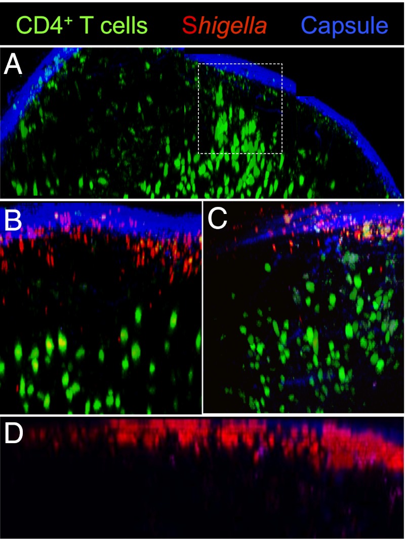
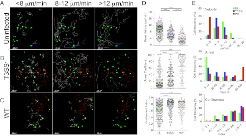
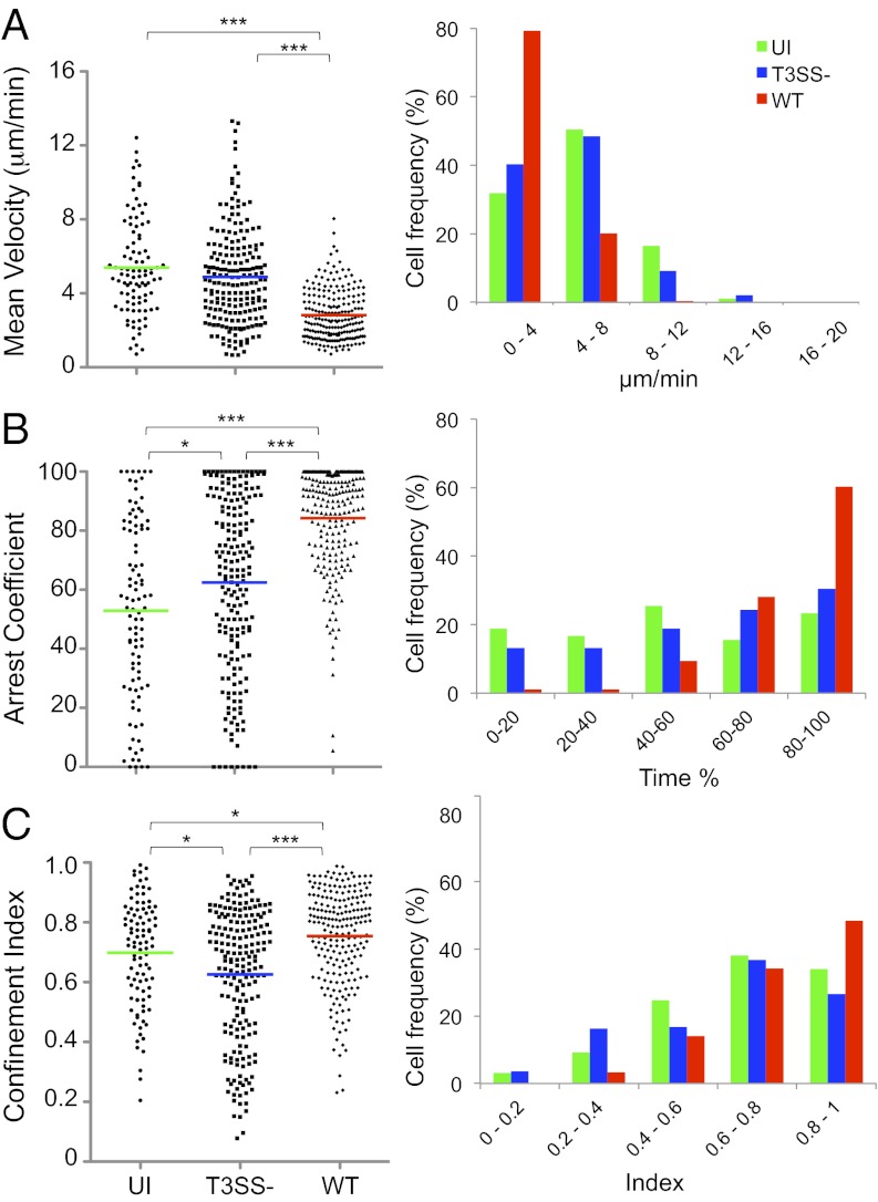
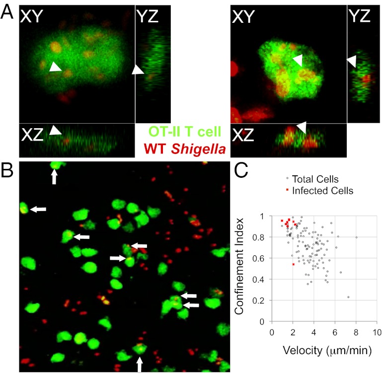
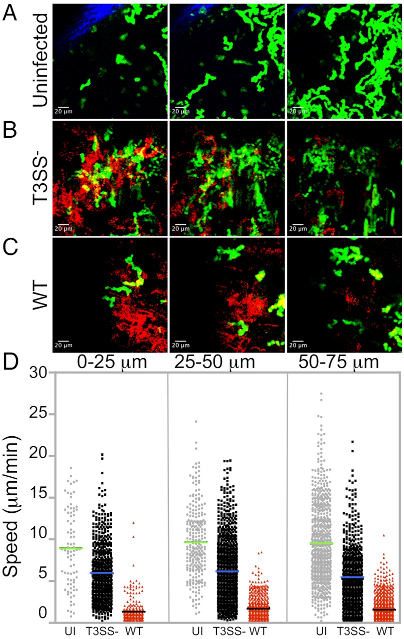
The Gram-negative enteroinvasive bacterium Shigella flexneri is responsible for the endemic form of bacillary dysentery, an acute rectocolitis in humans. S. flexneri uses a type III secretion system to inject effector proteins into host cells, thus diverting cellular functions to its own benefit. Protective immunity to reinfection requires several rounds of infection to be elicited and is short-lasting, suggesting that S. flexneri interferes with the priming of specific immunity. Considering the key role played by T-lymphocyte trafficking in priming of adaptive immunity, we investigated the impact of S. flexneri on T-cell dynamics in vivo. By using two-photon microscopy to visualize bacterium-T-cell cross-talks in the lymph nodes, where the adaptive immunity is initiated, we provide evidence that S. flexneri, via its type III secretion system, impairs the migration pattern of CD4(+) T cells independently of cognate recognition of bacterial antigens. We show that bacterial invasion of CD4(+) T lymphocytes occurs in vivo, and results in cell migration arrest. In the absence of invasion, CD4(+) T-cell migration parameters are also dramatically altered. Signals resulting from S. flexneri interactions with subcapsular sinus macrophages and dendritic cells, and recruitment of polymorphonuclear cells are likely to contribute to this phenomenon. These findings indicate that S. flexneri targets T lymphocytes in vivo and highlight the role of type III effector secretion in modulating host adaptive immune responses.


Conflict of interest statement

The authors declare no conflict of interest.

See QnAs on page 4437.

Figures

Fig. 1.
Fig. 1.
Visualizing S. flexneri and polyclonal CD4+ T-cell distribution in LNs. Two-photon microscopy reconstruction of the surface of uninfected and infected LNs, showing the capsule (blue, second harmonics), CFSE-labeled CD4+ T cells (green), and DsRed-expressing S. flexneri. (A) T-cell access to the SCS from the interfollicular regions in uninfected LNs (dashed square). T-cell proximity to the LN periphery in WT (B) or T3SS S. flexneri-infected LNs (C) 4 h post inoculation. (D) S. flexneri accumulates in the LN SCS 4 h post s.c. inoculation.
Fig. 2.
Fig. 2.
Polyclonal CD4+ T-cell migration patterns in the surface of the LN. (AE) Two-photon microscopy analysis of CD4+ T-cell dynamics in LNs infected or not with T3SS or WT S. flexneri. (AC) T-cell track projections depicted as white lines in uninfected (A), T3SS infected (B), or WT infected LNs (C). Tracks corresponding to T cells migrating at <8 μm/min (Left), 8 to 12 μm/min (Center), or >12 μm/min (Right) are overlaid on a one-time frame projection. (D) Three-dimensional quantification of mean velocity, confinement index, and arrest coefficient of T cells from individual tracks. Each symbol represents an individual cell. Colored horizontal lines indicate the mean value (***P < 0.0001, nonparametric one-way ANOVA, Kruskal–Wallis test; no P value indicates no statistical significance). Data are pooled from at least three independent experiments. (E) Distribution of T cells mean velocity, confinement index, and arrest coefficient under the various conditions tested.
Fig. 3.
Fig. 3.
OT-II T cells exhibit similar migration patterns as polyclonal CD4+ T cells. Two-photon microscopy analysis of OT-II T-cell dynamics in the surface of intact LNs infected or not with T3SS or WT S. flexneri. (A) Mean velocity, (B) arrest coefficient, and (C) confinement index of OT-II T cells calculated from individual tracks. Each symbol represents an individual cell. Colored horizontal lines indicate the mean value (*P < 0.02 and ***P < 0.0001, nonparametric one-way ANOVA, Kruskal–Wallis test; no P value indicates no statistical significance). Data are pooled from at least three independent experiments.
Fig. 4.
Fig. 4.
WT S. flexneri invades OT-II cells in vivo and infected cells are arrested. (A) Orthogonal views of OT-II cells infected with WT S. flexneri. (B) Two-photon microscopy image representative of LNs infected with S. flexneri. Arrows point to S. flexneri-invaded OT-II cells. (C) Confinement index and mean velocity of OT-II cells shown in B, underscoring the arrest of OT-II T cells invaded by S. flexneri.
Fig. 5.
Fig. 5.
The LN environment induced in the SCS area during infection dictates CD4+ T-cell speed and migration patterns. T-cell instantaneous speed determined at increasing depths from the capsule under infected or uninfected conditions. (AC) Maximum intensity projections in space and time of 25-μm sections, depicting the T-cell trajectories (green) at the infection foci (red). (D) Instantaneous speed of T cells in the three sections analyzed. Data are representative of at least three independent experiments.

Comment in

  • QnAs with Philippe J. Sansonetti.
    Gupta S. Gupta S. Proc Natl Acad Sci U S A. 2013 Mar 19;110(12):4437. doi: 10.1073/pnas.1302817110. Epub 2013 Mar 4. Proc Natl Acad Sci U S A. 2013. PMID: 23487802 Free PMC article. No abstract available.

References

    1. Mathan MM, Mathan VI. Morphology of rectal mucosa of patients with shigellosis. Rev Infect Dis. 1991;13(suppl 4):S314–S318. - PubMed
    1. Perdomo OJ, et al. Acute inflammation causes epithelial invasion and mucosal destruction in experimental shigellosis. J Exp Med. 1994;180(4):1307–1319. - PMC - PubMed
    1. Ashida H, Ogawa M, Mimuro H, Sasakawa C. Shigella infection of intestinal epithelium and circumvention of the host innate defense system. Curr Top Microbiol Immunol. 2009;337:231–255. - PubMed
    1. Parsot C, Sansonetti PJ. Invasion and the pathogenesis of Shigella infections. Curr Top Microbiol Immunol. 1996;209:25–42. - PubMed
    1. Phalipon A, Sansonetti PJ. Shigella’s ways of manipulating the host intestinal innate and adaptive immune system: A tool box for survival? Immunol Cell Biol. 2007;85(2):119–129. - PubMed

Publication types

Substances

LinkOut - more resources