. 2013 Sep;8(5):457-63.
doi: 10.1007/s12263-013-0333-y. Epub 2013 Feb 17.

Timed high-fat diet in the evening affects the hepatic circadian clock and PPARα-mediated lipogenic gene expressions in mice

Affiliations

Timed high-fat diet in the evening affects the hepatic circadian clock and PPARα-mediated lipogenic gene expressions in mice

Xiaoyan Wang et al. Genes Nutr. 2013 Sep.

Abstract

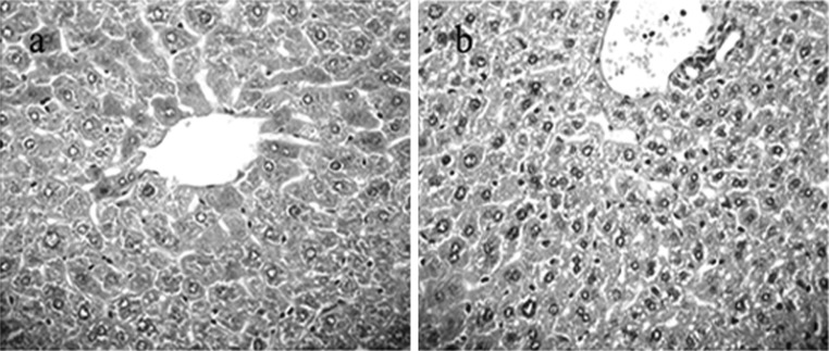
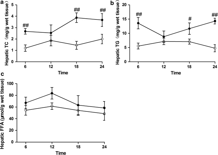
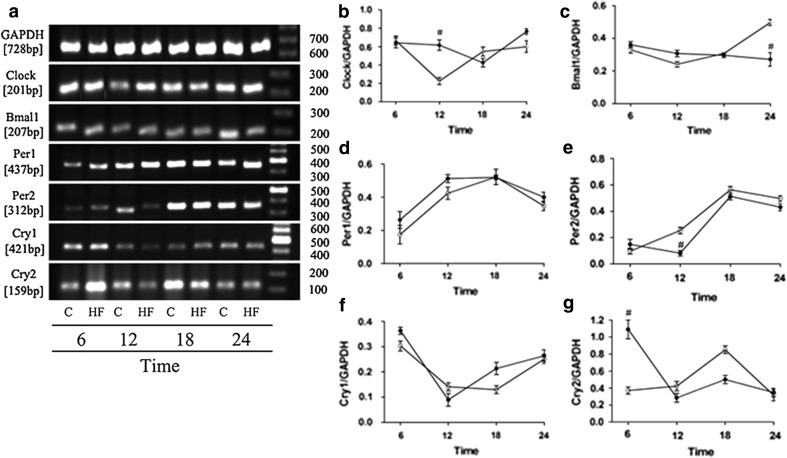
A long-term high-fat diet may result in a fatty liver. However, whether or not high-fat diets affect the hepatic circadian clock is controversial. The objective of this study is to investigate the effects of timed high-fat diet on the hepatic circadian clock and clock-controlled peroxisome proliferator-activated receptor (PPAR) α-mediated lipogenic gene expressions. Mice were orally administered high-fat milk in the evening for 4 weeks. The results showed that some hepatic clock genes, such as Clock, brain-muscle-Arnt-like 1 (Bmal1), Period 2 (Per2), and Cryptochrome 2 (Cry2) exhibited obvious changes in rhythms and/or amplitudes. Alterations in the expression of clock genes, in turn, further altered the circadian rhythm of PPARα expression. Among the PPARα target genes, cholesterol 7α-hydroxylase (CYP7A1), 3-hydroxy-3-methylglutaryl-coenzyme A reductase, low-density lipoprotein receptor, lipoprotein lipase, and diacylglycerol acyltransferase (DGAT) showed marked changes in rhythms and/or amplitudes. In particular, significant changes in the expressions of DGAT and CYP7A1 were observed. The effects of a high-fat diet on the expression of lipogenic genes in the liver were accompanied by increased hepatic cholesterol and triglyceride levels. These results suggest that timed high-fat diets at night could change the hepatic circadian expressions of clock genes Clock, Bmal1, Per2, and Cry2 and subsequently alter the circadian expression of PPARα-mediated lipogenic genes, resulting in hepatic lipid accumulation.


Figures

Fig. 1
Fig. 1
Histopathological changes in mouse hepatic tissues (HE staining, ×40). No steatosis was observed in matched control mice (a) and 1/3 to 2/3 of hepatocytes filled with empty lipid vacuoles were seen in high-fat milk-fed mice (b)
Fig. 2
Fig. 2
Hepatic TC, TG, and FFA contents in mice fed with high-fat milk in the evening for 4 weeks. White circles represent the matched control group, and black circles represent the high-fat milk-fed group. Values indicate the mean ± SD of n = 6 mice per group for each time point. #P < 0.05, ##P < 0.01 versus the matched control group at the same time point
Fig. 3
Fig. 3
Hepatic Clock, Bmal1, Per1, Per2, Cry1, and Cry2 mRNA expressions in mice fed with high-fat milk in the evening for 4 weeks. White circles represent the matched control group, and black circles represent the high-fat milk-fed group. C: the matched control group; HF: the high-fat milk group. Values indicate the mean ± SD of n = 6 mice per group for each time point. Gene bands were quantified relative to GAPDH. #P < 0.05 versus the matched control group at the same time point
Fig. 4
Fig. 4
Hepatic PPARα and lipogenic genes CYP7A1, HMGCR, LDLR, LPL, DGAT, and FAS mRNA expressions in mice fed with high-fat milk in the evening for 4 weeks. White circles represent the matched control group, and black circles represent the high-fat milk-fed group. C: the matched control group; HF: the high-fat milk group. Values indicate the mean ± SD of n = 6 mice per group for each time point. Gene bands were quantified relative to GAPDH. #P < 0.05, ##P < 0.01 versus the matched control group at the same time point

References

    1. Ando H, Yanagihara H, Hayashi Y, et al. Rhythmic messenger ribonucleic acid expression of clock genes and adipocytokines in mouse visceral adipose tissue. Endocrinology. 2005;146:5631–5636. doi: 10.1210/en.2005-0771. - DOI - PubMed
    1. Ando H, Takamura T, Matsuzawa-Nagata N, et al. The hepatic circadian clock is preserved in a lipid-induced mouse model of non-alcoholic steatohepatitis. Biochem Biophys Res Commun. 2009;380:684–688. doi: 10.1016/j.bbrc.2009.01.150. - DOI - PubMed
    1. Braissant O, Foufelle F, Scotto C, Dauca M, Wahli W. Differential expression of peroxisome proliferator-activated receptors (PPARs): tissue distribution of PPAR-alpha, -beta, and -gamma in the adult rat. Endocrinology. 1996;137:354–366. doi: 10.1210/en.137.1.354. - DOI - PubMed
    1. Desvergne B, Wahli W. Peroxisome proliferator activated receptors: nuclear control of metabolism. Endocr Rev. 1999;20:649–688. doi: 10.1210/er.20.5.649. - DOI - PubMed
    1. Farese RV, Cases S, Smith SJ. Triglyceride synthesis: insights from the cloning of diacylglycerol acyltransferase. Curr Opin Lipidol. 2000;11:229–234. doi: 10.1097/00041433-200006000-00002. - DOI - PubMed

LinkOut - more resources