. 2013 Apr 15;304(8):H1077-84.
doi: 10.1152/ajpheart.00956.2012. Epub 2013 Feb 15.

Microvascular permeability to water is independent of shear stress, but dependent on flow direction

Affiliations

Microvascular permeability to water is independent of shear stress, but dependent on flow direction

R H Adamson et al. Am J Physiol Heart Circ Physiol. .

Abstract

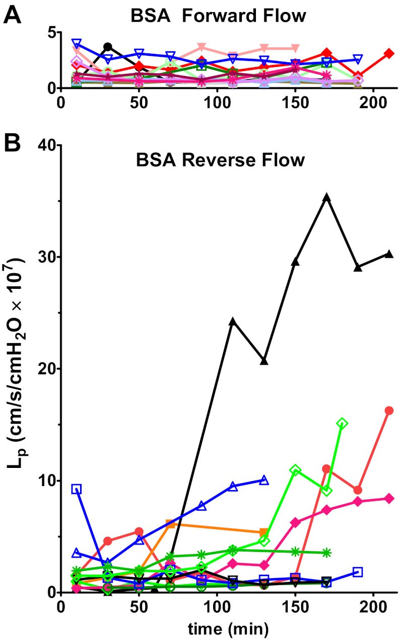
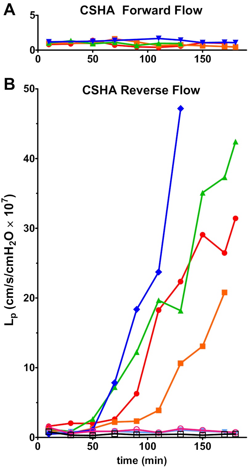
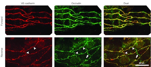
Endothelial cells in a cultured monolayer change from a "cobblestone" configuration when grown under static conditions to a more elongated shape, aligned with the direction of flow, after exposure to sustained uniform shear stress. Sustained blood flow acts to protect regions of large arteries from injury. We tested the hypothesis that the stable permeability state of individually perfused microvessels is also characteristic of flow conditioning. In individually perfused rat mesenteric venular microvessels, microvascular permeability, measured as hydraulic conductivity (Lp), was stable [mean 1.0 × 10(-7) cm/(s × cmH2O)] and independent of shear stress (3-14 dyn/cm(2)) for up to 3 h. Vessels perfused opposite to the direction of normal blood flow exhibited a delayed Lp increase [ΔLp was 7.6 × 10(-7) cm/(s × cmH2O)], but the increase was independent of wall shear stress. Addition of chondroitin sulfate and hyaluronic acid to perfusates increased the shear stress range, but did not modify the asymmetry in response to flow direction. Increased Lp in reverse-perfused vessels was associated with numerous discontinuities of VE-cadherin and occludin, while both proteins were continuous around the periphery of forward-perfused vessels. The results are not consistent with a general mechanism for graded shear-dependent permeability increase, but they are consistent with the idea that a stable Lp under normal flow contributes to prevention of edema formation and also enables physiological regulation of shear-dependent small solute permeabilities (e.g., glucose). The responses during reverse flow are consistent with reports that disturbed flows result in a less stable endothelial barrier in venular microvessels.


Figures

Fig. 1.
Fig. 1.
Hydraulic conductivity (Lp) plotted over the duration of the experiment for each vessel perfused with BSA/Ringer solution. A: vessels perfused in forward direction have a stable Lp for up to 3 h (n = 12). B: reverse-flow group was more variable; several vessels showed a gradual increase in Lp starting at about 90–120 min of perfusion (n = 11). Individual vessels are identified by a symbol and color.
Fig. 2.
Fig. 2.
Final ΔLp relative to baseline Lp plotted as a function of the average wall shear stress, τ. A: vessels perfused with BSA solutions. None of the vessels in the forward-flow (open symbols) group had a ΔLp higher than 4. The reverse-flow (closed symbols) group was more variable with five vessels having ΔLp below 4 and six vessels with ΔLp above 4. There was no clear relationship with wall shear stress. B: vessels perfused with CSHA solutions. None of the vessels in the forward-flow group (open symbols) had ΔLp higher than 4. The reverse-flow group (closed symbols) was variable with three vessels having ΔLp below 4 and four vessels with ΔLp above 4. There was no clear relationship with wall shear stress. CS, chondroitin sulfate; HA, hyaluronic acid. See results for description of CSHA solution.
Fig. 3.
Fig. 3.
Lp plotted over time for vessels perfused with CSHA solutions. A: vessels perfused in forward direction have a stable Lp for up to 3 h (n = 5). B: reverse-flow group was variable; several vessels showed a rapid increase in Lp starting at about 60–90 min of perfusion (n = 7). Individual vessels are identified by a symbol and color.
Fig. 4.
Fig. 4.
Representative segments of perfused microvessels labeled for VE-cadherin and occludin. The reverse-flow perfusions (bottom panels) showed numerous small gaps in both the occludin and VE-cadherin (arrowheads). Similar gaps were not seen in vessels perfused in the forward direction (top panels). A segment of tight junction occludin in the overlying mesothelium is visible (arrows) as a nonconnecting green shape. Scale bar, 20 μm for all images.

References

    1. Adamson RH, Lenz JF, Zhang X, Adamson GN, Weinbaum S, Curry FE. Oncotic pressures opposing filtration across non-fenestrated rat microvessels. J Physiol 557: 889–907, 2004 - PMC - PubMed
    1. Adamson RH, Sarai RK, Altangerel A, Thirkill TL, Clark JF, Curry FR. Sphingosine-1-phosphate modulation of basal permeability and acute inflammatory responses in rat venular microvessels. Cardiovasc Res 88: 344–351, 2010 - PMC - PubMed
    1. Adamson RH, Sarai RK, Clark JF, Altangerel A, Thirkill TL, Curry FE. Attenuation by sphingosine-1-phosphate of rat microvessel acute permeability response to bradykinin is rapidly reversible. Am J Physiol Heart Circ Physiol 302: H1929–H1935, 2012 - PMC - PubMed
    1. Adamson RH, Zeng M, Adamson GN, Lenz JF, Curry FE. PAF- and bradykinin-induced hyperpermeability of rat venules is independent of actin-myosin contraction. Am J Physiol Heart Circ Physiol 285: H406–H417, 2003 - PubMed
    1. Aird WC. Phenotypic heterogeneity of the endothelium. II. Representative vascular beds. Circ Res 100: 174–190, 2007 - PubMed

MeSH terms

LinkOut - more resources