. 2013 May 1;73(9):2782-94.
doi: 10.1158/0008-5472.CAN-12-3981. Epub 2013 Feb 15.

CSF1R signaling blockade stanches tumor-infiltrating myeloid cells and improves the efficacy of radiotherapy in prostate cancer

Affiliations

CSF1R signaling blockade stanches tumor-infiltrating myeloid cells and improves the efficacy of radiotherapy in prostate cancer

Jingying Xu et al. Cancer Res. .

Abstract

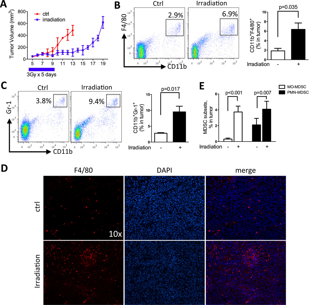
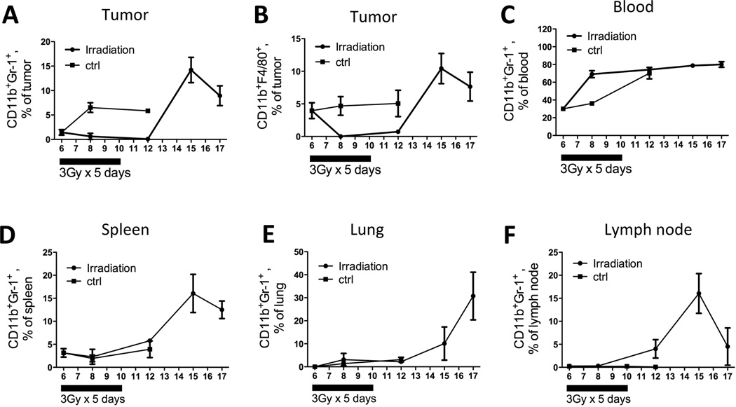
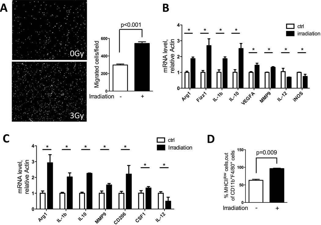
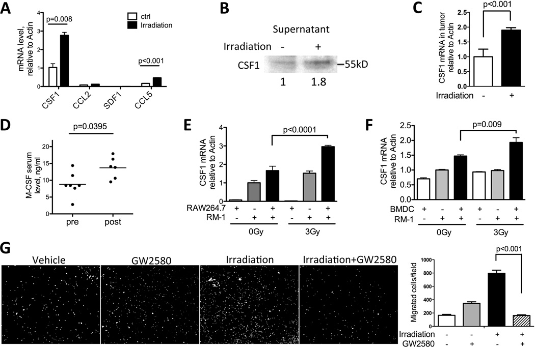
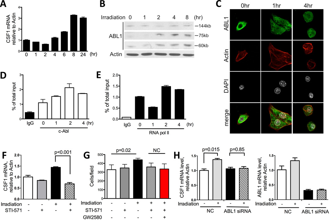
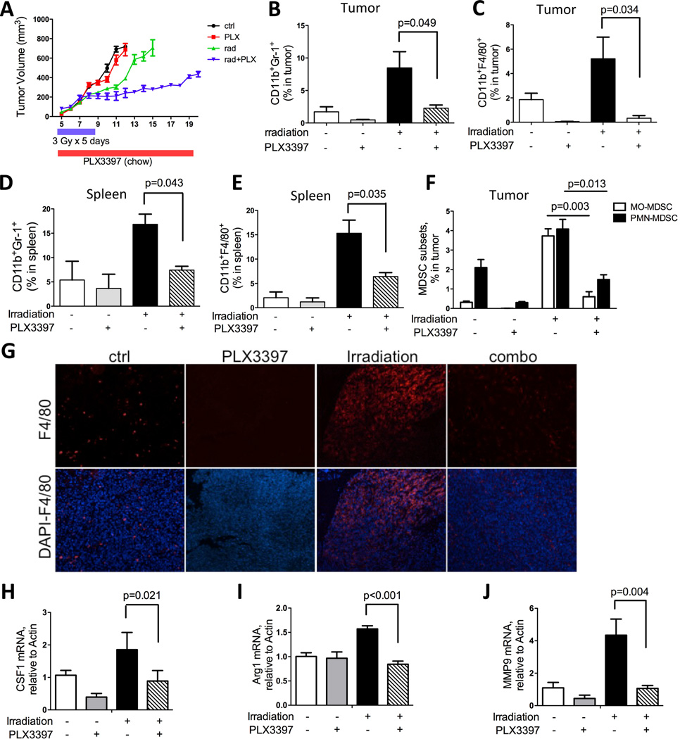
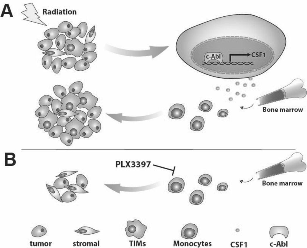
Radiotherapy is used to treat many types of cancer, but many treated patients relapse with local tumor recurrence. Tumor-infiltrating myeloid cells (TIM), including CD11b (ITGAM)(+)F4/80 (EMR1)+ tumor-associated macrophages (TAM), and CD11b(+)Gr-1 (LY6G)+ myeloid-derived suppressor cells (MDSC), respond to cancer-related stresses and play critical roles in promoting tumor angiogenesis, tissue remodeling, and immunosuppression. In this report, we used a prostate cancer model to investigate the effects of irradiation on TAMs and MDSCs in tumor-bearing animals. Unexpectedly, when primary tumor sites were irradiated, we observed a systemic increase of MDSCs in spleen, lung, lymph nodes, and peripheral blood. Cytokine analysis showed that the macrophage colony-stimulating factor CSF1 increased by two-fold in irradiated tumors. Enhanced macrophage migration induced by conditioned media from irradiated tumor cells was completely blocked by a selective inhibitor of CSF1R. These findings were confirmed in patients with prostate cancer, where serum levels of CSF1 increased after radiotherapy. Mechanistic investigations revealed the recruitment of the DNA damage-induced kinase ABL1 into cell nuclei where it bound the CSF1 gene promoter and enhanced CSF1 gene transcription. When added to radiotherapy, a selective inhibitor of CSF1R suppressed tumor growth more effectively than irradiation alone. Our results highlight the importance of CSF1/CSF1R signaling in the recruitment of TIMs that can limit the efficacy of radiotherapy. Furthermore, they suggest that CSF1 inhibitors should be evaluated in clinical trials in combination with radiotherapy as a strategy to improve outcomes.


Figures

Figure 1
Figure 1. Local irradiation enhances myeloid infiltration into tumors
Subcutaneous RM-1 tumors were collected, processed to single cell suspension and assayed by FACS and immunohistochemistry for CD11b+Gr-1+ MDSCs and CD11b+F4/80+ TAMs. A) Growth curve of RM-1 tumors with or without irradiation. B) FACS plots and quantification of TAMs in tumor. C) FACS plots and quantification for MDSCs in tumor. D) Representative F4/80 staining of RM-1 tumors from control and irradiation-treated mice. E) Effect of irradiation on the two subsets of MDSCs: MO-MDSC and PMN-MDSC. N=4 for each group.
Figure 2
Figure 2. Local irradiation enhances systemic myeloid cell expansion
Control mice were sacrifice on day 6, 8 and 12. Irradiated mice were sacrificed on day 6, 8, 12, 15 and 17. Tumors, blood, spleens, lungs and lymph nodes were collected for FACS analysis for MDSC and TAM population. N=4 for each time point.
Figure 3
Figure 3. Irradiation increases cell migration and induces protumorigenic genes in macrophages
A) RAW264.7 macrophages were seeded in 8-µm transwell inserts, and tumor conditioned media (collected 48hrs after 3Gy irradiation) was placed in the bottom. Cells were allowed to migrate toward bottom for 6 hrs. Then cells were fixed and stained with DAPI. Representative images of migrated cells are shown and they were quantified using ImageJ software (n=3). B) Effect of irradiation on bone marrow derived macrophages (BMDM). Bone marrows were collected and induced to macrophages by CSF1 (10ng/ml) for 6 days. Cells were counted, seeded and subjected to 3Gy irradiation. Cells were collected 24hrs later, and RT-PCR was performed to detect RNA for the protumorigenic and inflammatory genes noted. C) BMDMs as prepared above were cultured in 50% tumor conditioned media + 50% complete DMEM for 24hrs. Cells were collected and assayed by RT-PCR for the genetic markers noted. D) Tumors collected as shown in Fig 1A were analyzed by FACS for MHCII expression on CD11b+F480+ macrophages. An increase in MHCII low-expressing macrophage population was observed. (* indicates significant changes with P< 0.05)
Figure 4
Figure 4. CSF1 expression is increased by irradiation
A) MycCaP cells were plated overnight and irradiated with 3Gy. Cells were collected 24 hrs after, and assayed by RT-PCR for factors known to recruit myeloid cells: CSF1, CCL2, SDF1 and CCL5. CSF1 is shown to have the highest expression and most significant increase. B) Conditioned media from Myc-CaP cells 48hrs after irradiation were collected, concentrated by centrifugation, normalized and analyzed by SDS-PAGE. Secreted CSF1 was detected at 50kDa. Relative expression level quantified by ImageJ was shown below the blot. C) Tumors collected as shown in Fig 1A were assayed by RT-PCR for CSF1 mRNA expression. D) Serum samples from prostate cancer patients pre- and post-radiotherapy were analyzed by ELISA for CSF1. E) RM-1 cells and macrophage cell line RAW264.7 were co-cultured overnight and irradiated with 3Gy. Cells were collected 24hrs and analyzed by RT-PCR for CSF1 mRNA. F) RM-1 cells and BMDM were co-cultured for 4hrs and irradiated with 3Gy. Cells were collected 24hrs and analyzed by RT-PCR for CSF1 mRNA. G) RAW264.7 macrophage migration assay was performed by using conditioned media in lower chamber, collected as in panel B. GW2580 (1μM) was added to the top chamber. Quantification of 9 fields was summarized in panel F. Representative images of migrated cells were shown in G.
Figure 5
Figure 5. Irradiation induces CSF1 production through an ABL1-dependent mechanism
A) CSF1 mRNA expression in Myc-CaP cells 0, 1, 2, 4, 6, 8, and 24hrs after irradiation (3Gy). B) ABL1 is cleaved by irradiation in vitro. 4×10⁵ Myc-CaP cells were plated in 6-well plate overnight and irradiated with 3Gy the next day. Cells were collected at 1, 2, 4, and 8 hrs after irradiation. Cells lysates were normalized and assayed by SDS-PAGE. Western blot was probed with ABL1 (K-12) antibody for both full length and cleaved ABL1. Two cleavage products, 60kD and 75kD, were detected. C) Confocal images of Myc-CaP cells at 1 and 4hrs after irradiation. Green: ABL1; red: actin; white: nucleus (DAPI). D–E) Myc-CaP cells 0, 1, 2, 4 hrs after irradiation were fixed in 3% PFA and processed for ChIP assay using c-Abl antibody (D) and RNA polymerase II antibody (E) as described in Material and Methods section. Rabbit IgG was used as negative control. F) Myc-CaP cells were irradiated with 3Gy and STI-571 (5μM) was added right after. Cells were collected 24hrs later and analyzed by RT-PCR for CSF1 mRNA expression. G) Migration assay using conditioned media collected as in panel F. GW2580 was added to the top chamber to examine the additive effects between STI-571 and GW2580. H) CSF1-mRNA in human prostate cancer cells after ABL1 directed RNAi. CWR22Rv1 cells were transfected with ABL1 siRNA and negative control (NC: non-specific siRNA). Cells were irradiated 30hrs after siRNA treatment and collected 24hrs after irradiation. Expression level of ABL1 was reduced to 30% of control after specific siRNA treatment (right panel). CSF1 mRNA was analyzed by RT-PCR with and without irradiation.
Figure 6
Figure 6. CSF1/CSF1R blockade inhibits tumor growth after irradiation
A) Growth curve of subcutaneous RM-1 tumors treated with RT (3Gy×5 days), PLX3397 (in food chow) or combination as indicated. Tumors were measured daily by caliper. B–C) FACS analysis of CD11b+F4/80+ macrophages and CD11b+Gr-1+ MDSCs in tumor, collected at end points (tumor n=6 × for each cohort). D–E) FACS analysis of CD11b+F4/80+ macrophages and CD11b+Gr-1+ MDSCs in spleen, at the same termination time as above. F) FACS analysis for MDSC subsets, MO-MDSC and PMN-MDSC with single or combination treatment. G) Representative immunohistochemsitry staining for F4/80 on tumor sections with single or combination treatment. H–J) RT-PCR analysis of mRNA extracted from tumors for CSF1, MMP9 and Arg1.
Figure 7
Figure 7. Model of CSF1 expression induced by irradiation, promoting TIMs recruitment and tumor regrowth
A) Tumor irradiation activates ABL1, which translocates to the nucleus, binding to the promoter region of CSF1 and up-regulates its gene expression. Additional TIMs are recruited to tumor sites due to the increase in CSF1 and they can thus promote tumor growth. B) When tumor-bearing mice were treated with a small molecule CSF1R kinase inhibitor, CSF1/CSF1R signaling is inhibited resulting in decreased infiltration of TIMs, reducing their tumor growth promoting influences.

Comment in

References

    1. Delaney G, Jacob S, Featherstone C, Barton M. The role of radiotherapy in cancer treatment: estimating optimal utilization from a review of evidence-based clinical guidelines. Cancer. 2005;104(6):1129–1137. Epub 2005/08/05. PubMed PMID:16080176. - PubMed
    1. Agarwal PK, Sadetsky N, Konety BR, Resnick MI, Carroll PR. Treatment failure after primary and salvage therapy for prostate cancer: likelihood, patterns of care, and outcomes. Cancer. 2008;112(2):307–314. Epub 2007/12/01. PubMed PMID:18050294. - PubMed
    1. D'Amico AV, Renshaw AA, Sussman B, Chen MH. Pretreatment PSA velocity and risk of death from prostate cancer following external beam radiation therapy. Jama. 2005;294(4):440–447. Epub 2005/07/28. PubMed PMID:16046650. - PubMed
    1. Bianco FJ, Jr, Scardino PT, Stephenson AJ, Diblasio CJ, Fearn PA, Eastham JA. Long-term oncologic results of salvage radical prostatectomy for locally recurrent prostate cancer after radiotherapy. Int J Radiat Oncol Biol Phys. 2005;62(2):448–453. Epub 2005/05/14. PubMed PMID:15890586. - PubMed
    1. Kozin SV, Kamoun WS, Huang Y, Dawson MR, Jain RK, Duda DG. Recruitment of myeloid but not endothelial precursor cells facilitates tumor regrowth after local irradiation. Cancer Res. 2010;70(14):5679–5685. Epub 2010/07/16. PubMed PMID:20631066; PubMed Central PMCID: PMC2918387. - PMC - PubMed

Publication types

MeSH terms

Substances