Review
. 2012 Dec 15;125(Pt 24):5927-36.
doi: 10.1242/jcs.093724. Epub 2013 Feb 15.

The KMN protein network--chief conductors of the kinetochore orchestra

Affiliations
Review

The KMN protein network--chief conductors of the kinetochore orchestra

Dileep Varma et al. J Cell Sci. .

Abstract

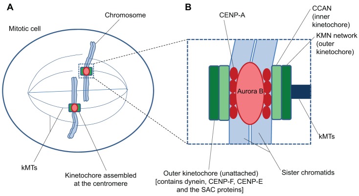
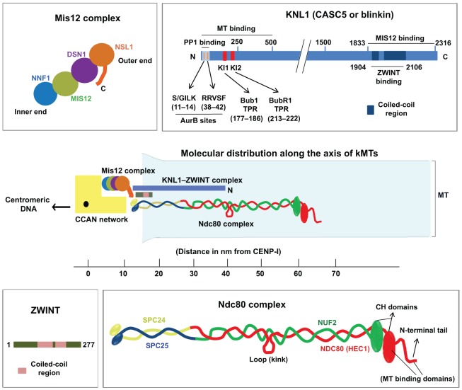
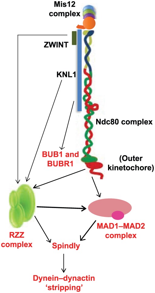
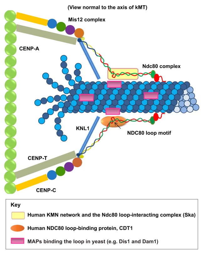
Successful completion of mitosis requires that sister kinetochores become attached end-on to the plus ends of spindle microtubules (MTs) in prometaphase, thereby forming kinetochore microtubules (kMTs) that tether one sister to one spindle pole and the other sister to the opposite pole. Sites for kMT attachment provide at least four key functions: robust and dynamic kMT anchorage; force generation that can be coupled to kMT plus-end dynamics; correction of errors in kMT attachment; and control of the spindle assembly checkpoint (SAC). The SAC typically delays anaphase until chromosomes achieve metaphase alignment with each sister kinetochore acquiring a full complement of kMTs. Although it has been known for over 30 years that MT motor proteins reside at kinetochores, a highly conserved network of protein complexes, called the KMN network, has emerged in recent years as the primary interface between the kinetochore and kMTs. This Commentary will summarize recent advances in our understanding of the role of the KMN network for the key kinetochore functions, with a focus on human cells.


Figures

Fig. 1.
Fig. 1.
Protein composition and domain structure of the vertebrate kinetochore in mitotic cells. (A) Kinetochores are assembled at the periphery of sister centromeres on mitotic chromosomes and attach to spindle microtubules. (B) The site of kinetochore assembly is specified by the presence of the modified histone H3 CENP-A. The CCAN protein network within the inner kinetochore domain links CENP-A-containing chromatin to the KMN network within the outer kinetochore domain. The KMN network is the major interface between kMTs and their associated proteins. The peripheral region of the outer domain contains long fibrous proteins, such as CENP-F, the microtubule motors CENP-E and dynein, as well as their associated proteins, particularly in the absence of kMT attachments at prometaphase. Protein components of the SAC are also found in the outer domain. Although primarily within the peripheral region, these proteins probably also extend into the inner parts of the outer domain because many of these are connected to the KMN network. See text for details.
Fig. 2.
Fig. 2.
Structure and composition of the KMN network. The molecular distribution of the components of the KMN network is shown in the centre of the figure and is projected along the axis of kMTs (shown in light blue). The CCAN (shown in yellow) forms the key connection between the KMN network at outer kinetochores and the centromeric chromatin. The human Mis12 complex (shown on the top left) consists of four subunits NNF1, MIS12, DSN1 and NSL1 that are apparently arranged linearly along the inside–outside axis of kinetochores in the sequence indicated (Petrovic et al., 2010). The C-terminal tail of the NSL1 subunit at the kinetochore-proximal outer end of the complex is important for interacting with the Ndc80 complex and KNL1, whereas the centromeric DNA-proximal inner end binds to the CCAN network. High-resolution two-color fluorescent imaging has shown that the Mis12 complex is located ∼50 nm inside of the outer end of the Ndc80 complex and ∼15 nm outside of CENP-I (part of the CCAN), and that MIS12 is probably oriented at an angle along the inner–outer kinetochore axis (Wan et al., 2009). Human KNL1 (top right) is a large multi-domain protein with the known functional domains and motifs indicated. KNL1 is elongated with its N-terminal region, which contains the proposed MT-binding site, located ∼40 nm away from CENP-I, and its C-terminal region, which is required for its kinetochore targeting (where it binds to MIS12 and its partner ZWINT), ∼15 nm away from CENP-I (Kiyomitsu et al., 2007; Petrovic et al., 2010; Wan et al., 2009). ZWINT (bottom left) is a relatively small coiled-coil rich kinetochore protein that binds the C-terminal coiled-coil domain of KNL1 and forms a tight complex with KNL1 (Petrovic et al., 2010). It has also been shown that ZWINT interacts with the Mis12 complex and the Ndc80 complex. The human Ndc80 complex (bottom right) is the major component of the core MT attachment module at kinetochores and consist of four subunits NDC80 (HEC1), NUF2, SPC24 and SPC25, which heterotetramerize through their coiled-coil regions as indicated. The N-terminal CH domain and charged unstructured tail regions of the NDC80 subunit form a bipartite MT-binding interface, whereas the globular C-terminal domains of SPC24 and SPC25 bind to CENP-T or the Mis12 complex to link the Ndc80 complex to the inner kinetochore. The CH domain of NDC80 has also been shown to be important for the retention of checkpoint proteins MAD1 and MAD2 at unattached kinetochores (Guimaraes et al., 2008; Martin-Lluesma et al., 2002; Miller et al., 2008). The conserved internal loop region of the Ndc80 complex is highly flexible, allowing the α-helical coiled-coil rod that ends in the MT-binding CH or head domains to rotate or twist at angles between 0 and 120°, with the loop region serving as a hinge. The N-terminal CH domain of NDC80 is ∼100 nm outside of CENP-A, which is bound to the centromere (not depicted), and about 65 nm outside of CENP-I within MT-bound metaphase kinetochores (Wan et al., 2009). It has also been observed that the distance between the MT-proximal NDC80 CH domain and the head domain of the centromere-proximal SPC24–SPC25 complex is only about 45 nm in human kinetochores (as compared to the 57-nm-long extended confirmation of the Ndc80 complex reported in yeast) suggesting that the MT-bound confirmation of the Ndc80 complex in human cells is slightly bent during metaphase (Wan et al., 2009).
Fig. 3.
Fig. 3.
Role of the KMN network in the SAC. The schematic illustration shows the known function of KMN network components in the recruitment of SAC proteins (red) and their recruitment of each other (indicated by arrows). ZWINT (KBP-5) and KNL-1 in C. elegans are required for the recruitment of the RZZ checkpoint complex. Both the Ndc80 complex and the RZZ complex have been shown to be required for the proper localization of the MAD1–MAD2 checkpoint complex to kinetochores that are unattached in prometaphase. KNL1 is important for the kinetochore recruitment of ZWINT and the BUB1–BUBR1 checkpoint complex. Spindly (recruited by the RZZ complex) serves as an adaptor between the dynein–dynactin motor complex and the MAD1–MAD2 and RZZ complexes, and is required for the dynein motor-driven stripping of these checkpoint complexes during SAC silencing. See text for details.
None
None

References

    1. Alushin G., Nogales E. (2011). Visualizing kinetochore architecture. Curr. Opin. Struct. Biol. 21, 661–669 10.1016/j.sbi.2011.07.009 - DOI - PMC - PubMed
    1. Alushin G. M., Ramey V. H., Pasqualato S., Ball D. A., Grigorieff N., Musacchio A., Nogales E. (2010). The Ndc80 kinetochore complex forms oligomeric arrays along microtubules. Nature 467, 805–810 10.1038/nature09423 - DOI - PMC - PubMed
    1. Alushin G. M., Musinipally V., Matson D., Tooley J., Stukenberg P. T., Nogales E. (2012). Multimodal microtubule binding by the Ndc80 kinetochore complex. Nat. Struct. Mol. Biol. 19, 1161–1167 10.1038/nsmb.2411 - DOI - PMC - PubMed
    1. Bock L. J., Pagliuca C., Kobayashi N., Grove R. A., Oku Y., Shrestha K., Alfieri C., Golfieri C., Oldani A., Dal Maschio M.et al. (2012). Cnn1 inhibits the interactions between the KMN complexes of the yeast kinetochore. Nat. Cell Biol. 14, 614–624 10.1038/ncb2495 - DOI - PMC - PubMed
    1. Bolanos–Garcia V. M., Lischetti T., Matak–Vinković D., Cota E., Simpson P. J., Chirgadze D. Y., Spring D. R., Robinson C. V., Nilsson J., Blundell T. L. (2011). Structure of a Blinkin-BUBR1 complex reveals an interaction crucial for kinetochore-mitotic checkpoint regulation via an unanticipated binding Site. Structure 19, 1691–1700 10.1016/j.str.2011.09.017 - DOI - PMC - PubMed

Publication types

MeSH terms