. 2013;8(2):e55890.
doi: 10.1371/journal.pone.0055890. Epub 2013 Feb 13.

Cells derived from the coelomic epithelium contribute to multiple gastrointestinal tissues in mouse embryos

Affiliations

Cells derived from the coelomic epithelium contribute to multiple gastrointestinal tissues in mouse embryos

Rita Carmona et al. PLoS One. 2013.

Abstract

Gut mesodermal tissues originate from the splanchnopleural mesenchyme. However, the embryonic gastrointestinal coelomic epithelium gives rise to mesenchymal cells, whose significance and fate are little known. Our aim was to investigate the contribution of coelomic epithelium-derived cells to the intestinal development. We have used the transgenic mouse model mWt1/IRES/GFP-Cre (Wt1(cre)) crossed with the Rosa26R-EYFP reporter mouse. In the gastrointestinal duct Wt1, the Wilms' tumor suppressor gene, is specific and dynamically expressed in the coelomic epithelium. In the embryos obtained from the crossbreeding, the Wt1-expressing cell lineage produces the yellow fluorescent protein (YFP) allowing for colocalization with differentiation markers through confocal microscopy and flow cytometry. Wt1(cre-YFP) cells were very abundant throughout the intestine during midgestation, declining in neonates. Wt1(cre-YFP) cells were also transiently observed within the mucosa, being apparently released into the intestinal lumen. YFP was detected in cells contributing to intestinal vascularization (endothelium, pericytes and smooth muscle), visceral musculature (circular, longitudinal and submucosal) as well as in Cajal and Cajal-like interstitial cells. Wt1(cre-YFP) mesenchymal cells expressed FGF9, a critical growth factor for intestinal development, as well as PDGFRα, mainly within developing villi. Thus, a cell population derived from the coelomic epithelium incorporates to the gut mesenchyme and contribute to a variety of intestinal tissues, probably playing also a signaling role. Our results support the origin of interstitial cells of Cajal and visceral circular muscle from a common progenitor expressing anoctamin-1 and SMCα-actin. Coelomic-derived cells contribute to the differentiation of at least a part of the interstitial cells of Cajal.


Conflict of interest statement

Competing Interests: The authors have declared that no competing interests exist.

Figures

Figure 1
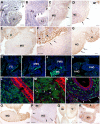
Figure 1. Expression of Wt1 and cytokeratins in early and middle intestinal development and early localization of Wt1cre-YFP cells.
A–D. Wt1 immunolocalization in consecutive sections from the same E10 embryo. Wt1 is expressed in a few cells of the coelomic epithelium of the foregut (FG) and in some mesenchymal cells (arrows in A and B). As shown in C and D, the expression becomes more abundant in the midgut (MG) and is weak and restricted to the coelomic epithelium in the hindgut (arrows in D). The Wt1-positive liver bud (LI), parietal coelomic epithelium (P) and intermediate mesoderm (IM) are positive controls. E–G. Wt1 immunolocalization in an E10.5 embryo. Wt1 immunoreactivity is present throughout the coelomic epithelium of the midgut (MG). F shows a higher magnification of the dorsal area of the midgut. Wt1 expressing epithelium is shown by the arrows. Expression in the hindgut has increased as compared with the earlier stage shown in D (arrows in G). Developing kidneys (K) are positive controls. H–K. Wt1cre-YFP cells in consecutive sections of the same E10.5 embryo. Wt1cre-YFP cells are abundant in the foregut (FG) and anterior midgut (AMG), becoming scarcer in the posterior midgut (PMG). Abundant Wt1cre-YFP cells are present in the mesentery (MS) at this level. Ony a few cells are present in the anterior hindgut (AHG) while the posterior hindgut (PHG) lacks of Wt1cre-YFP cells. L,M. Wt1cre-YFP cells in the midgut mesenchyme of an E10.5 embryo. YFP positive and negative mesenchymal cells show a dot-like cytokeratin immunostaining by E10.5 (arrows and arrowheads, respectively in M). The endoderm is strongly cytokeratin immunoreactive. N. Cytokeratin immunostaining has disappeared from the mesenchymal cells by the stage E13.5, and it is restricted to the coelomic epithelium of the intestine (arrows). O–P. Wt1 immunolocalization in an E12.5 embryo. The midgut (MG) intestinal loops contained in the physiological umbilical hernia show a strong Wt1 immunoreactivity (H). However, the hindgut mesothelium is Wt1-negative by this stage (arrows in P). Note the strong Wt1 immunoreactivity of the posterior intermediate mesoderm (IM). Q–R. Wt1 immunolocalization in an E14.5 embryo. All the digestive tract mesothelium is Wt1-immunoreactive, including the small intestine (SI) and the hindgut (HG). K: kidney. Scale bars: A–G, O–R = 50 µm; H–K = 100 µm; L–N = 25 µm.
Figure 2
Figure 2. Vascular contribution of the Wt1-expressing cell lineage.
A–C. Colocalization of the endothelial markers Pecam-1 (A, B) and CD34 (C) with YFP in intestinal vessels at the stage E11.5. D. Colocalization of the pericyte marker NG2 with YFP at the stage E13.5. E. Immunolocalization of the fibroblastic marker 5B5 at the stage E18.5. This marker does not colocalize with YFP. F. Immunolocalization of the fibroblastic marker FSP1 in a neonate. Colocalization with YFP is not observed. G. Analysis of dissociated intestines from an E11.5 embryo by flow cytometry. In this representative experiment, colocalization of PECAM-1 with YFP (arrow) was found in a 0.3% of the total cells, and in 2.9% of the Wt1cre-YFP cells. Scale bars A, E, F = 50 µm; B,C,D = 25 µm.
Figure 3
Figure 3. Contribution of the Wt1cre-YFP cells to the visceral smooth muscle and Cajal-like interstitial cells.
A. Colocalization of SMCα-actin with YFP in cells ventral to the endoderm at the stage E11.5 (arrows). SMCα-actin+/Wt1cre-YFP cells can be seen in the wall of a large vessel (arrowhead). B,C. Colocalization of SMCα-actin with YFP in visceral circular muscle layer (CL) at E15.5, in transverse and longitudinal section, respectively. A submesothelial cell, positive for both markers, is shown in C by the arrowhead. Note the YFP-positive vessel wall (arrow in C). D, E. The intestinal coelomic epithelium is RALDH2 immunoreactive by E13.5 (D) and E15.5 (E). Note the presence of SMCα-actin immunoreactive cells, probably progenitors of the longitudinal muscle layer, lying behind the RALDH2+ mesothelium in E. F. In this neonate, colocalization of YFP with SMCα-actin is observed in the circular muscular layer (arrows). CD34+ cells are abundant in the submucosal layer, showing thin prolongations. Some of these cells are also Wt1cre-YFP (arrowheads). Note the presence of submucosal SMCα-actin cells forming the innermost muscle layer. CD34 does not colocalize with SMCα-actin. Scale bars = 25 µm.
Figure 4
Figure 4. Contribution of Wt1cre-YFP cells to the differentiation of the interstitial cells of Cajal.
A. ANO1 immunoreactivity is present within the circular muscle layer and in submesothelial cells from neonates. Colocalization with YFP is observed (arrows). B. Immunolocalization of SMCα-actin and ANO1 in an E18.5 embryo. Colocalization is only detected in the circular muscle layer (CL). However both, the longitudinal (LL) and the submucosal (SML) layers are SMCα-actin positive and ANO1 negative. ANO1 positive, SMCα-actin negative cells (arrows) are present between circular and longitudinal layers, probably representing ICC. C. Immunolocalization of SMCα-actin with ANO1 in a neonate, longitudinal section. Colocalization of ANO1 with YFP is shown (arrows). These cells are negative for SMCα-actin. D,E. Immunolocalization of c-Kit in two neonates. By this stage, c-Kit immunoreactivity labels ICC. Some of these cells are also Wt1cre-YFP (arrows). A c-Kit+/Wt1cre-YFP putative ICC connected to a c-Kit+/YFP- cell is shown in the insert. F. Flow cytometry analysis of cells obtained from the dissociation of an intestine from an E15.5 embryo. Colocalization of c-Kit with YFP (arrow) was found only in 0.03% of all the cells, but the percentage of the Wt1cre-YFP cells that were also c-Kit+ was 3.8% in this representative experiment. Scale bars 25 µm.
Figure 5
Figure 5. RT-PCR analysis of c-Kit expression in the intestinal Wt1cre-YFP cell fraction.
This fraction was purified by cell sorting of the dissociated intestine of three E15.5 embryos. Positive control: cDNA from a whole E15.5 embryo. Negative control: water instead of cDNA.
Figure 6
Figure 6. Presence of Wt1cre-YFP cells in the endoderm.
A. Fibronectin immunoreactivity is high in the prospective submucosal area. Note the presence of Wt1cre-YFP cells inside the endodermal mucosa, as well as into the intestinal lumen. Many luminal cells show pycnotic nuclei suggesting cell death. B-D. Sections obtained from an E13.5 embryo. Wt1cre-YFP cells seem to be migrating into the endoderm through discontinuities of the basal lamina revealed by lack of laminin immunoreactivity (arrows in B and D). However, at more posterior levels of the same embryo (C) the basal lamina of the endoderm (arrows) is continuous and no Wt1cre-YFP cells are present in the mucosa. E,F. Laminin immunoreactivity shows a continuous endodermal basal lamina in areas lacking of Wt1cre-YFP cells within the mucosa (F, neonate). However, where Wt1cre-YFP cells are still present in the endoderm, the lamina basal seems to be still discontinuous (arrows in E, E18.5 embryo). Scale bars = 25 µm.
Figure 7
Figure 7. Localization of FGF9 and PDGFRα in the developing intestine.
A,B. FGF9 immunoreactivity in the intestine of E15.5 embryos. Colocalization with YFP is observed in cells of the circular muscle layer as well as in Wt1cre-YFP cells within the endodermal mucosa (white arrows). Note the large FGF9+ cells, probably neural, between the circular and the longitudinal muscle layers (arrowheads). The coelomic epithelium is also FGF9+ (yellow arrow in B. C,D. Immunolocalization of PDGFRα in the small intestine of an E18.5 embryo. Positive mesenchymal cells are located within the developing villi (arrows in C). Colocalization of PDGFRα with YFP is shown at higher magnification in D (arrows). Scale bars A–C = 25 µm; D = 10 µm.

References

    1. Rolle U, Piaseczna-Piotrowska A, Puri P (2007) Interstitial cells of Cajal in the normal gut and in intestinal motility disorders of childhood. Pediatr Surg Int 23: 1139–1152. - PubMed
    1. Lavine KJ, Yu K, White AC, Zhang X, Smith C, et al. (2005) Endocardial and epicardial derived FGF signals regulate myocardial proliferation and differentiation in vivo. Dev Cell 8: 85–95. - PubMed
    1. Merki E, Zamora M, Raya A, Kawakami Y, Wang J, et al. (2005) Epicardial retinoid X receptor alpha is required for myocardial growth and coronary artery formation. Proc Natl Acad Sci USA 102: 18455–18460. - PMC - PubMed
    1. Martinez-Estrada OM, Lettice LA, Essafi A, Guadix JA, Slight J, et al. (2010) Wt1 is required for cardiovascular progenitor cell formation through transcriptional control of Snail and E-cadherin. Nat Genet 42: 89–93. - PMC - PubMed
    1. Que J, Wilm B, Hasegawa H, Wang F, Bader D, et al. (2008) Mesothelium contributes to vascular smooth muscle and mesenchyme during lung development. Proc Natl Acad Sci USA 105: 16626–166230. - PMC - PubMed

Publication types

MeSH terms