. 2013;8(2):e56738.
doi: 10.1371/journal.pone.0056738. Epub 2013 Feb 13.

The nonconventional MHC class II molecule DM governs diabetes susceptibility in NOD mice

Affiliations

The nonconventional MHC class II molecule DM governs diabetes susceptibility in NOD mice

Marc A J Morgan et al. PLoS One. 2013.

Abstract

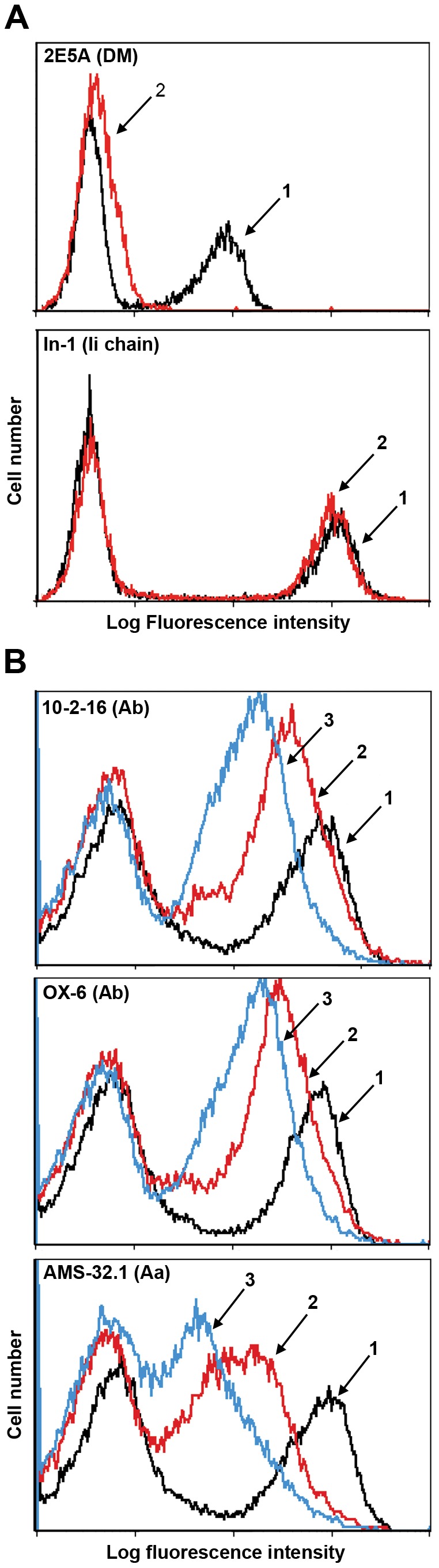
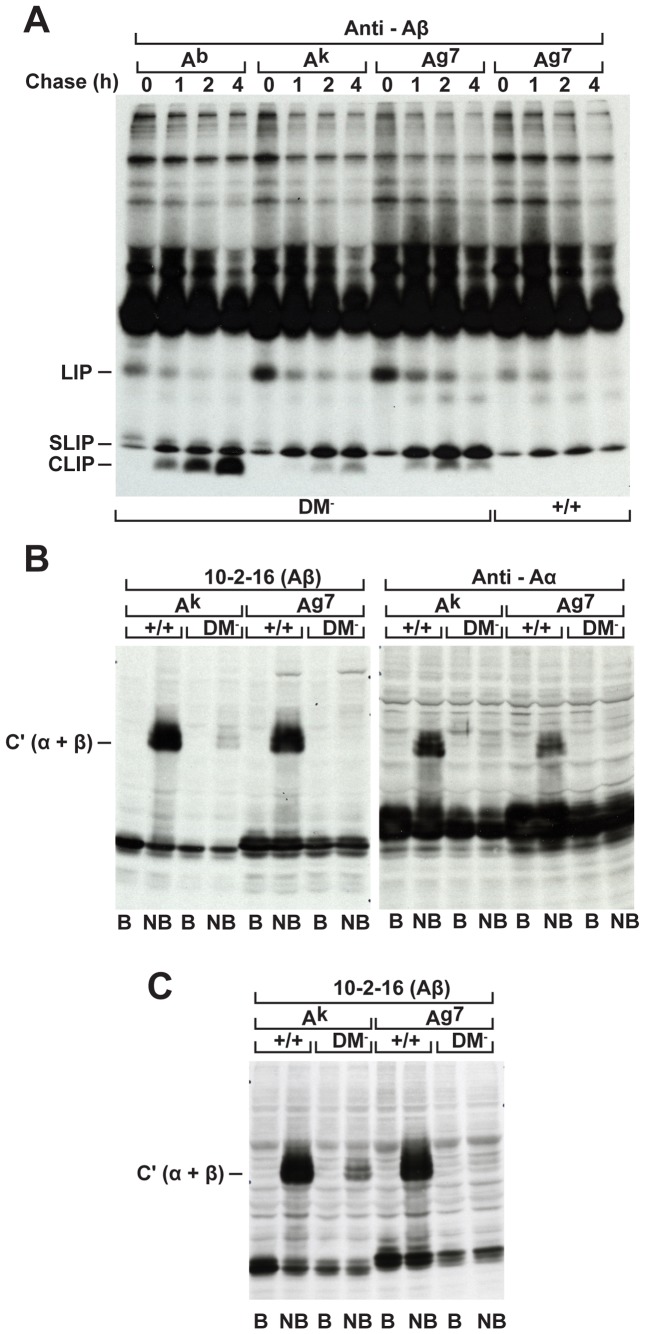
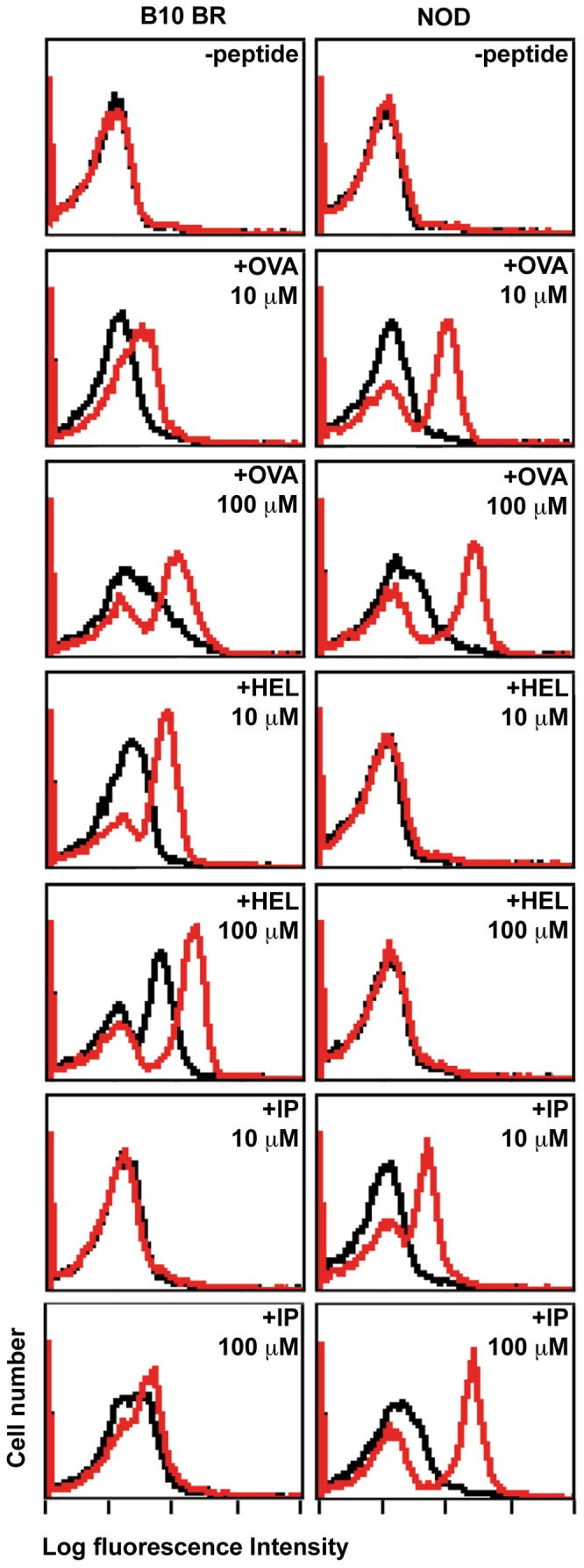
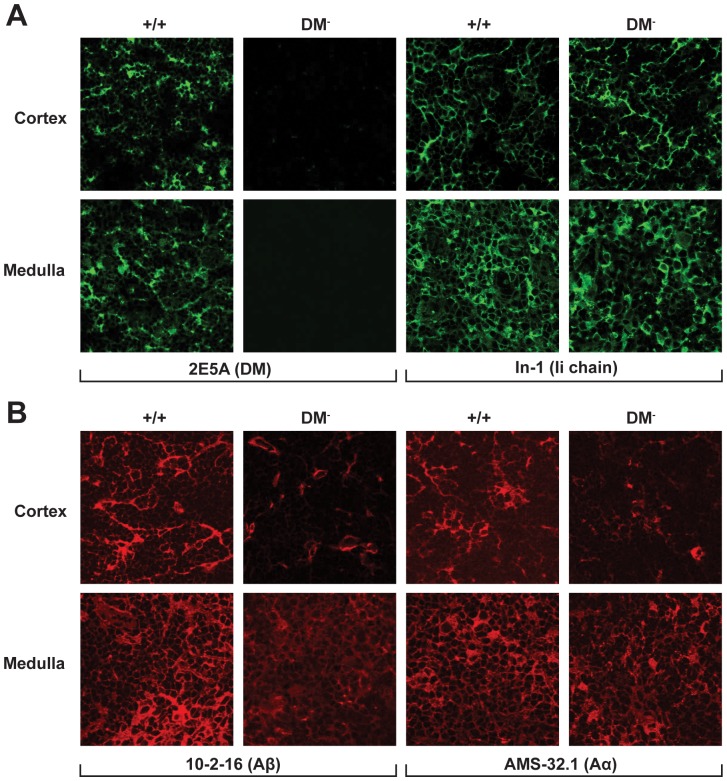
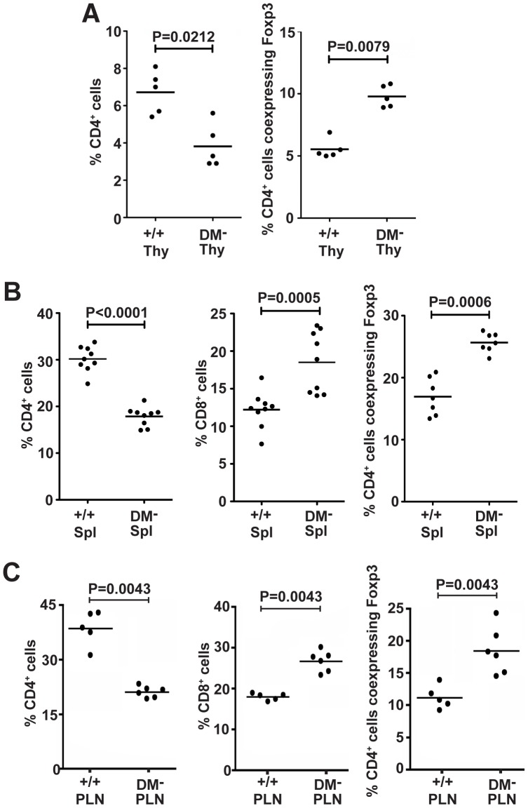
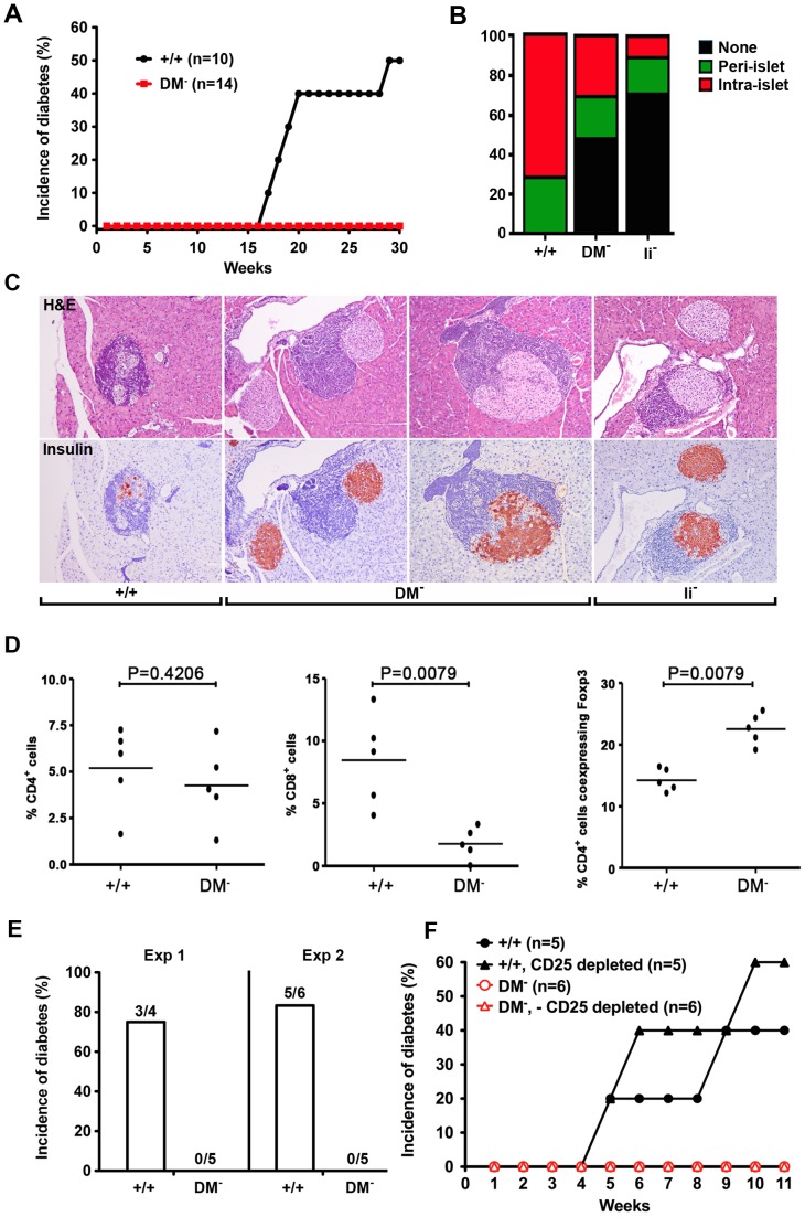
The spontaneous destruction of insulin producing pancreatic beta cells in non-obese diabetic (NOD) mice provides a valuable model of type 1 diabetes. As in humans, disease susceptibility is controlled by the classical MHC class II genes that guide CD4(+) T cell responses to self and foreign antigens. It has long been suspected that the dedicated class II chaperone designated HLA-DM in humans or H-2M in mice also makes an important contribution, but due to tight linkage within the MHC, a possible role played by DM peptide editing has not been previously tested by conventional genetic approaches. Here we exploited newly established germ-line competent NOD ES cells to engineer a loss of function allele. DM deficient NOD mice display defective class II peptide occupancy and surface expression, and are completely protected against type 1 diabetes. Interestingly the mutation results in increased proportional representation of CD4(+)Foxp3(+) regulatory T cells and the absence of pathogenic CD4(+) T effectors. Overall, this striking phenotype establishes that DM-mediated peptide selection plays an essential role in the development of autoimmune diabetes in NOD mice.


Conflict of interest statement

Competing Interests: The authors have declared that no competing interests exist.

Figures

Figure 1
Figure 1. DM loss in NOD mice results in decreased I-Ag7 surface expression.
(A) Saponin-permeabilized splenocytes from wildtype (1) or DM mutant (2) NOD mice were incubated with anti-DM or anti-Ii chain mAb and FITC-conjugated anti-rat IgG (H+L) and analyzed by FACS. The DMα mutation eliminates DM and has no effect on Ii chain expression. (B) Splenocytes from (1) wildtype, (2) DM-deficient, or (3) Ii chain mutant NOD mice were stained with biotin-conjugated mAbs as indicated, followed by FITC-conjugated avidin. The shifts were detectable with conformationally dependent mAbs directed against distinct epitopes contributed by both chains and thus reflect decreased expression rather than a serological change. Representative data from one of three identical experiments with similar outcomes are shown.
Figure 2
Figure 2. DM mutant NOD mice weakly express Ag7/CLIP complexes and entirely lack mature compact Ag7 dimers.
(A) To evaluate occupancy by the Ii chain-derived CLIP peptide, DM mutant spleen cells expressing either the b or k haplotype, DM loss of function and wildtype NOD splenocytes were compared. Lysates were prepared from spleen cells pulsed for 40 mins with 35S-methionine and chased for different times as indicated and extensively pre-cleared with Rabbit anti-mouse IgG (H+L) Abs before immunoprecipitation with beta chain-specific Rabbit Ab. Complexes were boiled for 10 mins and analyzed on 10% polyacrylamide gels. (B) Spleen cell lysates were boiled (B) or kept on ice (NB) and samples resolved on 10% gels under reducing conditions, transferred to nitrocellulose membranes, and Western blots probed with 10-2-16 or alpha chain-specific Rabbit Ab as indicated. (C) Western blot analysis of lysates prepared from LPS-stimulated BMDCs.
Figure 3
Figure 3. DM mutants display functionally empty I- Ag7 molecules with markedly enhanced peptide-binding capabilities.
DM mutant (shown in red) and control wildtype (shown in black) splenocytes from B10.BR or NOD mouse strains were cultured for 5 hrs at 37°C with biotin-conjugated peptides or medium alone as indicated, stained with FITC-labelled avidin, and analysed by FACS. OVA 323–339 preferentially binds to I- Ag7, HEL 46–61 selectively binds to Ak molecules, whereas NOD DM mutants gain reactivity towards the lambda repressor cI peptide P12–26 (IP).
Figure 4
Figure 4. DM functional loss results in decreased Ag7 expression within the thymic cortical epithelium and medullary regions.
Acetone-fixed cryostat sections of thymic tissue were stained with the indicated mAbs.
Figure 5
Figure 5. DM functional loss favours development of the CD4+ Foxp3+ T cell subset.
Thymus (A), spleen (B), and pancreatic lymph node (C) cell suspensions were stained for CD4, CD8, and Foxp3 expression and analysed by flow cytometry.
Figure 6
Figure 6. DM mutant NOD mice protected against autoimmune diabetes fail to develop pathogenic CD4+ T effectors.
(A) The onset of diabetes was evaluated by measurement of blood glucose levels in age-matched DM mutant and wildtype females. The incidence of diabetes has been absolutely zero over the past 2 years with parents in homozygous mutant breeding cages routinely tested at 6 mo of age. (B) The percentage of islets with a given degree of infiltration at 5 mo of age. (C) Typical examples of islet architecture assessed by H & E and insulin staining demonstrate substantial destruction by intra-islet infiltrates in wildtype mice, the normal islets predominantly seen in Ii chain mutants, and benign peri-islet infiltrates present in DM deficient NOD mice. (D) The representation of CD4, CD8, and Foxp3+ T cell subsets in pancreatic infiltrates was analyzed by flow cytometry. (E) DM-deficient NOD mice are resistant to cyclophosphamide-induced type 1 diabetes. Blood glucose levels were measured in age-matched wildtype and mutant females injected i.p. with cyclophosphamide at a dose of 200 mg/kg. (F) Depletion of CD25+ T cells fails to reveal the presence of pathogenic CD4+ T effectors in DM mutants. NOD.scid females were reconstituted with the indicated T cell populations.

Similar articles

Cited by

References

    1. Todd JA, Bell JI, McDevitt HO (1987) HLA-DQbeta gene contributes to susceptibility and resistance to insulin-dependent diabetes mellitus. Nature 329: 599–604. - PubMed
    1. Serreze DV, Leiter EH (2001) Genes and cellular requirements for autoimmune diabetes susceptibility in nonobese diabetic mice. Curr Dir Autoimmun 4: 31–67. - PubMed
    1. Todd JA, Wicker LS (2001) Genetic protection from the inflammatory disease type diabetes in humans and animal models. Immunity 15: 387–395. - PubMed
    1. Corper AL, Stratmann T, Apostolopoulos V, Scott CA, Garcia KC, et al. (2000) A structural framework for deciphering the link between I-Ag7 and autoimmune diabetes. Science 288: 505–511. - PubMed
    1. Latek RR, Suri A, Petzold SJ, Nelson CA, Kanagawa O, et al. (2000) Structural basis of peptide binding and presentation by the type 1 diabetes-associated MHC class II molecule of NOD mice. Immunity 12: 699–710. - PubMed

Publication types

MeSH terms

Substances