. 2013 Feb;6(1):66-74.
doi: 10.1593/tlo.12349. Epub 2013 Feb 1.

Changes in the Secretory Profile of NSCLC-Associated Fibroblasts after Ablative Radiotherapy: Potential Impact on Angiogenesis and Tumor Growth

Affiliations

Changes in the Secretory Profile of NSCLC-Associated Fibroblasts after Ablative Radiotherapy: Potential Impact on Angiogenesis and Tumor Growth

Turid Hellevik et al. Transl Oncol. 2013 Feb.

Abstract

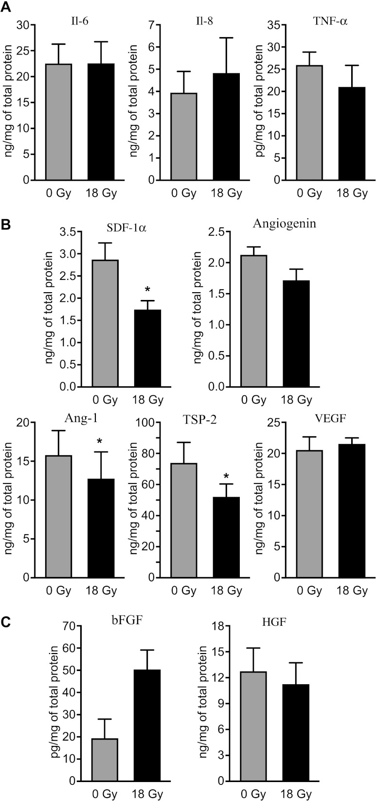
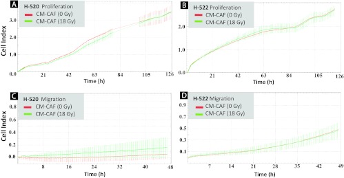
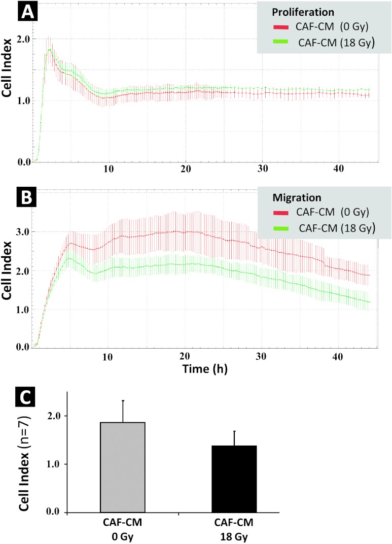
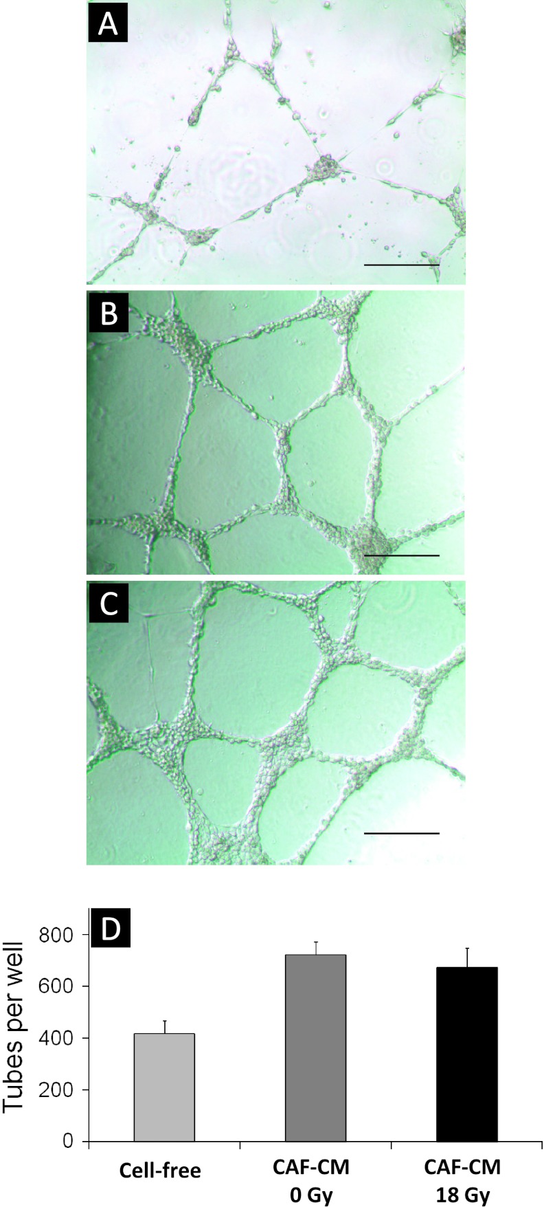
In the context of radiotherapy, collateral effects of ablative doses of ionizing radiation (AIR) on stromal components of tumors remains understudied. In this work, cancer-associated fibroblasts (CAFs) isolated from freshly resected human lung tumors were exposed to AIR (1x 18 Gy) and analyzed for their release of paracrine factors. Inflammatory mediators and regulators of angiogenesis and tumor growth were analyzed by multiplex protein assays in conditioned medium (CM) from irradiated and non-irradiated CAFs. Additionally, the profile of secreted proteins was examined by proteomics. In functional assays, effects of CAF-CM on proliferative and migratory capacity of lung tumor cells (H-520/H-522) and human umbilical vein endothelial cells (HUVECs) and their tube-forming capacity were assessed. Our data show that exposure of CAFs to AIR results in 1) downregulated release of angiogenic molecules such as stromal cell-derived factor-1, angiopoietin, and thrombospondin-2 (TSP-2); 2) upregulated release of basic fibroblast growth factor from most donors; and 3) unaffected expression levels of hepatocyte growth factor, interleukin-6 (IL-6), IL-8, IL-1β, and tumor necrosis factor-α. CM from irradiated and control CAFs did not affect differently the proliferative or migratory capacity of tumor cells (H-520/H-522), whereas migratory capacity of HUVECs was partially reduced in the presence of irradiated CAF-CM. Overall, we conclude that AIR mediates a transformation on the secretory profile of CAFs that could influence the behavior of other cells in the tumor tissue and hence guide therapeutic outcomes. Downstream consequences of the changes observed in this study merits further investigations.


Figures

Figure 1
Figure 1
Secretion of tumor regulatory molecules by irradiated and non-irradiated CAFs. Quantitative determinations of growth factors and cytokines in culture supernatants from five different donors were measured by multiplex protein arrays. Only factors giving values above the detection limit of the assay are represented. Panel (A) demonstrates expression of inflammatory molecules. The following cytokines were included in the array: IL-1β, IL-6, IL-8, and TNF-α. Panel (B) shows array of angiogenic factors including SDF-1α, angiogenin, angiopoietin, TSP-2, and VEGF. Finally, (C) presents array of detectable tumor growth regulators, comprising HGF, bFGF, and PDGF. Mean values from five different donor samples are shown; statistical analyses were performed by applying paired samples Student's t test. Statistically significant differences are indicated by asterisk (P < .05).
Figure 2
Figure 2
Effect of CAF-CM on proliferative and migratory capacity of lung tumor cell lines H520 and H522. CAFs cultured in T-75 flasks were treated with 1x 18 Gy and medium was conditioned in a serum-free setting for 4 to 6 days post-irradiation. Proliferative capacity of two lung carcinoma cell lines, H520 (squamous cell carcinoma) (A) and H522 (adenocarcinoma) (B), was monitored on E-plates located in the xCELLigence machine, whereas corresponding migratory capacity of H520 and H522 in double-chambered CIM plates is presented in (C) and (D), respectively. For both experiments, H520 and H522 were exposed to (1:1) non-irradiated CAF-CM (red line) and (1:1) irradiated CAF-CM (green line) from a randomly selected donor. Similar profiles were obtained after running the assay with CAF-CM from three different donors.
Figure 3
Figure 3
Effect of CAF-CM on the proliferative and migratory capacity of HUVECs. CAFs were treated with 1x 18 Gy and incubation medium was conditioned in serum-free conditions for 4 to 6 days post-irradiation. Proliferative capacity of HUVECs was monitored on E-plates (A) and migratory capacity in double-chambered CIM plates (B), with readout by the xCELLigence System for both types of plates. For both experiments, HUVECs were exposed to (1:1) non-irradiated CAF-CM (red line), and (1:1) 1x 18 Gy irradiated CAF-CM (green line) from one randomly selected donor. Similar profiles were obtained after running the assay with CAF-CM from seven different donors. Columns in (C) represent mean and SEs of CI values after a 20-hour observation of HUVEC migratory behavior, averaged from seven independent experiments using CAF-CM from seven randomly selected donors. Error bars represent SD.
Figure 4
Figure 4
Effect of CAF-CM on endothelial cell tube formation. CAFs were treated with 1x 18 Gy and incubation medium was conditioned in serum-free conditions between days 4 and 6 post-irradiation. HUVECs were plated in Matrigel-coated multiwell plates, and after an attachment phase of 2 hours, HUVECs were exposed to CM from irradiated (18 Gy) and non-irradiated CAFs (0 Gy) and incubated for another 20 hours. Images show tube formation by HUVECs after 20 hours of growth in Matrigel. In (A), tube formation by HUVECs exposed to standard (cell-free) incubation medium is shown, whereas (B) and (C) demonstrate tube-forming HUVECs exposed to CM from non-irradiated CAFs (CAF-CM 0 Gy) and irradiated CAFs (CAF-CM 18 Gy), respectively. Bars, 320 µm. Panel (D) reflects quantification of HUVEC forming tubes/well. Data represent average values from three independent experiments (error bars represent SD).

References

    1. Ferlay J, Shin HR, Bray F, Forman D, Mathers C, Parkin DM. Estimates of worldwide burden of cancer in 2008: GLOBOCAN 2008. Int J Cancer. 2010;127:2893–2917. - PubMed
    1. Powell JW, Dexter E, Scalzetti EM, Bogart JA. Treatment advances for medically inoperable non-small-cell lung cancer: emphasis on prospective trials. Lancet Oncol. 2009;10:885–894. - PubMed
    1. Kavanagh BD, Miften M, Rabinovitch RA. Advances in treatment techniques: stereotactic body radiation therapy and the spread of hypofractionation. Cancer J. 2011;17:177–181. - PubMed
    1. Heinzerling JH, Kavanagh B, Timmerman RD. Stereotactic ablative radiation therapy for primary lung tumors. Cancer J. 2011;17:28–32. - PubMed
    1. Lanni TB, Jr, Grills IS, Kestin LL, Robertson JM. Stereotactic radiotherapy reduces treatment cost while improving overall survival and local control over standard fractionated radiation therapy for medically inoperable non-small-cell lung cancer. Am J Clin Oncol. 2011;34:494–498. - PubMed

LinkOut - more resources