. 2013 Jan:295:130-9.
doi: 10.1016/j.heares.2012.03.009.

The cerebellum as a novel tinnitus generator

Affiliations

The cerebellum as a novel tinnitus generator

Carol A Bauer et al. Hear Res. 2013 Jan.

Abstract

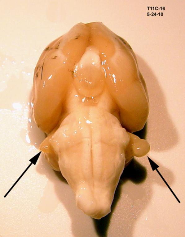
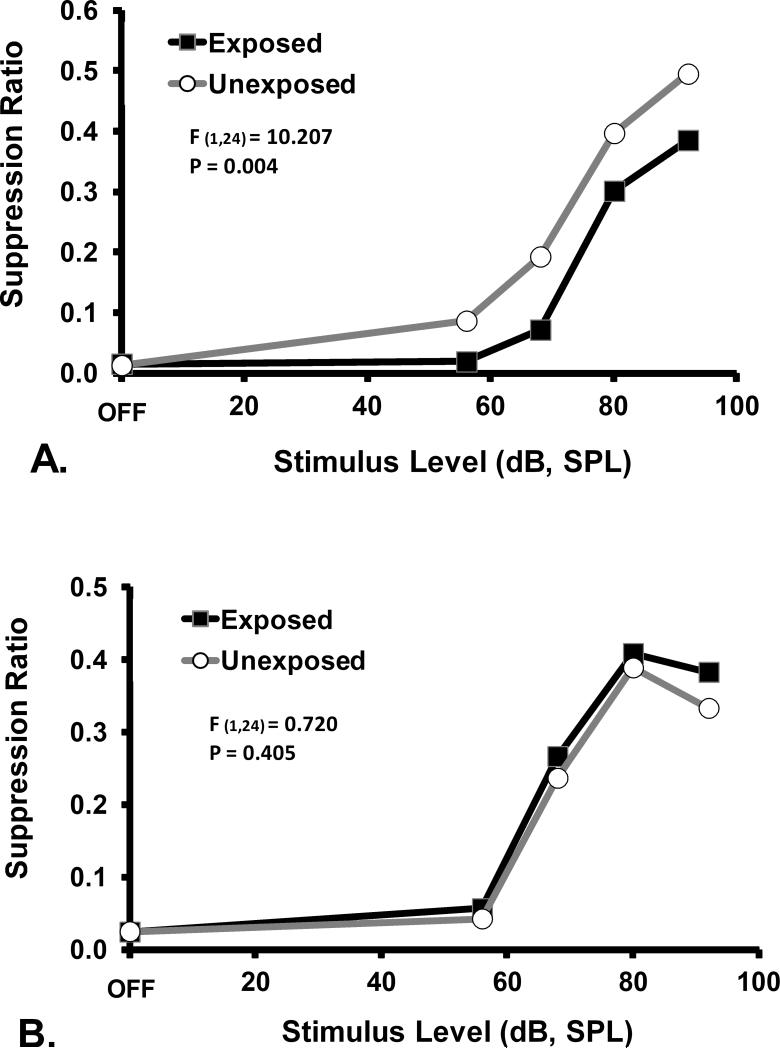
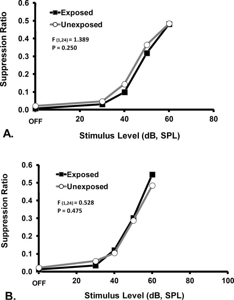
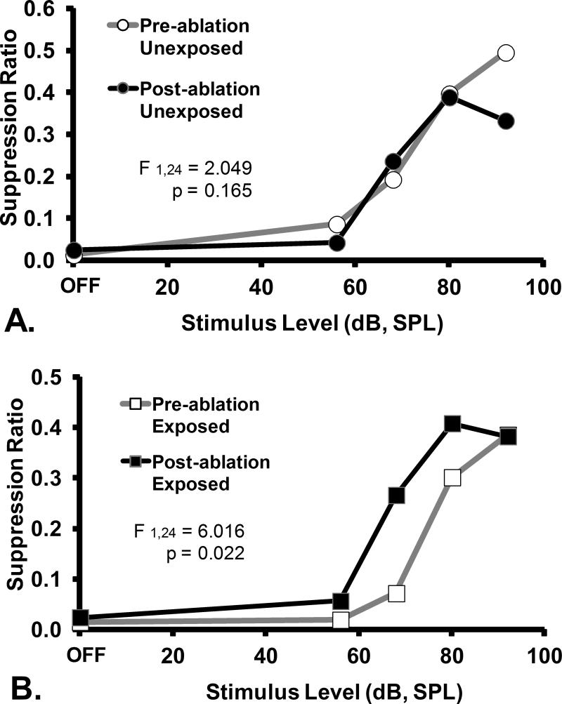
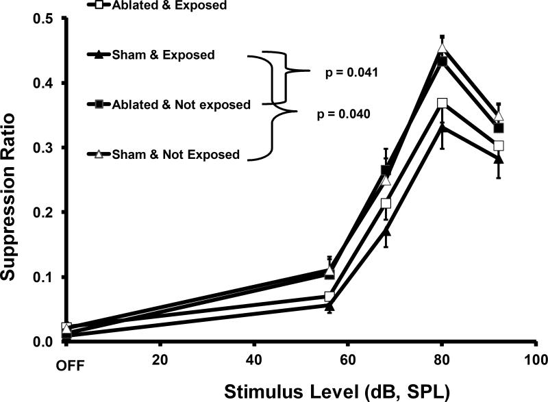
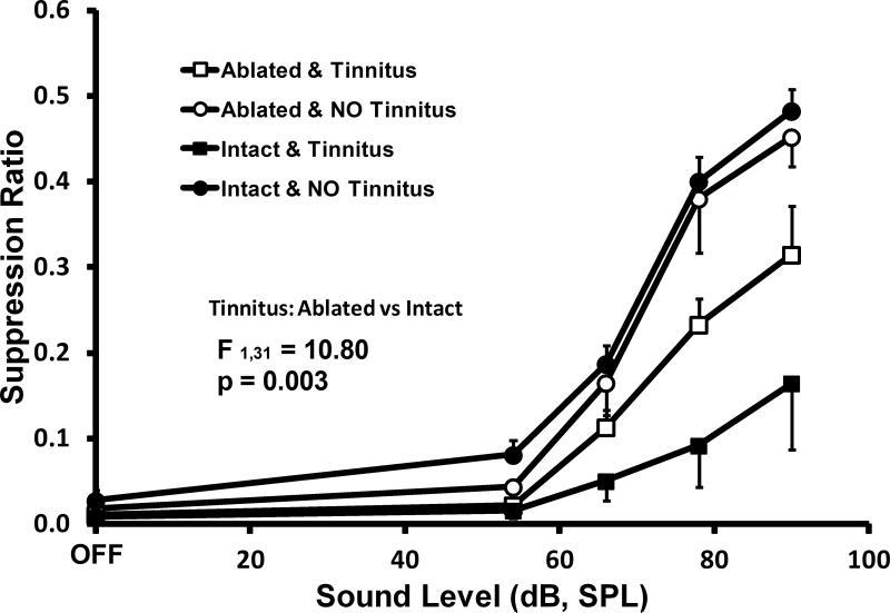
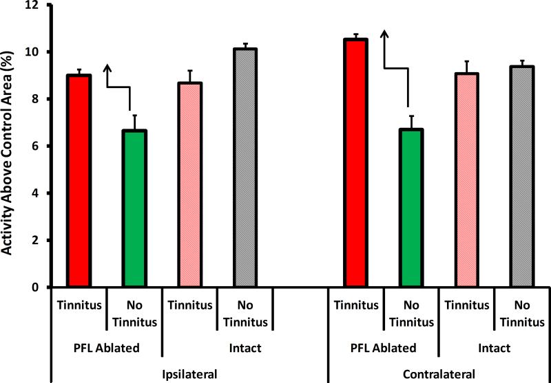
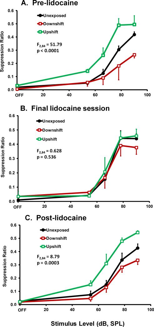
The role of the cerebellum in auditory processing is largely unknown. Recently it was shown that rats with psychophysical evidence of tinnitus had significantly elevated neural activity in the paraflocculus of the cerebellum (PFL), as indicated by functional imaging. It was further shown that PFL activity was not elevated in normal rats listening to a tinnitus-like sound. This suggests that plastic changes in the PFL may underpin chronic tinnitus, i.e., it may serve as a tinnitus generator. Using a rat model of acoustic trauma-induced tinnitus, the role of the cerebellum was further examined in a series of experiments:The PFL was surgically ablated in animals with established tinnitus; the PFL was surgically ablated in animals before induction of tinnitus; the PFL was reversibly inactivated by chronic lidocaine infusion into the subarcuate fossa of animals with established tinnitus. It was found that PFL ablation eliminated established tinnitus without altering auditory discrimination. Similar to the ablation results, PFL inactivation with lidocaine reversibly eliminated existing tinnitus. In contrast however, PFL ablation before tinnitus induction attenuated, but did not completely eliminate, tinnitus. In a rat model of noise-induced chronic tinnitus, the cerebellar PFL may serve as a sufficient but non-obligatory generator of tinnitus.


Figures

Figure 1
Figure 1
A typical unilateral PFL ablation. The remnant of the right PFL stalk is evident in this ventral view of an excised brain, 3 months after thermal ablation of the PFL.
Figure 2
Figure 2
Psychophysical functions for the tinnitus diagnostic stimulus of 16 kHz tones, prior to (A) and after (B) ablation of the PFL ipsilateral to noise exposure. Discrimination performance, as indicated by the suppression ratio (R) is plotted on the y axis. The significant downshift of the exposed group, prior to ablation, is indicative of tinnitus. The statistical summary in each panel shows the difference between groups at stimulus levels above OFF. Ablation abolished the indication of tinnitus.
Figure 3
Figure 3
Psychophysical functions for the non-diagnostic stimulus of broad-band noise, prior to (A) and after (B) ablation of PFL ipsilateral to noise exposure. Subjects and statistical tests as in Fig. 2. Ablation had no significant general effect on psychophysical performance.
Figure 4
Figure 4
A within group comparison of the effects of ipsilateral PFL ablation on the diagnostic stimulus (16 kHz tones) performance of A. unexposed and B. exposed rats. Data depicted in Fig. 2 have been re-plotted to show that there was no ablation effect on the unexposed animals (A), but significant effect on the exposed animals (B). Statistics and axes as in preceding figures.
Figure 5
Figure 5
A within group comparison of the effects of ipsilateral PFL ablation on the non-diagnostic stimulus (broad-band noise) performance of A. unexposed and B. exposed rats. Data depicted in Fig. 3 have been re-plotted to show that there was no ablation effect on the general psychophysical performance of either group. Statistics and axes as in preceding figures.
Figure 6
Figure 6
The effect of ipsilateral PFL ablation prior to high-level noise exposure. Psychophysical performance for the diagnostic stimulus of 16 kHz tones. Intact exposed animals showed the function downshift typical of animals with tinnitus (sham exposed, solid triangular data points). Unexposed animals, whether or not ablated, did not show evidence of tinnitus. Ipsilateral ablation did not completely protect animals exposed to high-level noise after the ablation (ablated exposed, open square data points). The ablated exposed animals were not significantly different statistically than the other positive or negative control groups. Significance levels were for uncorrected independent t tests. Error bars show the standard error of the mean.
Figure 7
Figure 7
Tinnitus diagnostic performance for 20 kHz test tones of exposed animals selected from Experiment 2. All animals were exposed; 8 ablated and 8 intact (sham ablated). Each of these groups were equally divided two groups of 4 each, that showed either maximum tinnitus (square data points) or minimum tinnitus (circular data points). Not all exposed rats developed significant tinnitus (filled vs solid circular points). Of animals developing tinnitus, intact consistently showed greater evidence (filled square point) than ablated (open square points). At the conclusion of testing, all animals were functionally brain imaged using MEMRI. Error bars show the standard error of the mean. The statistical summary shows the difference between ablated and intact groups at stimulus levels above OFF
Figure 8
Figure 8
Summary of the MEMRI imaging results of Experiment 2 animals depicted in Fig. 7. The only consistent activity pattern differences, tinnitus vs. no-tinnitus, that appeared between intact and ablated animals, was evident in the MGB. Ablated rats that developed tinnitus had elevated MGB activity with respect to ablated rats that did not develop tinnitus (dark red vs. dark green bars). In contrast, intact rats that developed tinnitus had the same level of MGB activity as intact rats without tinnitus (light red vs gray bars). The activity pattern of other auditory areas was similar across all four groups (not shown, see text). Error bars shown the mean deviation.
Figure 9
Figure 9
Reversible attenuation of tinnitus and hyperacusis (potentially with tinnitus) by chronic infusion of 4% lidocaine into the subarcuate fossa. The diagnostic test stimulus was 20 kHz tones. Group psychophysical functions are shown before infusion (A), at the conclusion of infusion (B) and after a washout period (C). Statistical summary and axes as in previous figures.
Figure 10
Figure 10
Mean 20 kHz time series performance across lidocaine treatment sessions, of the groups depicted in Fig. 9. Downshifted data reflected tinnitus, while upshifted data indicated hyperacusis with tinnitus. Error bars show the standard error of the mean. Broken lines show the least-mean-square regression line for each function. As treatment progressed, the psychophysical evidence of both tinnitus and hyperacusis, progressively decreased.

References

    1. Alberti PW. Tinnitus in occupational hearing loss: nosological aspects. J Otolaryngol. 1987;16:34–5. - PubMed
    1. Azizi SA, Woodward DJ. Interactions of visual and auditory mossy fiber inputs in the paraflocculus of the rat: a gating action of multimodal inputs. Brain Res. 1990;533:255–62. - PubMed
    1. Azizi SA, Burne RA, Woodward DJ. The auditory corticopontocerebellar projection in the rat: inputs to the paraflocculus and midvermis. An anatomical and physiological study. Exp Brain Res. 1985;59:36–49. - PubMed
    1. Bauer CA, Brozoski TJ. Assessing tinnitus and prospective tinnitus therapeutics using a psychophysical animal model. J of the Assoc for Res in Otolaryngol. 2001;2:54–64. - PMC - PubMed
    1. Bauer CA, Brozoski TJ, Rojas R, Boley J, Wyder M. Behavioral model of chronic tinnitus in rats. Otolaryngol Head Neck Surg. 1999;121:457–62. - PubMed

Publication types

MeSH terms