

A boy with homozygous microdeletion of NEUROG1 presents with a congenital cranial dysinnervation disorder [Moebius syndrome variant]
- PMID: 23419067
- PMCID: PMC3599919
- DOI: 10.1186/1744-9081-9-7
A boy with homozygous microdeletion of NEUROG1 presents with a congenital cranial dysinnervation disorder [Moebius syndrome variant]
Abstract
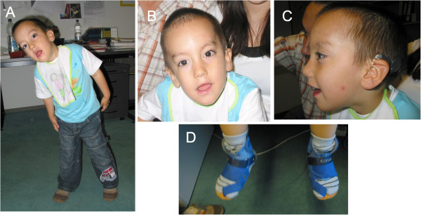
Background: We report on a 6-year-old Turkish boy with profound sensorineural deafness, balance disorder, severe disorder of oral motor function, and mild developmental delay. Further findings included scaphocephaly, plagiocephaly, long palpebral fissures, high narrow palate, low-set posteriorly rotated ears, torticollis, hypoplastic genitalia and faulty foot posture. Parents were consanguineous.
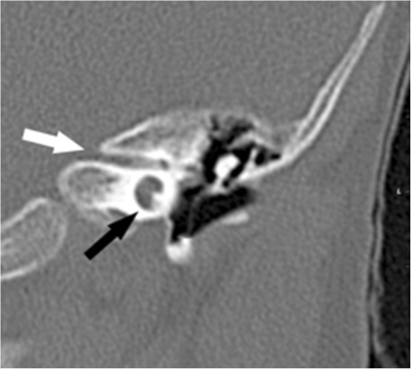
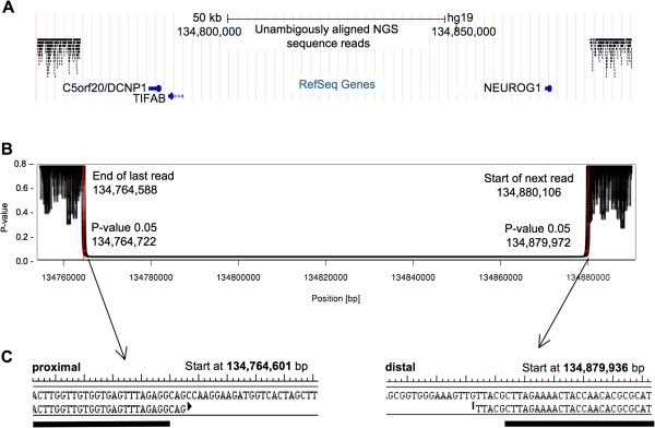
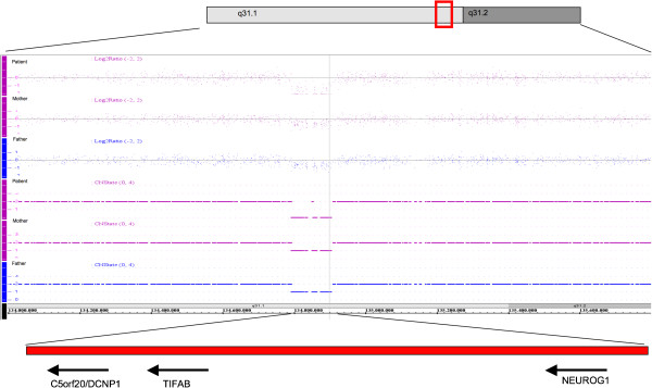
Methods and results: Computed tomography and magnetic resonance imaging showed bilateral single widened cochlear turn, narrowing of the internal auditory canal, and bilateral truncation of the vestibulo-cochlear nerve. Microarray analysis and next generation sequencing showed a homozygous deletion of chromosome 5q31.1 spanning 115.3 kb and including three genes: NEUROG1 (encoding neurogenin 1), DCNP1 (dendritic cell nuclear protein 1, C5ORF20) and TIFAB (TIFA-related protein). The inability to chew and swallow, deafness and balance disorder represented congenital palsies of cranial nerves V (trigeminal nerve) and VIII (vestibulo-cochlear nerve) and thus a congenital cranial dysinnervation disorder.
Conclusions: Based on reported phenotypes of neurog1 null mutant mice and other vertebrates, we strongly propose NEUROG1 as the causative gene in this boy. The human NEUROG1 resides within the DFNB60 locus for non-syndromic autosomal recessive deafness on chromosome 5q22-q31, but linkage data have excluded it from being causative in the DFNB60 patients. Given its large size (35 Mb, >100 genes), the 5q22-q31 area could harbor more than one deafness gene. We propose NEUROG1 as a new gene for syndromic autosomal recessive hearing loss and congenital cranial dysinnervation disorder including cranial nerves V and VIII.
Figures
References
-
- Webb JF, Noden DM. Ectodermal placodes: contributions to the development of the vertebrate head. Am Zool. 1993;33:434–447.
-
- Schlosser G. Making senses development of vertebrate cranial placodes. Int Rev Cell Mol Biol. 2010;283:129–234. - PubMed
MeSH terms
Substances
LinkOut - more resources
Full Text Sources
Other Literature Sources
Molecular Biology Databases
Research Materials
Miscellaneous
