. 2013:3:1316.
doi: 10.1038/srep01316.

In vitro engineering of vascularized tissue surrogates

Affiliations

In vitro engineering of vascularized tissue surrogates

Katsuhisa Sakaguchi et al. Sci Rep. 2013.

Abstract

In vitro scaling up of bioengineered tissues is known to be limited by diffusion issues, specifically a lack of vasculature. Here, we report a new strategy for preserving cell viability in three-dimensional tissues using cell sheet technology and a perfusion bioreactor having collagen-based microchannels. When triple-layer cardiac cell sheets are incubated within this bioreactor, endothelial cells in the cell sheets migrate to vascularize in the collagen gel, and finally connect with the microchannels. Medium readily flows into the cell sheets through the microchannels and the newly developed capillaries, while the cardiac construct shows simultaneous beating. When additional triple-layer cell sheets are repeatedly layered, new multi-layer construct spontaneously integrates and the resulting construct becomes a vascularized thick tissue. These results confirmed our method to fabricate in vitro vascularized tissue surrogates that overcomes engineered-tissue thickness limitations. The surrogates promise new therapies for damaged organs as well as new in vitro tissue models.


Conflict of interest statement

T. Shimizu and M. Yamato are consultants for CellSeed, Inc. T. Okano is an investor in CellSeed, Inc., and is an inventor/developer designated on the patent for temperature-responsive culture surfaces.

Figures
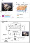
Figure 1
Figure 1. Culture device and perfusion culture system.
(a) The collagen-gel base with microchannels imitates the conditions of a subcutaneous structure. In the right-hand expanded view, the blue area shows collagen-gel that can imitate a subcutaneous extracellular matrix, the pink arrows indicate the direction of culture medium flow imitate blood and tissue fluid. The culture medium can diffuse into the collagen gel and provide oxygen and nutrients to the cell sheet. The left picture shows a photograph of the culture device. (b) The circuit of culture system allows the culture medium to flow in the microchannels and monitors the pH and oxygen conditions of the culture medium as well as using partial pressure sensors. The culture medium was perfused into the culture device at 0.5 mL/min by a syringe pump. The oxygen and pH were monitored with an optic sensor to verify the conditions of the culture medium.
Figure 2
Figure 2. Cell migration from the cell sheet and tissue viability.
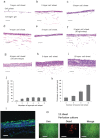
The specimens were stained with AZAN. In the photographs (a)~(d), the upper layers are triple-layered cell sheets, and the lower part is collagen gel. The dotted-line circles are the microchannels. (a) Histological observation shows a triple-layered cell sheet on the collagen-gel cultured for 5 days without perfusion. After 5 days of static cultivation, the cell sheet had necrosed throughout. (b) A triple-layered cell sheet was cultured with perfusion at 0.5 mL/min for 5 days. The cell sheet construct was able to survive and several cells migrated into the collagen gel. (c) With the addition of vascular endothelial growth factor (VEGF), AZAN section showed that many cells had migrated and can be seen between the cell sheets and the microchannels. (d) VEGF and basic fibroblast growth factor (bFGF) stimulated triple-layered cell sheets on the collagen-gel. A large number of cells were found to migrate and create lumens in the collagen-gel base. (Scale bar, 100 μm) (e) Migrating cell areas were counted under the conditions in (a)~(d). The areas in perfusion culture systems were significantly larger than that in static culture. The areas in perfusion culture containing both VEGF and bFGF were significantly larger than those in perfusion cultures without two growth factors and only with VEGF. (*P < 0.05, n = 3) (f) Live/Dead assay was performed in the static culture and perfusion culture for the 3-layer cardiac cell sheet. Fluorescence micrograph shows cells stained by 5 μmol/L calcein-AM (green = live; top images) and 0.5 μmol/L ethidium homodimer-1 (red = dead; middle images). Bottom images are a merged version of the Live/Dead images. Most of the cells died in the static condition, consistent with the AZAN staining image. On the other hand, most of cells survived in the perfusion culture (Scale bar, 20 μm).
Figure 3
Figure 3. Geometry of endothelial cells of the cell sheet.
(a)~(c) GFP-expressing endothelial cells were isolated from GPF neonatal rat heart and replaced with normal endothelial cells of the cardiac cell sheet by a magnetic cell sorter technique. These images are the top views of a 3-layer cardiac cell sheet after 5 days in perfusion culture under a microscope. GFP-expressing endothelial cells are seen as green. (a) Low magnification of the center of the construct. (b) Meddle magnification of edge in the construct. (c) High magnification of the center of the construct. (d) The specimen shows that GFP-positive cells exist at the lumen surface. Blue signals indicated nuclei. (Scale bar, 100 μm). (e) The right photograph shows the expanded view of the square area in (d) (Scale bar, 20 μm).
Figure 4
Figure 4. Red blood cells and resin perfusion into the endothelial lumen.
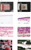
(a) When the device has no endothelial cells, the flow line of red blood cells is clearly observed to follow on the microchannels (Scale bar 1 cm). (b) When endothelial cells are included, rat blood cells spread throughout the cell sheets like real subcutaneous vessels (Scale bar, 1 cm). (c) HE stained section shows no migrating cells in the construct without endothelial cells (Scale bar, 200 μm). (d) HE stained section of the construct containing endothelial cells shows a lot of migrating cells and a vascular formation. The red blood cells flowed into the newly created vascular network (Scale bar, 200 μm). (e) In the high magnification image, arrowheads indicate that red blood cells locate consistently to the lumens of the vascular networks between the microchannel and the cell sheet. The dotted line indicates the collagen-based microchannel (Scale bar, 50 μm). (f)Red blood cells locate in the cell sheet capillaries (Scale bar 20 μm). Arrow heads indicate the red blood cells. (g) The newly created vessel shape was cast by epoxy resin (Scale bar, 500 μm). (h) Expanded view of the resin cast of the newly created microvessels (Scale bar, 300 μm).
Figure 5
Figure 5. Fabrication of layered cardiac cell sheets on a perfusion bioreactor.
In photographs (a)~(f), the upper layers are 1~6-layered cell sheets, and the lower parts are collagen gel. (a) The thickness single-layered cell sheet shows 5 ± 0.5 μm in the bioreactor after 5 days cultivation; (b) double-layered cell sheet is 17 ± 4 μm; (c) triple-layered cell sheet is 24 ± 4 μm; (d) quadruple-layered cell sheet is 27 ± 2 μm; (e) quintuple-layered cell sheet is 26 ± 2 μm; (f) sextuple-layered cell sheet is 27 ± 2 μm. (g) The thickness of the sextuple-layered cell sheet is 35 ± 2 μm. (h) The thickness of quintuple-layered cell sheet is 65 ± 8 μm. (i) Four triple-layered cell sheets layered up to four times at 5-day intervals produced a twelve layer sheet. The thickness of the twelve-layered cell sheet is 110 ± 4 μm. (a)~(i) (Scale bar, 50 μm). (j) Graph shows tissue thickness of the constructed grafts with a single-step layered procedure. The sheet thickness increased linearly up to triple-layered cell sheet and reached a plateau in quadruple to sextuple-layered cell sheets. (k) Graph shows that the tissue thickness of layered cell sheets increased up to a twelve-layered cell sheet with a multi-layered procedure. (l) Troponin T staining demonstrates the stratified cardiac muscle in a 6-layer cardiac cell sheet by the double-step procedure. Troponin T, Green; blue, nuclei (Scale bar, 20 μm). (m) Live/Dead assay was performed on the perfusion culture for the 12-layer cardiac cell sheet. Fluorescence micrographs show cells stained with calcein-AM (green-live; top images) and ethidium homodimer-1 (red = dead; middle images). Bottom images are merged from the Live/Dead images. A Live/Dead assay showed that most of the cells survived, but that focal cell death was observed (Scale bar, 20 μm).
Figure 6
Figure 6. Fabrication process of vascularized cardiac tissue in vitro.
The illustrations show the process of vascularization of multi-layer cardiac cell sheets on collagen-based microchannels. (a) A triple-layered cardiac cell sheet on a collagen-gel base with microchannels. (b) After 5 days cultivation with perfusion through the microchannels, endothelial cells migrated into the collagen gel and formed a lumen structure. (c) New lumen-like vascular network was able to connect to the collagen-based microchannels. Fresh medium could flow into the new vascular network and to the vessels in the triple-layer cardiac cell sheets. (d) After new microvessels formed connecting with the collagen-gel microchannels, another triple-layered cardiac cell sheet was placed on the existing cell sheet. (e) The newly-layered cell sheet spontaneously integrated with the existing cell sheet and was also rapidly infiltrated with budding vasculature extending from the previously vascularized cell sheets in the bioreactor. By repeating the sheet layering process, the subsequently added cell sheets were perfused with fresh medium through the newly-created vessels.

References

    1. Levenberg S. et al. Engineering vascularized skeletal muscle tissue. Nature Biotechnology 23, 879–884 (2005). - PubMed
    1. Melero-Martin J. M. et al. Engineering robust and functional vascular networks in vivo with human adult and cord blood-derived progenitor cells. Circulation Research 103, 194–202 (2008). - PMC - PubMed
    1. Morritt A. N. et al. Cardiac tissue engineering in an in vivo vascularized chamber. Circulation 115, 353–360 (2007). - PubMed
    1. Shimizu T. et al. Fabrication of pulsatile cardiac tissue grafts using a novel 3-dimensional cell sheet manipulation technique and temperature-responsive cell culture surfaces. Circulation Research 90, e40–e48 (2002). - PubMed
    1. Shimizu T. et al. Polysurgery of cell sheet grafts overcomes diffusion limits to produce thick, vascularized myocardial tissues. The FASEB Journal 20, 708 (2006). - PubMed

Publication types