. 2012 Jun;4(3):343-357.
doi: 10.2217/iim.12.27.

Biomarkers for the clinical evaluation of the cognitively impaired elderly: amyloid is not enough

Affiliations

Biomarkers for the clinical evaluation of the cognitively impaired elderly: amyloid is not enough

Linda K McEvoy et al. Imaging Med. 2012 Jun.

Abstract

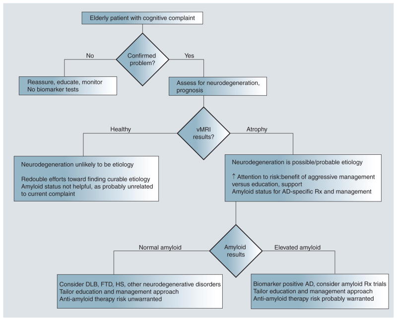
The number of elderly patients seeking clinical treatment for memory problems will rise sharply in coming years as our population ages. These patients present a challenge for diagnosis and prognosis since cognitive problems in older patients can arise from many etiologies, some of which are curable. With the development of clinically available biomarkers for detecting Alzheimer's disease pathology in living patients, evaluation of cognitively impaired elderly patients is about to undergo a major paradigm shift. This article describes the two classes of biomarkers available for assessing Alzheimer's disease risk: those that indicate presence of amyloid pathology and those that provide evidence of neuronal injury and neurodegeneration. We argue that, currently, incorporation of biomarkers of neurodegeneration can help in patient prognosis whereas tests for amyloid, if used in isolation, have potential for harm. Amyloid tests are clinically useful only when evidence suggests progressive cognitive decline or neurodegeneration.

Keywords: Alzheimer’s disease; MCI; MRI; PET; amyloid imaging; biomarker; florbetapir; mild cognitive impairment.


Figures

Figure 1
Figure 1. Coronal section of a T1-weighted volumetric MRI from an Alzheimer’s Disease Neuroimaging Initiative mild cognitive impairment patient
Data have been automatically segmented into different tissue types and brain regions using NeuroQuant® software [101]. The hippocampus, which is highly vulnerable to Alzheimer’s disease, but also affected in other neurodegenerative disorders, is shown in gold. This mild cognitive impairment patient, despite testing positive for cerebrospinal fluid Aβ42 had age-appropriate hippocampal and inferior lateral ventricle volumes (circled in yellow on the left) at baseline and at all follow-ups. This patient retained the mild cognitive impairment diagnosis through 3 years of follow-up.
Figure 2
Figure 2. Risk of developing dementia in patients with mild cognitive impairment as a function of individual and combined biomarker status
(A–C) Red shows risk of developing AD in MCI patients testing positive for cerebrospinal fluid (CSF) Aβ42 for the CSF tau/A ratio and for medial temporal atrophy (HOC), respectively. Blue lines indicate negative results on these tests. (D) Mild cognitive impairment patients are stratified on the basis of CSF A β42 and atrophy risk. Those testing negative for both (green line) are at very low risk of developing dementia within 3 years, those testing positive for both (red line) are at very high risk. Those testing positive for atrophy, even in the presence of a negative CSF Aβ42 test (purple line), are also at very high risk of developing Alzhemer’s disease. HOC: Hippocampal occupancy score. Adapted with permission from [60].
Figure 3
Figure 3. Example NeuroQuant® report for one Alzheimer’s Disease Neuroimaging Initiative mild cognitive impairment patient
This patient tested positive for cerebrospinal fluid Aβ42 and showed smaller hippocampal volumes and larger inferior lateral ventricle than expected for his age at baseline. Follow-up scans indicated progressive neurodegeneration. This patient was diagnosed with dementia at the 24-month follow-up visit.
Figure 4
Figure 4. Recommended decision tree for clinical evaluation of the elderly patient with cognitive complaints
AD: Alzheimer’s disease; DLB: Dementia with Lewy bodies; FTD: Frontotemporal dementia; HS: Hippocampal sclerosis; Rx: Clinical treatment; vMRI: Volumetric MRI.

References

    1. Klunk WE, Engler H, Nordberg A, et al. Imaging brain amyloid in Alzheimer’s disease with Pittsburgh compound-B. Ann Neurol. 2004;55(3):306–319. First study to report imaging of amyloid in the living human brain. - PubMed
    1. Rowe CC, Ackerman U, Browne W, et al. Imaging of amyloid β in Alzheimer’s disease with 18F-BAY94–9172, a novel PET tracer: proof of mechanism. Lancet Neurol. 2008;7(2):129–135. - PubMed
    1. Wong DF, Rosenberg PB, Zhou Y, et al. In vivo imaging of amyloid deposition in Alzheimer disease using the radioligand 18F-AV-45 (florbetapir [corrected] F 18) J Nucl Med. 2010;51(6):913–920. - PMC - PubMed
    1. Herholz K, Ebmeier K. Clinical amyloid imaging in Alzheimer’s disease. Lancet Neurol. 2011;10(7):667–670. - PubMed
    1. Department of Economic and Social Affairs. World population ageing. United Nations Publications; UN, NY, USA: 2001. Population Division.

Website

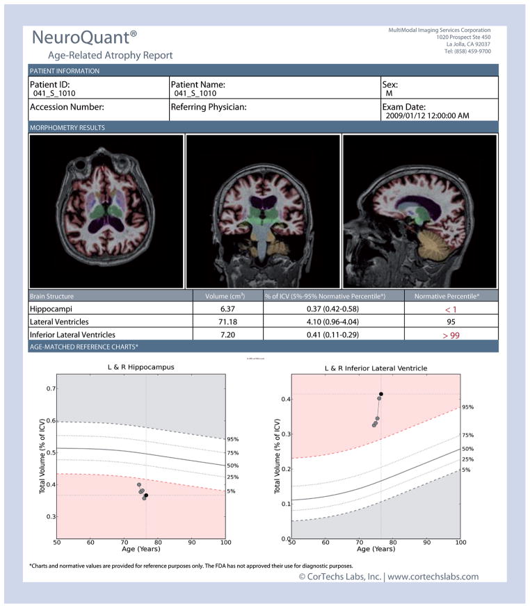
    1. NeuroQuant®. www.cortechslabs.com.

LinkOut - more resources