. 2013 Apr 5;288(14):10101-10109.
doi: 10.1074/jbc.M112.448399. Epub 2013 Feb 18.

A new way to degrade heme: the Mycobacterium tuberculosis enzyme MhuD catalyzes heme degradation without generating CO

Affiliations

A new way to degrade heme: the Mycobacterium tuberculosis enzyme MhuD catalyzes heme degradation without generating CO

Shusuke Nambu et al. J Biol Chem. .

Abstract

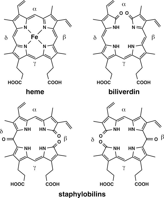
MhuD is an oxygen-dependent heme-degrading enzyme from Mycobacterium tuberculosis with high sequence similarity (∼45%) to Staphylococcus aureus IsdG and IsdI. Spectroscopic and mutagenesis studies indicate that the catalytically active 1:1 heme-MhuD complex has an active site structure similar to those of IsdG and IsdI, including the nonplanarity (ruffling) of the heme group bound to the enzyme. Distinct from the canonical heme degradation, we have found that the MhuD catalysis does not generate CO. Product analyses by electrospray ionization-MS and NMR show that MhuD cleaves heme at the α-meso position but retains the meso-carbon atom at the cleavage site, which is removed by canonical heme oxygenases. The novel tetrapyrrole product of MhuD, termed "mycobilin," has an aldehyde group at the cleavage site and a carbonyl group at either the β-meso or the δ-meso position. Consequently, MhuD catalysis does not involve verdoheme, the key intermediate of ring cleavage by canonical heme oxygenase enzymes. Ruffled heme is apparently responsible for the heme degradation mechanism unique to MhuD. In addition, MhuD heme degradation without CO liberation is biologically significant as one of the signals of M. tuberculosis transition to dormancy is mediated by the production of host CO.


Figures

FIGURE 1.
FIGURE 1.
Structures of heme, biliverdin, and staphylobilins.
FIGURE 2.
FIGURE 2.
Heme environment structures of N7A IsdG (A), IsdI (B), rat HO-1 (C), and MhuD (D). Protein Data Bank (PDB) codes are: 2ZDO, 3QGP, 1N45, and 3HX9, respectively.
FIGURE 3.
FIGURE 3.
Resonance Raman analysis of monoheme-MhuD complex in 0.1 m HEPES, pH 7.0. A, resonance Raman spectra of ferrous CO-bound monoheme-MhuD complex prepared with 12C16O (top) and 13C16O (middle). The bottom trace is a difference spectrum (12C16O − 13C16O). The spectra were obtained with 405 nm excitation and a laser power of ∼3 milliwatts. B, a correlation plot for the Fe–C and C–O frequencies of the CO-bound ferrous monoheme-MhuD complex. Data points are for His-ligated heme proteins (circles), Cys-ligated heme proteins (squares), and MhuD (filled circle). C, resonance Raman spectrum for the ferrous heme-MhuD complex without exogenous ligands (442 nm excitation with a laser power of ∼13 milliwatts).
FIGURE 4.
FIGURE 4.
EPR and absorption spectra of monoheme-MhuD complexes in 0. 1 m HEPES, pH 7.0. A, EPR spectra of the 15NO-bound ferrous monoheme-MhuD complexes of wild type (top) and the H75A variant (bottom). The spectra were recorded at 25 K with a microwave power of 0.2 milliwatt with 0.1-millitesla field modulation at 100 kHz. B, absorption spectra of the ferric monoheme-MhuD complexes of wild type (solid line), the H75A variant (dashed line), and the ferric CN-bound complex of wild type (dotted line) at 20 °C.
FIGURE 5.
FIGURE 5.
Heme degradation by MhuD in the presence of NADPH and cytochrome P450 reductase. A, absorption spectral change recorded before (solid line) and 10, 20, 30 (dotted line), and 50 (red line) min after initiation of the reaction by adding cytochrome P450 reductase. Directions of absorbance changes are indicated by arrows. B, normalized decrease in Soret absorbance upon heme degradation by wild type, H75A, and N7A monoheme-MhuD (black, blue, and red lines, respectively). C, HPLC chromatograms of the MhuD reaction products (red line) and authentic biliverdin (blue line) monitored at 360 nm. The two purple pigments eluted at 40 and 45 min are designated mycobilin-a and mycobilin-b, respectively. D, absorption spectra of mycobilin-a and -b and biliverdin (red, blue, and black lines, respectively). Inset shows colors of the isolated solutions of mycobilin-a (left) and mycobilin-b (right). E, CO quantification in the heme degradation. Spectral changes of ferrous H64L Mb were calculated from the spectra taken before and after the heme degradation by 5 μm heme complexes of rat HO-1 (blue line) and MhuD (black line), respectively. The red line represents difference spectrum of 5 μm ferrous H64L Mb upon the saturation with CO.
FIGURE 6.
FIGURE 6.
Electrospray ionization-MS spectra of mycobilins. A, high resolution mass spectra for mycobilin-a (top) and mycobilin-b (bottom) prepared in air-saturated buffer. B and C, mycobilin-a and mycobilin-b produced under 18O2 atmosphere (top), incubated with H218O (middle) and produced under 18O2 followed by incubation with H218O (bottom), respectively.
FIGURE 7.
FIGURE 7.
1H NMR spectra and structures of mycobilins. A and B represent molecular structures of mycobilin-a and mycobilin-b, respectively. C, NOESY spectra of mycobilin-a.
FIGURE 8.
FIGURE 8.
Superimposed structures of heme complexes of CN-heme-IsdI (green) and diheme-MhuD (gray), PDB codes: 3QGP and 3HX9, respectively. A, overall structures. B, hemes in top view. C, hemes in side view.
FIGURE 9.
FIGURE 9.
Possible reaction pathways of HO and MhuD. meso-Hydroxyhemes including its complex with the HO enzyme are highly reactive with O2 to afford verdoheme with release of CO. Although the hydroxyheme formation is also postulated for MhuD catalysis, its reactivity appears to be drastically modified by heme ruffling so that it does not cleave its macrocycle through the verdoheme intermediate.

References

    1. Maines M. D. (1997) The heme oxygenase system: A regulator of second messenger gases. Annu. Rev. Pharmacol. Toxicol. 37, 517–554 - PubMed
    1. Poss K. D., Tonegawa S. (1997) Reduced stress defense in heme oxygenase 1-deficient cells. Proc. Natl. Acad. Sci. U.S.A. 94, 10925–10930 - PMC - PubMed
    1. Yoshida T., Kikuchi G. (1978) Features of the reaction of heme degradation catalyzed by the reconstituted microsomal heme oxygenase system. J. Biol. Chem. 253, 4230–4236 - PubMed
    1. Tenhunen R., Marver H. S., Schmid R. (1969) Microsomal heme oxygenase: characterization of the enzyme. J. Biol. Chem. 244, 6388–6394 - PubMed
    1. Ortiz de Montellano P. R. (1998) Heme oxygenase mechanism: Evidence for an electrophilic, ferric peroxide species. Acc. Chem. Res. 31, 543–549

Publication types

MeSH terms