. 2013 Apr 5;288(14):9915-9923.
doi: 10.1074/jbc.M112.434910. Epub 2013 Feb 18.

Diacylglycerol acyltransferase-1 localizes hepatitis C virus NS5A protein to lipid droplets and enhances NS5A interaction with the viral capsid core

Affiliations

Diacylglycerol acyltransferase-1 localizes hepatitis C virus NS5A protein to lipid droplets and enhances NS5A interaction with the viral capsid core

Gregory Camus et al. J Biol Chem. .

Erratum in

Abstract

The triglyceride-synthesizing enzyme acyl CoA:diacylglycerol acyltransferase 1 (DGAT1) plays a critical role in hepatitis C virus (HCV) infection by recruiting the HCV capsid protein core onto the surface of cellular lipid droplets (LDs). Here we find a new interaction between the non-structural protein NS5A and DGAT1 and show that the trafficking of NS5A to LDs depends on DGAT1 activity. DGAT1 forms a complex with NS5A and core and facilitates the interaction between both viral proteins. A catalytically inactive mutant of DGAT1 (H426A) blocks the localization of NS5A, but not core, to LDs in a dominant-negative manner and impairs the release of infectious viral particles, underscoring the importance of DGAT1-mediated translocation of NS5A to LDs in viral particle production. We propose a model whereby DGAT1 serves as a cellular hub for HCV core and NS5A proteins, guiding both onto the surface of the same subset of LDs, those generated by DGAT1. These results highlight the critical role of DGAT1 as a host factor for HCV infection and as a potential drug target for antiviral therapy.


Figures
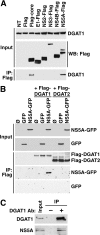
FIGURE 1.
FIGURE 1.
NS5A interacts specifically with DGAT1. A, shown is co-immunoprecipitation of endogenous DGAT1 with FLAG-tagged HCV proteins core, E1, NS2, NS3, NS4B, and NS5A, in Huh7 cells. After immunoprecipitation with α-FLAG M2 affinity gel, the endogenous DGAT1 and FLAG-tagged proteins were detected by Western blots (WB) with respective antibodies. The input control was 12% of the whole-cell lysate used for each immunoprecipitation. NT, not tranfected. B, shown is co-IP of NS5A-GFP in Huh7 cells transfected with expression vectors for FLAG-DGAT1, FLAG-DGAT2, NS5A-GFP, and GFP proteins. The input control was 15% of the whole-cell lysate used for each immunoprecipitation. C, co-IP of NS5A with endogenous DGAT1 from HCV Jc1-infected Huh7.5 is shown. Normal rabbit IgG (Invitrogen) was used as a negative control (−). The input control was 4% of the whole-cell lysate used for each immunoprecipitation.
FIGURE 2.
FIGURE 2.
DGAT1 is required for interaction of NS5A and core and forms a tripartite complex with these viral proteins. A, shown are sequential co-IP experiments of DGAT1, core, and NS5A. Top panel, 293T cells were transfected with plasmids expressing NS5A-GFP, FLAG-DGAT1, and HA-core. After 24 h, cells were lysed and subjected to Western blotting with α-GFP, α-core, and α-FLAG antibodies. Middle panel, immunoprecipitation was performed with α-HA antibody-conjugated agarose and subjected to Western blotting. Bottom panel, tandem immunoprecipitations were performed with α-FLAG M2 affinity gel and α-HA antibody-conjugated agarose. α-FLAG M2 affinity gel was eluted with FLAG peptide, and the eluates were incubated with α-HA antibody-conjugated agarose. Bound proteins were subjected to Western blotting. The input control was 12% of the whole-cell lysate used for the single immunoprecipitation and 6% of that used for the tandem immunoprecipitation. B, immunoprecipitation of HA core in shRNA-expressing Huh7 cells transfected with NS5A-GFP and HA-core is shown. Cell lysates and bound proteins were analyzed by Western blot with α-GFP, α-DGAT1, and α-core antibodies. NT, not transduced. The input control is 6% of the whole-cell lysate used for each immunoprecipitation. C, shown is real-time RT-PCR analysis of DGAT1 or DGAT2 mRNA expression levels in Huh7 cells transduced with the corresponding shRNAs. Data are the mean ± S.E.; n = 3 independent experiments.
FIGURE 3.
FIGURE 3.
Intracellular localization of HCV proteins expressed individually and validation of the DGAT1 inhibitors. A, shown are representative images of Huh7-Lunet cells transfected with individual FLAG-tagged HCV proteins (FLAG-core, E1-FLAG, E2-FLAG, p7-FLAG, NS2-FLAG, NS4A-FLAG, NS4B-FLAG, and NS5A-FLAG). Cells were fixed and stained using α-FLAG antibodies (green), LipidTox Red (LDs, red), and Hoechst (nucleus, blue) before imaging via epifluorescence microscopy (scale bar = 10 μm). B, shown is quantification of triglycerides extracted from shRNA-transduced and inhibitor-treated Huh7-Lunet cells previously depleted of LDs. Lunet cells were transduced with shRNAs targeting DGAT1, DGAT2, or luciferase and then treated sequentially with Triacsin C for 18 h and DGAT1 inhibitors for 24 h. After depletion of intracellular LDs by triacsin C treatment, DGAT1 is the most potent enzyme in synthesizing triglyceride using exogenous fatty acid sources (11, 35). Significant inhibition of this DGAT1 activity was observed with concentrations of 20 and 75 μm iA and iB, respectively. C, Huh7.5 cells transfected with HCV Luc-Jc1 viral RNA were treated with the DGAT1 inhibitors iA (20 μm) or iB (75 μm) or DMSO. After 48 h of treatment, viral particles secreted in the supernatants were used to infect naive Huh7.5 cells (Infected), and the producing cells were lysed (transfected). Shown are luciferase values expressed as percentage of DMSO control (mean ± S.E., n = 3). NT, not treated. D, shown is quantification of triglycerides extracted from hepatoma cells treated with DMSO, 20 μm iA, or 75 μm iB for 48 h or 2.5 mg/ml of oleic acid-albumin for 16 h. In normal conditions, DGAT2 can compensate for the loss of DGAT1 activity, preventing any change in the triglyceride content of the cells.
FIGURE 4.
FIGURE 4.
NS5A localization at LDs is dependent on DGAT1 activity. Shown are representative images (scale bar = 10 μm) and quantification of Huh7-Lunet cells transfected with HCV Jc1 RNA (A and B), NS5A-GFP expression vector (C and D), or NS5A-FLAG expression vector (E and F) and treated with DGAT1 inhibitors (iA at 20 μm or iB at 75 μm) or DMSO control for 48 h. Cells were fixed and stained with α-NS5A or α-FLAG antibodies (A and E, green), LipidTox Red (A and C) or far red (E) (LDs, red), and Hoechst (nucleus, blue). Green fluorescence in C arises directly from NS5A-GFP signal. Cells were analyzed by epifluorescence microscopy in A–D and by confocal microscopy in E–F. Colocalization of NS5A and LDs per cell was quantified by the automatic measurement program of the Zeiss axiovision software (mean of 40 cells ± S.E.) (B, D, and F).
FIGURE 5.
FIGURE 5.
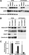
NS5A association with LDs is dependent on DGAT1 activity. A, shown are Western blots of cell extracts (left panels) or isolated LD fractions (right panels) from Huh7-Lunet cells transfected with NS5A-GFP and incubated with oleate, DMSO, 20 μm DGAT1 inhibitor iA described in Ref. (16), 75 μm DGAT1 inhibitor iB (TOCRIS Bioscience). NT, not transfected. B, shown are Western blots analysis of cell extracts (left panels) or isolated LD fractions (right panels) from shRNA-expressing Huh7-Lunet cells. C, shown is real-time RT-PCR analysis of DGAT1 or DGAT2 mRNA expression levels in Huh7-Lunet cells transduced with the corresponding shRNAs (mean ± S.E.; n = 3 independent experiments).
FIGURE 6.
FIGURE 6.
Overexpression of DGAT1 catalytically inactive mutant suppresses NS5A trafficking to LDs. A, shown is co-IP of NS5A-GFP with FLAG-DGAT1, FLAG-DGAT1-H426A, or FLAG-DGAT2 in Huh7 cells (Ø, empty vector). The input control is ∼15% of the whole-cell lysate used for each immunoprecipitation. B, shown are Western blots of the LD fraction from NS5A-GFP-transfected Huh7-Lunet cells expressing FLAG-DGAT1, FLAG-DGAT1-H426A, or FLAG-DGAT2. C, shown are Western blots of the LD fraction purified from FLAG-core-transfected Huh7-Lunet cells expressing FLAG-DGAT1, FLAG-DGAT1-H426A, or FLAG-DGAT2. D and E, Huh7-Lunet cells were cotransfected with an expression vector for FLAG-DGAT1 wild type or FLAG-DGAT1-H426A, and NS5A-GFP or HA core. Cells were fixed and stained using α-FLAG and α-core antibodies, LipidTox Red (LDs) and Hoechst (nucleus) before epifluorescence microscopy (scale bar = 10 μm).
FIGURE 7.
FIGURE 7.
Overexpression of DGAT1 catalytically inactive mutant inhibits viral replication. A, shown are infectious titers released from Huh7.5 cells transfected with HCV Luc-Jc1 viral RNA and vectors expressing wild-type FLAG-DGAT1 or mutant FLAG-DGAT1-H426A or with an empty control vector. Naive Huh7.5 cells were infected with cell supernatants of transfected cells and lysed 48 h after infection to analyze luciferase activity. The relative light units (RLU) are expressed as percentage relative to the empty vector-transfected control; mean ± S.E.; n = 4; *, p < 0.0002). B, shown are Western blots of cell extracts, lysed on day 6 after HCV RNA transfection.
FIGURE 8.
FIGURE 8.
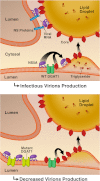
Model of HCV NS5A and core recruitment to DGAT1-generated lipid droplets. DGAT1 interacts with core and NS5A proteins and co-loads them onto DGAT1-generated LDs. This process is crucial for assembly of HCV virions. When NS5A access to LDs is dominantly suppressed by overexpressing a catalytically inactive mutant of DGAT1, infectious particles cannot form despite the presence of core around LDs (right panel).

References

    1. Wasley A., Alter M. J. (2000) Epidemiology of hepatitis C. Geographic differences and temporal trends. Semin. Liver Dis. 20, 1–16 - PubMed
    1. Feld J. J., Hoofnagle J. H. (2005) Mechanism of action of interferon and ribavirin in treatment of hepatitis C. Nature 436, 967–972 - PubMed
    1. Masaki T., Suzuki R., Murakami K., Aizaki H., Ishii K., Murayama A., Date T., Matsuura Y., Miyamura T., Wakita T., Suzuki T. (2008) Interaction of hepatitis C virus nonstructural protein 5A with core protein is critical for the production of infectious virus particles. J. Virol. 82, 7964–7976 - PMC - PubMed
    1. Blight K. J., Kolykhalov A. A., Rice C. M. (2000) Efficient initiation of HCV RNA replication in cell culture. Science 290, 1972–1974 - PubMed
    1. Miyanari Y., Atsuzawa K., Usuda N., Watashi K., Hishiki T., Zayas M., Bartenschlager R., Wakita T., Hijikata M., Shimotohno K. (2007) The lipid droplet is an important organelle for hepatitis C virus production. Nat. Cell Biol. 9, 1089–1097 - PubMed

Publication types

MeSH terms