. 2013 Feb 18;200(4):493-504.
doi: 10.1083/jcb.201204106.

Grb2 controls phosphorylation of FGFR2 by inhibiting receptor kinase and Shp2 phosphatase activity

Affiliations

Grb2 controls phosphorylation of FGFR2 by inhibiting receptor kinase and Shp2 phosphatase activity

Zamal Ahmed et al. J Cell Biol. .

Abstract

Constitutive receptor tyrosine kinase phosphorylation requires regulation of kinase and phosphatase activity to prevent aberrant signal transduction. A dynamic mechanism is described here in which the adaptor protein, growth factor receptor-bound protein 2 (Grb2), controls fibroblast growth factor receptor 2 (FGFR2) signaling by regulating receptor kinase and SH2 domain-containing protein tyrosine phosphatase 2 (Shp2) phosphatase activity in the absence of extracellular stimulation. FGFR2 cycles between its kinase-active, partially phosphorylated, nonsignaling state and its Shp2-dephosphorylated state. Concurrently, Shp2 cycles between its FGFR2-phosphorylated and dephosphorylated forms. Both reciprocal activities of FGFR2 and Shp2 were inhibited by binding of Grb2 to the receptor. Phosphorylation of Grb2 by FGFR2 abrogated its binding to the receptor, resulting in up-regulation of both FGFR2's kinase and Shp2's phosphatase activity. Dephosphorylation of Grb2 by Shp2 rescued the FGFR2-Grb2 complex. This cycling of enzymatic activity results in a homeostatic, signaling-incompetent state. Growth factor binding perturbs this background cycling, promoting increased FGFR2 phosphorylation and kinase activity, Grb2 dissociation, and downstream signaling. Grb2 therefore exerts constitutive control over the mutually dependent activities of FGFR2 and Shp2.


Figures

Figure 1.
Figure 1.
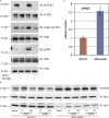
Knockdown of Grb2 and Shp2 reveal that the level of WTFGFR2 phosphorylation is controlled by Grb2. (A) Total HEK293T cell lysates were immunoblotted with anti-pFGFR antibody (top), and reprobed for total FGFR (middle) and Grb2 (bottom). Anti-pFGFR2 antibody is specific for A loop residues Y653 and Y654. (B) Cell lysates of overnight serum-staved stable A431 cells containing control shRNA (Ci), Grb2-shRNA (Grb2i), or Shp2-shRNA (Shp2i) were analyzed for FGFR2 phosphorylation as above. Only the nonstimulated state is shown (i.e., each lane is duplicated). Numbers on pFGFR2 panel are normalized intensity pFGFR2/total FGFR2. (C) Analysis of FGFR2 phosphorylation in Rat-1 fibroblast cells with control shRNA (Ci) and Grb2-shRNA (Grb2i) as above. Only the nonstimulated state was investigated (i.e., each lane is duplicated). (D) Inhibition of Shp2 in Grb2 knockdown cells restores basal receptor phosphorylation. Serum-starved WT-Ci and WT-Grb2i cells were incubated with 50 µM NSC87877 for 4 h and the resultant cell lysates were analyzed by Western blotting with anti-pFGFR2 antibody, Shp2 pY542-specific antibody, and anti-Grb2 antibody. The immunoblot was stripped and reprobed for total FGFR2 and Shp2 as the loading control. The numbers on the pFGFR2 panel represent normalized intensity pFGFR2/total FGFR2.
Figure 2.
Figure 2.
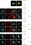
Grb2 inhibits the interaction of Shp2 with FGFR2. (A) FLIM analysis of the FRET between the FGFR2-GFP and RFP-Shp2. In the control (WT-Ci) serum-starved cells no interaction between FGFR2 and Shp2 was observed in the basal state. The mean FRET lifetime is ∼2.0 ns (line in right-hand panel), which corresponds to the mean lifetime for isolated GFP. No apparent interaction between FGFR2 and Shp2 in WT-Grb2i cells (B), or WT-Ci cells transfected with the RFP-tagged substrate-trapping C459S Shp2 mutant (C). Interaction between FGFR2 and Shp2 is observed in the Grb2i the substrate-trapping C459S mutant (D). Stimulating cells that contain WTShp2 or C459S mutant Shp2 with FGF9 results in clear binding between FGFR2 and Shp2 after 15 min (E and F), respectively. Bar, 10 µm.
Figure 3.
Figure 3.
Shp2 phosphorylation by FGFR2 is inhibited by Grb2. (A) Serum-starved HEK293T cells were incubated with 30 µM FGFR inhibitor (SU5402) for 2 h and then either stimulated with 10 ng/ml FGF9 for 15 min or left untreated. Cell lysates were prepared and analyzed by Western blotting with the indicated antibody. Anti-pFGFR and anti-Y542 on Shp2 antibodies were used to evaluate phosphorylation of proteins. The immunoblot was stripped and reprobed with a pan-antibody to determine total protein level. (B) Comparison of ligand-stimulated Shp2 phosphorylation between A431-Ci and A431-Grb2i cells in nonstimulated and on stimulation by FGF2 or FGF9 for 1 h. Shp2 phosphorylation was detected with anti-pY542 antibody (top). The immunoblot was reprobed for total Shp2 as a loading control (middle) and with Grb2 (bottom). (C) Densitometric quantification of basal state Shp2 phosphorylation levels in A431 cells in control shRNA (A431-Ci) and Grb2-shRNA (A431-Grb2i). Error bars represent SD, n = 7.
Figure 4.
Figure 4.
Shp2 dephosphorylates Grb2. (A) Wild-type or FGFR2 stably transfected HEK293T were starved overnight, then stimulated using 10 ng/ml FGF9 for either 15 or 60 min. Cells were lysed in the presence of protease and phosphatase inhibitors. 50 µg of total cell lysate were used for immunoblotting studies. Phosphorylation of FGFR2 was examined using anti-pFGFR2 (first panel). To examine the Grb2 phosphorylation states in the absence or presence of FGFR2 expression, 1 mg of total cell lysates were used for immunoprecipitation using an anti-Grb2 antibody, and probed with an anti-Grb2 antibody. The immunoprecipitated Grb2 from FGFR2-overexpressing cells show multiple bands (both serum starved and FGF9 stimulated), suggesting the high molecular weight species is tyrosine-phosphorylated Grb2, which is only phosphorylated in the presence of FGFR2. (B) Recombinant Grb2 C-SH3 mutants (Y160F, left; Y209F, right) were expressed and purified from E. coli and incubated with pure FGFR2 cytoplasmic domain in a 1:1 molar ratio in the presence of ATP and MgCl2 at room temperature for 1 h. Recombinant GST-fused pShp2 was obtained via the same protocol. A general anti-pY antibody was used to examine the phosphorylation state of FGFR2-phosphorylated Shp2 (lanes 4, 6, 10, and 12; panel 1) and Grb2 C-SH3 domains (lanes 2 and 8; panel 4). A specific anti-pY542 Shp2 antibody was also used to confirm that Y542 of Shp2 is phosphorylated. A pool of both proteins was dephosphorylated by mixing phosphatase (either pShp2 or Shp2) with phosphorylated protein substrates (either pGrb2 C-SH3 Y160F or phospho-Grb2 C-SH3 Y209F) at 4°C overnight. The anti-pY blot shows only the pGrb2 C-SH3 Y160F can be dephosphorylated by both pShp2 and Shp2 (lanes 5 and 6; panel 4). However, the phosphorylation state of pGrb2 C-SH3 Y209F is not affected by Shp2, suggesting that the Y209 is the target of Shp2. A total Shp2 antibody (panel 3) and total Grb2 antibody (panel 5) were used to confirm equal protein loading. (C) HEK293T cells were cotransfected with FGFR2-GFP and Grb2-strep-tag. After 48 h cells were starved for 4 h and incubated with either FGFR-specific inhibitor (50 µM SU5402) or Shp2-specific inhibitor (100 µM NSC87877) for 1 h. Cell lysates were subjected to affinity purification using strep-tactin agarose beads and immunoblotted with anti-pY antibody (top) followed by anti-Grb2 antibody (bottom).
Figure 5.
Figure 5.
Interaction between Grb2 and Shp2. CFP-Grb2 and RFP-Shp2 colocalization and direct interaction measurement using FLIM in A431 cells. (A) Control lifetime measurement for CFP alone. (B) Interaction of CFP-Grb2 with RFP-tagged wild-type Shp2 (RFP-WTShp2) at basal and after 20 ng/ml FGF9 stimulation. (C) Co-localization and direct interaction of Y542F mutant Shp2 with CFP-Grb2 at basal and after FGF9 stimulation. (D) Constitutive interaction of the C459S substrate-trapping Shp2 mutant with Grb2. A left-shifted peak relative to the line drawn along 2.2 ns indicates a binding. A peak centered on the 2.2 ns line indicates nonbinding. Bar, 20 µM.
Figure 6.
Figure 6.
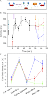
In vitro demonstration of catalytic cycling of FGFR2 and Shp2 in the presence of Grb2. (A) Schematic of interactions performed in vitro to demonstrate catalytic activity of FGFR2 and Shp2 on Grb2. Mixing FGFR2cyto (blue) with Grb2 (red) promotes the formation of a heterotetrameric complex (Lin et al., 2012). Addition of ATP and MgCl2 to this results in phosphorylation of FGFR2 and Grb2 (green circle). Addition of Shp2 (orange) results in dephosphorylation of FGFR2 and Grb2 (blue line). The heterotetrameric complex is recovered under these conditions. (B) Fluorescence lifetime measurement between GFP-FGFR2cyto and RFP-Grb2 as a function of time. The first point corresponds to the fluorescence lifetime for isolated GFP-FGFR2 (black arrow). On addition of Grb2 (red arrow) a heterotetrameric complex between Grb2 and FGFR2 forms. This results in FRET between the GFP and RFP and the concomitant reduction in fluorescence lifetime. On addition of ATP/Mg2+ (purple arrow) up-regulation of the RTK ensues and Y209 on Grb2 becomes phosphorylated and the FGFR2–Grb2 complex dissociates. The lifetime increases, reflecting reduction in complex concentration and the accumulation of pGrb2. After 80 min Shp2 was added (orange arrow). At this point clear reassociation of Grb2 and FGFR2 is observed as Grb2 is dephosphorylated in the presence of Shp2 and consequently the fluorescence lifetime decreases (blue line on graph). Replacing WTShp2 with the Y542F (red line) or C459S (green line) mutant results in no immediate reduction in lifetime, confirming that the FGFR2–Grb2 complex is not rescued by adding these compromised phosphatases. (C) Measurement of FRET between GFP-FGFR2cyto (Cyto) and RFP-Grb2 in solution using FLIM. Cyto alone is GFP-FGFR2cyto and represents the background false-positive percentage FRET readout. Cyto+Grb2 is the population of molecules undergoing FRET when RFP-Grb2 is present. Cyto+Grb2+ATP is the population of GFP-FGFR2cyto undergoing FRET with RFP-Grb2 when the FGFR2 kinase was activated. Shp2 30 min and 18 h represent the reestablishment of GFP-FGFR2cyto/RFP-Grb2 complex in the presence of wild-type (blue line), Y542F (red line), and C459S (green line) mutant Shp2 as a function of time.
Figure 7.
Figure 7.
Grb2 inhibits Shp2 activity toward FGFR2. (A) WT-Ci and WT-Grb2i cells were incubated with serum-free media with or without 50 µM Shp2 inhibitor NSC87877 overnight, and then either stimulated with 10 ng/ml FGF9 for 15 min or left untreated. Total cell lysates were analyzed by Western blotting with the indicated antibody. (B and C) Densitometric quantification of bands from experiments as described in A. Histogram values correspond to normalized bands for pFGFR2 against total FGFR2 (B) and pErk against total Erk (C) of three independent experiments. Error bars represent SD. (D) A431-Ci and A431-Grb2i cells were serum starved with 50 µM Shp2 inhibitor overnight, then either stimulated with FGF9 or left untreated. 50 µg total cell lysates were immunoblotted with indicated antibody. (E) Densitometric quantification of bands from experiments as described in D, where the ratio of pERK/total Erk is plotted from three independent experiments. Error bars represent SD. The A431-Ci FGF9/NSC87877 ratio was fixed as 1.0 for each experiment. Arrows highlight the comparison of the level of pErk in the control cells after FGF-ligand stimulation and the recovery of this level in the Grb2 knockdown cells only when stimulated in the presence of Shp2 inhibitor. (F) Shp2 inhibition does not affect EGF-stimulated MAP kinase response in A431 cells. The experimental procedure is as above except 50 ng/ml EGF was used to stimulate cells for 5 min.
Figure 8.
Figure 8.
Schematic diagram of cycle of enzymatic activity under the control of Grb2 in the absence of extracellular stimulation. (A) FGFR2 (blue) is stabilized in a basally phosphorylated state in the form of a heterotetramer in which a dimer of Grb2 recruits two receptor molecules. In this complex the receptors are able to autophosphorylate the activation loop tyrosines. The partially phosphorylated, nonsignaling state is represented by inclusion of the green circle with dashed border. (B) Active Shp2 is able to dephopshorylate FGFR2. This phosphatase activity is inhibited by Grb2 (red oval) when it is bound to the receptor. (C) Basally activated FGFR2 is able to phosphorylate Shp2 phosphatase (orange). The phosphorylation of Shp2 is represented by a solid green line. On phosphorylation of Y542 Shp2 is enhanced. This catalytic activity is inhibited in the presence of Grb2 bound to FGFR2. The phosphorylated Shp2 is represented by inclusion of the green circle. (D) Grb2 is phosphorylated by FGFR2. In this phosphorylated state Grb2 is no longer able to bind to the receptor and hence its inhibitory properties are lost. (E) Shp2 is able to dephosphorylate Grb2. This restores the adaptor protein to a state competent of binding FGFR2. Key: straight lines between proteins represent the change from one state of that protein to another. Green lines, phosphorylation. Blue lines, dephosphorylation. Green dashed line, autophosphorylation. Red lines, inhibition. Curved arrows, enzymatic activity, e.g., blue line from Shp2 intercepting dephosphorylation blue line between pFGFR and FGFR indicates that Shp2 is the active enzyme for that change of state.

References

    1. Agazie Y.M., Hayman M.J. 2003. Development of an efficient “substrate-trapping” mutant of Src homology phosphotyrosine phosphatase 2 and identification of the epidermal growth factor receptor, Gab1, and three other proteins as target substrates. J. Biol. Chem. 278:13952–13958 10.1074/jbc.M210670200 - DOI - PubMed
    1. Ahmed Z., Schüller A.C., Suhling K., Tregidgo C., Ladbury J.E. 2008. Extracellular point mutations in FGFR2 elicit unexpected changes in intracellular signalling. Biochem. J. 413:37–49 10.1042/BJ20071594 - DOI - PubMed
    1. Ahmed Z., George R., Lin C.C., Suen K.M., Levitt J.A., Suhling K., Ladbury J.E. 2010. Direct binding of Grb2 SH3 domain to FGFR2 regulates SHP2 function. Cell. Signal. 22:23–33 10.1016/j.cellsig.2009.08.011 - DOI - PubMed
    1. Araki T., Nawa H., Neel B.G. 2003. Tyrosyl phosphorylation of Shp2 is required for normal ERK activation in response to some, but not all, growth factors. J. Biol. Chem. 278:41677–41684 10.1074/jbc.M306461200 - DOI - PubMed
    1. Barford D., Neel B.G. 1998. Revealing mechanisms for SH2 domain mediated regulation of the protein tyrosine phosphatase SHP-2. Structure. 6:249–254 10.1016/S0969-2126(98)00027-6 - DOI - PubMed

Publication types

MeSH terms

Substances